




Abstract
Red and white wine polyphenols have been reported to provide substantial health benefits. In this study, the cytotoxic activity of red and white wine polyphenolic extracts and of resveratrol was evaluated against different cancer cell lines—human cervix adenocarcinoma HeLa, human breast adenocarcinoma MDA-MB-361, and human breast carcinoma MDA-MB-453—and normal human peripheral blood mononuclear cells (PBMCs). Qualitative and quantitative compositions of wine polyphenolic extracts obtained by fractional vacuum distillation of corresponding wines were determined using spectrophotometric methods and high-performance liquid chromatography with diode array detection and liquid chromatography with electrospray ionization-time of flight mass spectrometry analysis. It was demonstrated that wine polyphenolic extracts and resveratrol exerted higher cytotoxic activity against HeLa and MDA-MB-453 cells in comparison to MDA-MB-361 cells and unstimulated and stimulated PBMCs. Furthermore, white wine polyphenolic extract exhibited a significantly higher antiproliferative action on cancer cell lines than red wine extract. The presence of condensed or fragmented nuclei in HeLa cells, pretreated with extract of white wine and stained with a mixture of acridine orange and ethidium bromide, pointed to the morphological signs of apoptosis. In addition, HeLa cells in late stages of apoptosis or secondary necrosis were also observed. Results from our study suggest that polyphenolic extracts from red and white wine may have anticarcinogenic potential.
Introduction
C
Among the various fruits, berries and grapes contain a variety of bioactive phytochemicals, which may provide substantial health benefits. 2 –6 Polyphenols are prominent dietary constituents of fruits. The grape is one of the fruits with the highest content of phenolic antioxidants, which are present in skin, pulp, and seeds. Grape polyphenols are partially extracted during the winemaking procedures, and for that reason, wine is considered to be one of the richest sources of bioactive polyphenols. 7,8 Phenolic compounds from wine could be classified into two groups: flavonoids (flavanols, flavonols, and anthocyans) and non-flavonoids (phenolic acids and stilbenes). 7 –9 It is well established that specific classes of polyphenols determine the special properties of specific grape varieties and the corresponding wines. 8 There are essential differences in qualitative and quantitative compositions of red and white wines, which could be attributed to the phytochemical composition of specific grape varieties, environmental factors, and winemaking techniques. 10 –14
The anticarcinogenic potential of wine polyphenolic extracts and their individual phenolic constituents is based on their antiproliferative activity and ability to induce apoptosis in various cancer cell lines. Wine polyphenols scavenge free radicals and thus reduce and repair oxidative damage of DNA, proteins, and lipids. Phenolic compounds also regulate carcinogen-activating and -detoxifying enzymes, cytokines involved in inflammatory reactions, and growth and transcription factors, as well as molecular targets of intracellular signaling pathways of cell growth, proliferation, and apoptosis. 3,15
One of the most extensively studied polyphenols is resveratrol (trans-3,4′,5-trihydroxystilbene), a natural phytoalexin found in berries, grapes, red and white wines, peanuts, and polygonium roots. Resveratrol has been shown to exhibit several health beneficial effects, including antioxidant and pro-oxidant, antimutagen, anti-inflammatory, cardioprotective, and anticarcinogenic activities. It is considered that the antioxidant activity of resveratrol plays a crucial role in chemoprevention, inhibiting the initiation of malignant transformation. In addition, this polyphenol exerts an inhibitory effect on promotion and progression of cancer. Results from numerous in vitro and in vivo studies have demonstrated the cancer chemopreventive and therapeutic potential of resveratrol. 16,17
The aim of the present research was to determine the cytotoxic activity of red and white wine extracts rich in polyphenols against various cancer cell lines. In order to assess the sensitivity of healthy human immunocompetent cells, which are known to be included in the antitumor immunity, the cytotoxic effect on normal PBMCs was also tested. The cytotoxic action of resveratrol was investigated as well. Furthermore, red and white wine polyphenolic extracts were characterized in order to elucidate the role of chemical composition in the observed differences in cytotoxic effects.
Materials and Methods
Red and white wine polyphenolic extracts
Red and white wine polyphenolic extracts were obtained by fractional vacuum distillation of corresponding wines. The wines used were Vranac red wine (Serbia) and Smederevka white wine (Serbia).
Characterization of red and white wine polyphenolic extracts
Determination of total phenolic content
The total phenolic contents of the wine extracts investigated were determined according to the Folin-Ciocalteu method. 18 In brief, 200 μL of each polyphenolic extract (diluted 1:5 with distilled water) was added to a volume of 1:10 diluted Folin-Ciocalteu reagent and maked up 1 mL. After 4 minutes, 800 μL of sodium carbonate (75 g/L) was added in the reaction mixture, which was incubated for 2 hours at room temperature. Absorbance (A) was measured at the wavelength of 765 nm. Gallic acid was used as a calibration standard, and results were expressed as milligrams of gallic acid equivalents/100 mL of extracts. The measurements were repeated three times, and values were averaged.
Determination of total anthocyanin content
Total anthocyanin content was determined according to the procedure in the European Pharmacopoeia 6.0, 19 with slight modifications. In brief, 95 mL of methanol was added to 5 g of extracts, and the mixture was mechanically stirred for 30 minutes and then filtered into a 100-mL volumetric flask. The filter was rinsed, and the mixture was diluted to 100 mL with methanol. A 50-fold dilution of this solution in a 0.1% (vol/vol) solution of hydrochloric acid in methanol was prepared. The A value of the solution was measured at 528 nm, using a 0.1% (vol/vol) solution of hydrochloric acid in methanol as the compensation liquid.
The percentage content of anthocyanins, expressed as cyanidin-3-glucoside chloride, was calculated from the following formula: (A × 5,000/718) × m, where A is the A value at 528 nm, 718 is the specific A value of cyanidin-3-glucoside chloride at 528 nm, and m is the mass of the substance to be examined (in g).
Determination of gallic acid content
Red and white wine extracts were analyzed by high-performance liquid chromatography for gallic acid content. Samples were diluted 1:10 with distilled water and filtered prior to injection using cellulose filters (pore size, 0.45 μm). Analyses were carried out on an HP 1090 liquid chromatograph (Agilent Technologies, Palo Alto, CA, USA) with a diode-array detector, on a reverse-phase Zorbax SB-C18 analytical column (150 × 4.6 mm; particle size, 5 μm; Agilent Technologies). The flow rate was 1.0 mL/minute. The mobile phases were acetonitrile (phase A) and H2O containing 1% H3PO4 (phase B). Elution was performed according to the following scheme: 95% B, 0–3 minutes; 95–0% B, 3–20 minutes. Detection was carried out at 255 nm. An external calibration method using gallic acid as the standard was used. Triplicate measurements were taken, and mean values were calculated.
Liquid chromatography/mass spectrometry analysis
Liquid chromatography/mass spectrometry analysis was performed on an Agilent Technologies MSD time of flight instrument coupled to an Agilent 1200 series high-performance liquid chromatograph, using an RR Zorbax Eclipse Plus C18 column (particle size, 1.8 μm; 150 × 4.6 mm). Mobile phase A was 0.2% formic acid in water, and mobile phase B was acetonitrile. The injection volume was 1 μL, and elution rate was set at 0.95 mL/minute with a gradient program as follows: 0–20 minutes, 5–16% B; 20–28 minutes, 16–40% B; 28–32 minutes, 40–70% B; 32–36 minutes, 70–99% B; 36–45 minutes, 99% B; 45–46 minutes, 99–5% B. Ultraviolet-visible detection was carried out at 280 and 254 nm. Mass spectra were acquired using an Agilent electrospray ionization MSD time of flight instrument. Drying gas (N2) flow rate was 12 L/minute, nebulizer pressure was 45 psig, and drying gas temperature was 350°C. For electrospray ionization analysis, the parameters were as follows: capillary voltage, 4,000 V; fragmentor, 140 V; skimmer, 60 V; and Oct RF V 250 V, for negative modes. The mass range was from 100 to 2,000 m/z. Data processing was carried out using Molecular Feature Extractor and Mass Profiler software (Agilent).
Cell culture
Human cervix adenocarcinoma HeLa, human breast adenocarcinoma MDA-MB-361, and human breast carcinoma MDA-MB-453 cells were cultured as monolayers in nutrient medium, which consisted of RPMI 1640 medium, supplemented with
Preparation of PBMCs
PBMCs were separated from whole heparinized blood of two healthy volunteers by Lymphoprep™ (Nycomed, Oslo, Norway) gradient centrifugation. Interface cells, washed three times with Haemacel® (TheraSelect GmbH, Marburg, Germany) (aqueous solution supplemented with 145 mM Na+, 5.1 mM K+, 6.2 mM Ca2+, 145 mM Cl-, and 35 g/L gelatin polymers, pH 7.4), were counted and resuspended in nutrient medium.
Treatment of cancer cell lines
HeLa (2,000 cells per well), MDA-MB-361 (10,000 cells per well), and MDA-MB-453 (3,000 cells per well) cells were seeded into 96-well microtiter plates, and 20 hours later, after cell adherence, five different concentrations of the investigated agents were added to the wells. Only nutrient medium was added to the cells in the control wells. Stock solutions of wine polyphenolic extracts were diluted with nutrient medium and applied to target cells to various final concentrations, ranging from 0.0625% to 1% for the white wine extract and from 0.125% to 3% for the red wine extract. Stock solution of resveratrol was made at a concentration of 10 mM in dimethyl sulfoxide, and this solution was diluted with nutrient medium. Final concentrations of resveratrol ranged from 6.25 μM to 100 μM. Resveratrol (>99%) was purchased from Sigma-Aldrich (St. Louis, MO, USA). All experiments were done in triplicate.
Treatment of PBMCs
PBMCs (150,000 cells per well) were seeded into nutrient medium or in nutrient medium enriched with phytohemagglutinin (PHA) (5 μg/mL) in 96-well microtiter plates. After 2 hours, the investigated agents were added to the wells, in triplicate, to five final concentrations, except to the control wells where nutrient medium only was added to the cells. Final concentrations of the investigated agents were the same for PBMCs as for the cancer cell lines. However, the maximal applied final concentration of white wine extract was 2%.
Determination of target cell survival
Survival of target cancer cells was determined indirectly by measuring total cellular protein using the Kenacid Blue Dye binding (KBR) test. After 72 hours of continuous action of investigated agents, medium was discarded, and target cells were washed twice with warm (37°C) phosphate-buffered saline. Cancer cells were always centrifuged for 10 minutes at 2,000 rpm (centrifuge from Tehtnica, Zelezniki, Slovenia), and supernatant was aspirated, leaving a small amount of medium in order to avoid cell disturbance in the pellet. Then target cells were fixed for 20 minutes with 150 μL of a mixture of methanol and acetic acid (3:1 vol/vol), stained for 2–3 hours with 0.04% Coomassie Brilliant Blue R-250 in 25% ethanol and 12% glacial acetic acid, and then washed. The bound dye was dissolved in desorbing solution (1 M potassium acetate, 70% ethanol). The A value at 570 nm was measured 2 hours later.
In comparison to the proliferation of cancer cells and of PBMCs stimulated for propagation by PHA, proliferation of nonstimulated PBMCs during the experiment was shown to be negligible. With the aim of enhancing the accuracy of data on PBMCs survival, which if obtained by KBR test could be slightly inadequate, because the KBR test is based on the determination of total protein content in live and in dead cells in the samples and because there is a small possibility that dead cells might not be totally detached from the wells during performance of the KBR test, we measured the decrease in survival of target PBMCs 72 hours after the addition of the investigated compounds by the 3-(4,5-dimethylthiazol-2-yl)-2,5-diphenyltetrazolium bromide (MTT) test, which is based on the determination of only live cells, and it is accepted as one of the main tests for measuring cell survival. In brief, the medium was discarded, and PBMCs were washed twice with warm phosphate-buffered saline. PBMCs were always centrifuged. Fresh nutrient medium was added to the wells, and PBMCs were incubated for at least 40 minutes at 37°C in a humidified atmosphere with 5% CO2. After this procedure, 20 μL of MTT solution (5 mg/mL in phosphate-buffered saline) was added to each well. Samples were incubated for a further 4 hours, and then 100 μL of 10% sodium dodecyl sulfate was added to the wells. The A value at 570 nm was measured the next day.
To calculate cell survival (percentage), the A value of a sample with cells grown in the presence of various concentrations of the investigated agents was divided with control optical density (the A value of control cells grown only in the nutrient medium) and multiplied by 100. It was implied that the A value of the blank was always subtracted from the A value of the corresponding sample with target cells. The IC50 value was defined as the concentration of an agent inhibiting cell survival by 50%, compared to a vehicle-treated control.
Morphological evaluation of HeLa cell death
In order to evaluate whether red and white wine polyphenolic extracts could also exert pro-apoptotic activity in HeLa cells, morphological analysis by microscopic examination of acridine orange/ethidium bromide-stained cells was performed. HeLa cells were seeded overnight on coverslips (100,000 cells) in 2 mL of complete medium. The next day cells were treated with the investigated wine extracts for 24 and 48 hours. Applied concentrations corresponded to double IC50 values, which were obtained after 72 hours of continuous action of extracts. After this period, the target cells were stained with 18 μL of a mixture of the DNA dyes acridine orange and ethidium bromide (3 mg/mL acridine orange and 10 mg/mL ethidium bromide in phosphate-buffered saline) and visualized under a fluorescence microscope using an fluorescin isothiocyanate filter set.
Results
Chemical characterization of red and white wine polyphenolic extracts
Qualitative and quantitative compositions of red and white wine polyphenolic extracts affect their antioxidant activity, which might be related to the cytotoxic activity of the extracts. In general, there is a direct correlation between phenolic content and antioxidant activity. 20 –22 Therefore, the total phenolic contents of the wine extracts were determined, and these results are presented in Table 1.
GAE, gallic acid equivalents.
Total phenolic content is higher in red wine extract than in white wine extract. This observation indicates that this extract probably has a higher antioxidant potential. It is noteworthy that each polyphenolic constituent of the extract has a specific antioxidant capacity, which is structure dependent. Determination of the antioxidant capacity of extracts should consider the concentrations and composition of various phenolic antioxidants. Levels of total anthocyanins are significantly higher in red wine extract (Table 1). In contrast, anthocyanins were found in traces in white wine extract, as was expected. However, no anthocyanins were detected in most of the white wines examined. 10,23
Liquid chromatography-mass spectrometry analysis revealed considerable differences in phenolic composition between the red and the white wine extracts (Table 2). Red wine extract contains predominantly gallic acid (2.24 mg/mL), caftaric acid, coutaric acid, and syringic acid, whereas white wine extract contains caftaric acid, coutaric acid, caffeic acid, and fertaric acid. It is well known that white wines exhibit lower polyphenol concentration and lower antioxidant capacity in comparison to red wines. Some data indicate that hydroxycinnamic acids and their esters with tartaric acid, found in abundance in investigated wine polyphenolic extracts, are dominant non-flavonoids in red wines and also dominant phenolic compounds in white wines. 12 It should be stressed that phenolic compounds from wine are labile substances that can undergo oxidation, condensation, and polymerization reactions, responsible for wine color changes during storage and aging. 24
Quantification was done using high-performance liquid chromatography.
tR, retention time; DAD, diode array detection; MS, mass spectrometry; R, red wine; W, white wine; sh, shoulder.
In vitro cytotoxic activity
On cancer cell lines
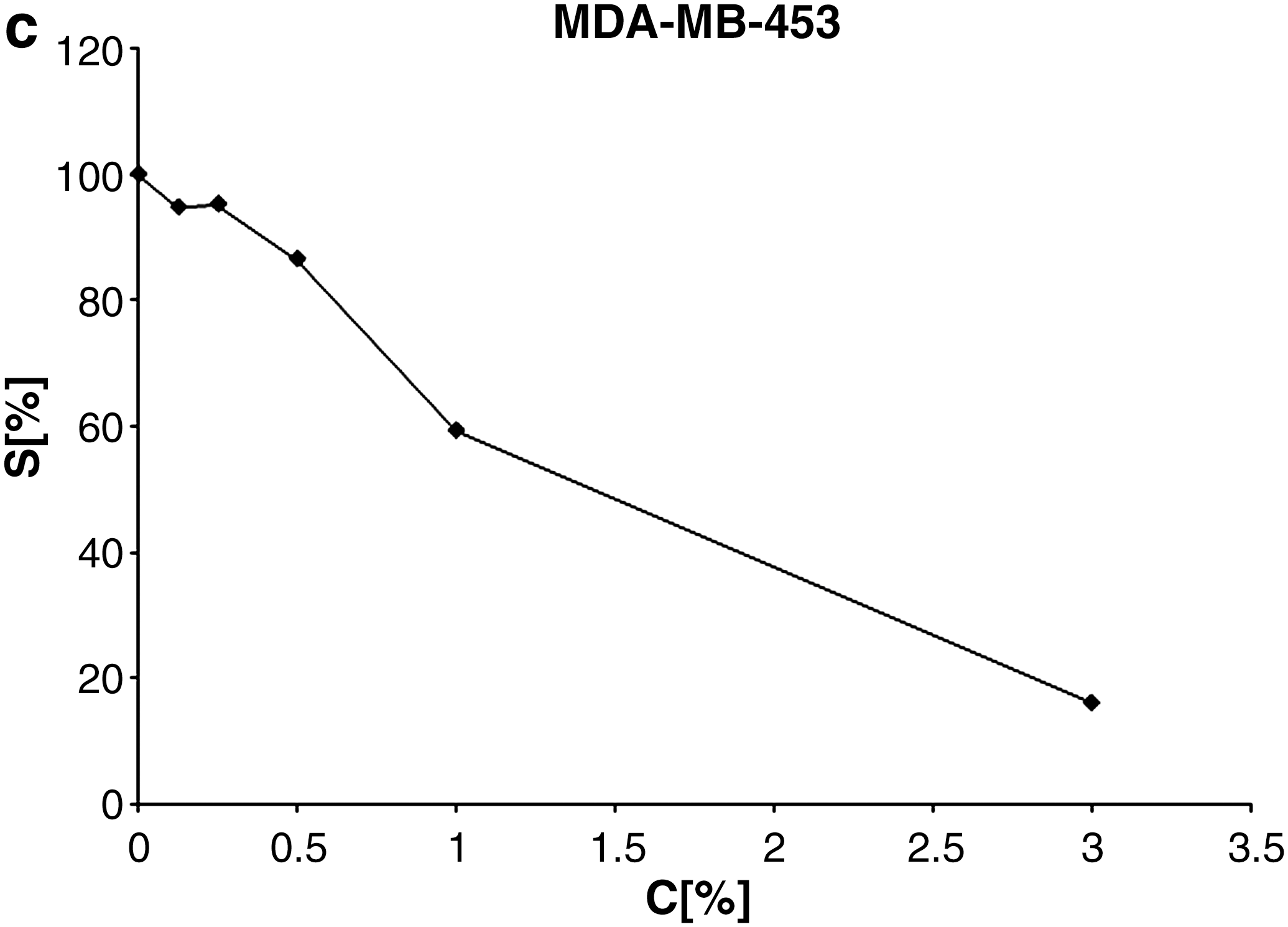
In vitro cytotoxic action was determined against malignant cell lines: human cervix adenocarcinoma HeLa, human breast adenocarcinoma MDA-MB-361 and human breast carcinoma MDA-MB-453 cells. Results from this study show that red and white wine polyphenolic extracts and resveratrol exert a cytotoxic effect on human malignant cell lines. However, cytotoxicities of these cancer-suppressive agents are significantly different. The decrease in cancer cell survival induced by wine polyphenolic extracts and the polyphenolic antioxidant resveratrol is presented in Figures 1 –4 and Table 3.

Survival of (

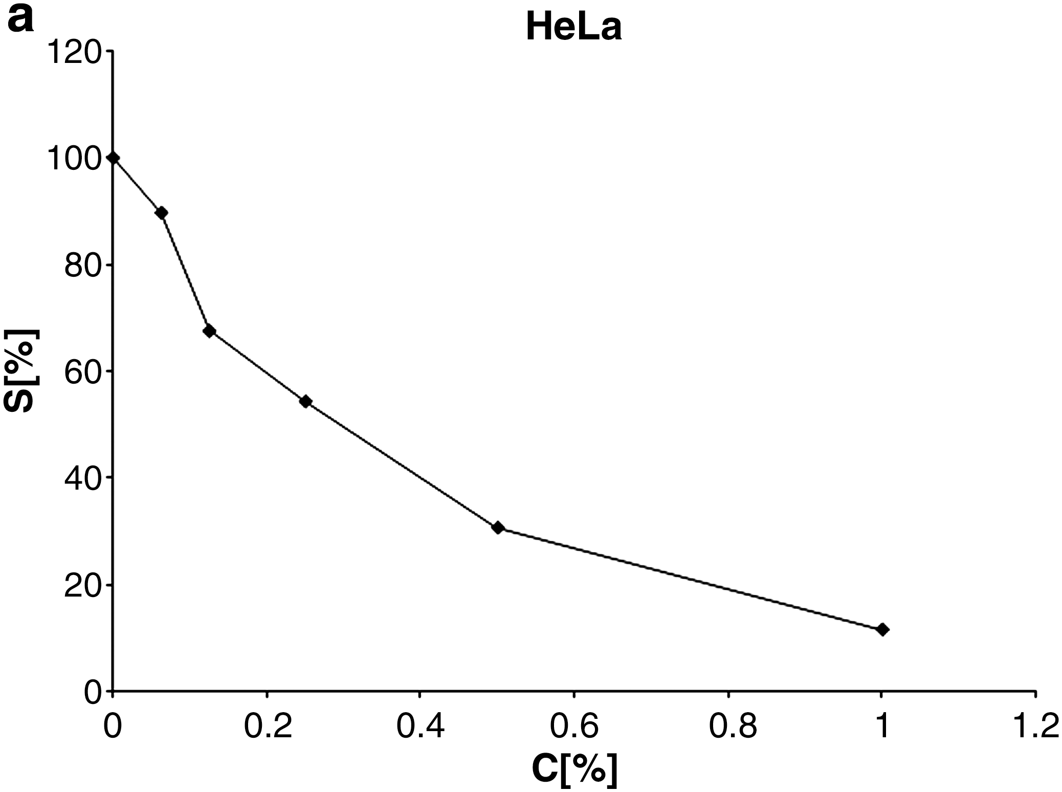
Survival of (
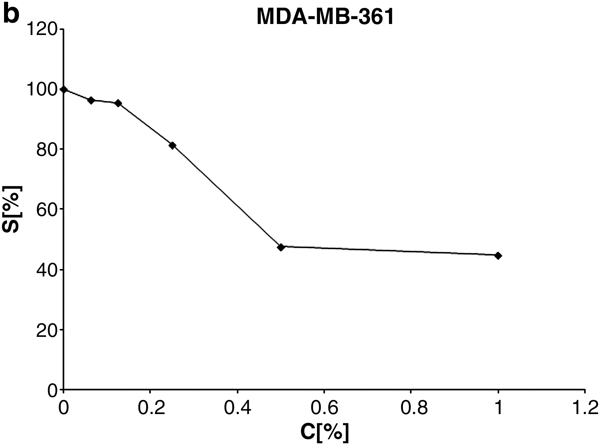


Survival of (


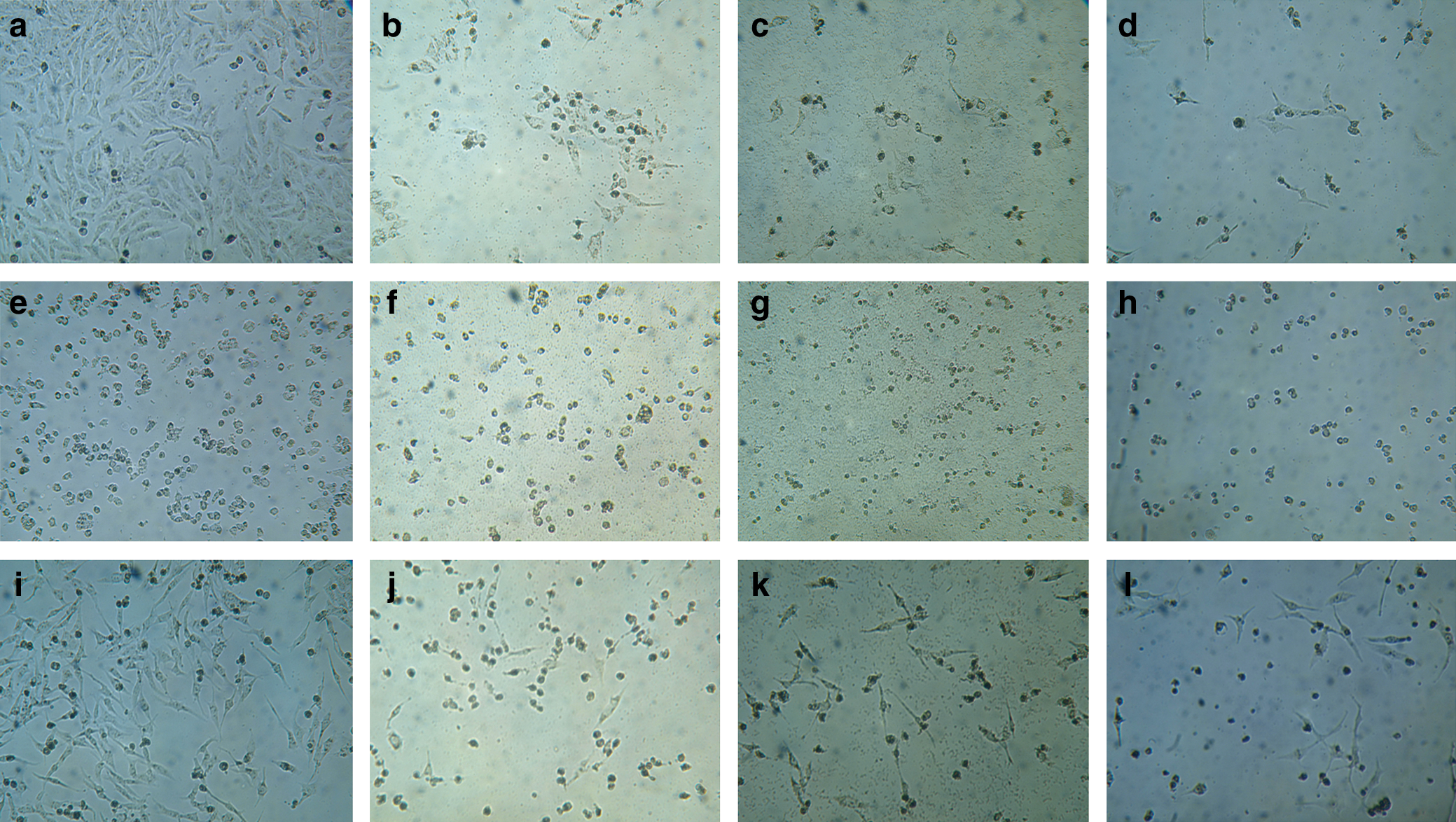
Photomicrographs of (
IC50 data are mean ± SD values of three independent experiments. Red and white wine extract values are percentages; resveratrol and cisplatin values are μM.
n = 1.
Photomicrographs of HeLa, MDA-MB-361, and MDA-MB-453 cells obtained 72 hours after continuous action of investigated agents clearly demonstrate that red and white wine polyphenolic extracts and resveratrol as well cause a decrease in the number of survived cells in relation to cells in the control sample (Fig. 4).
Red wine polyphenolic extract induced a dose-dependent decrease in the number of survived cells in comparison to cells grown in nutrient medium. It is noted that red wine extract exhibits the highest cytotoxic effect against the HeLa cell line. Furthermore, the cytotoxic activity of the extract is lower against the MDA-MB-453 cell line. On the other hand, the MDA-MB-361 cell line is the least sensitive to the cytotoxicity of this extract. It is clearly seen that the white wine polyphenolic extract exerts a strong dose-dependent cytotoxic action against all the cell lines investigated. White wine polyphenols are more active against HeLa and MDA-MB-453 cells than against MDA-MB-361 cells. Resveratrol, a polyphenol found in both red and white wine, has pronounced cytotoxic activity on cancer cell lines. The cytotoxic effect is the highest against MDA-MB-453 cell line. A lower effect is observed against HeLa cells. Moreover, resveratrol exerts the lowest activity on the MDA-MB-361 cell line. Cytotoxicity of resveratrol could be attributed to its antiproliferative activity, which is concentration dependent.
On PBMCs
A large and growing number of reports demonstrate the cytotoxic effect of polyphenolic compounds on a wide variety of malignant cell lines. In contrast, little is known about the effects of wine polyphenols on healthy immunocompetent cells. In order to determine the anticancer potential of red and white wine polyphenolic extracts and of resveratrol, the activity of these agents was tested against human PBMCs, unstimulated and stimulated to proliferate with a mitogen, PHA (results shown in Figs. 5 –7 and Table 4).
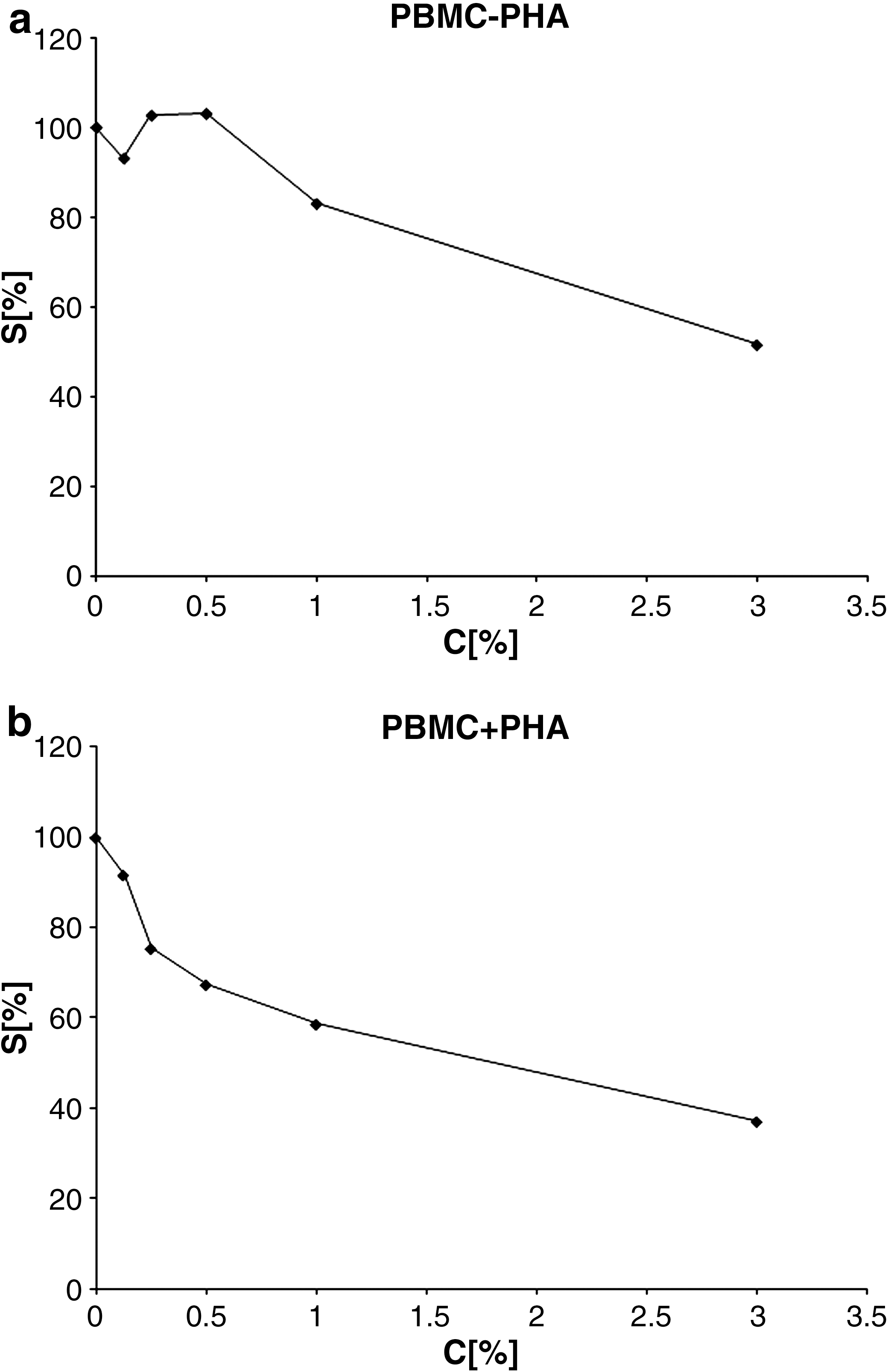
Survival of (

Survival of (
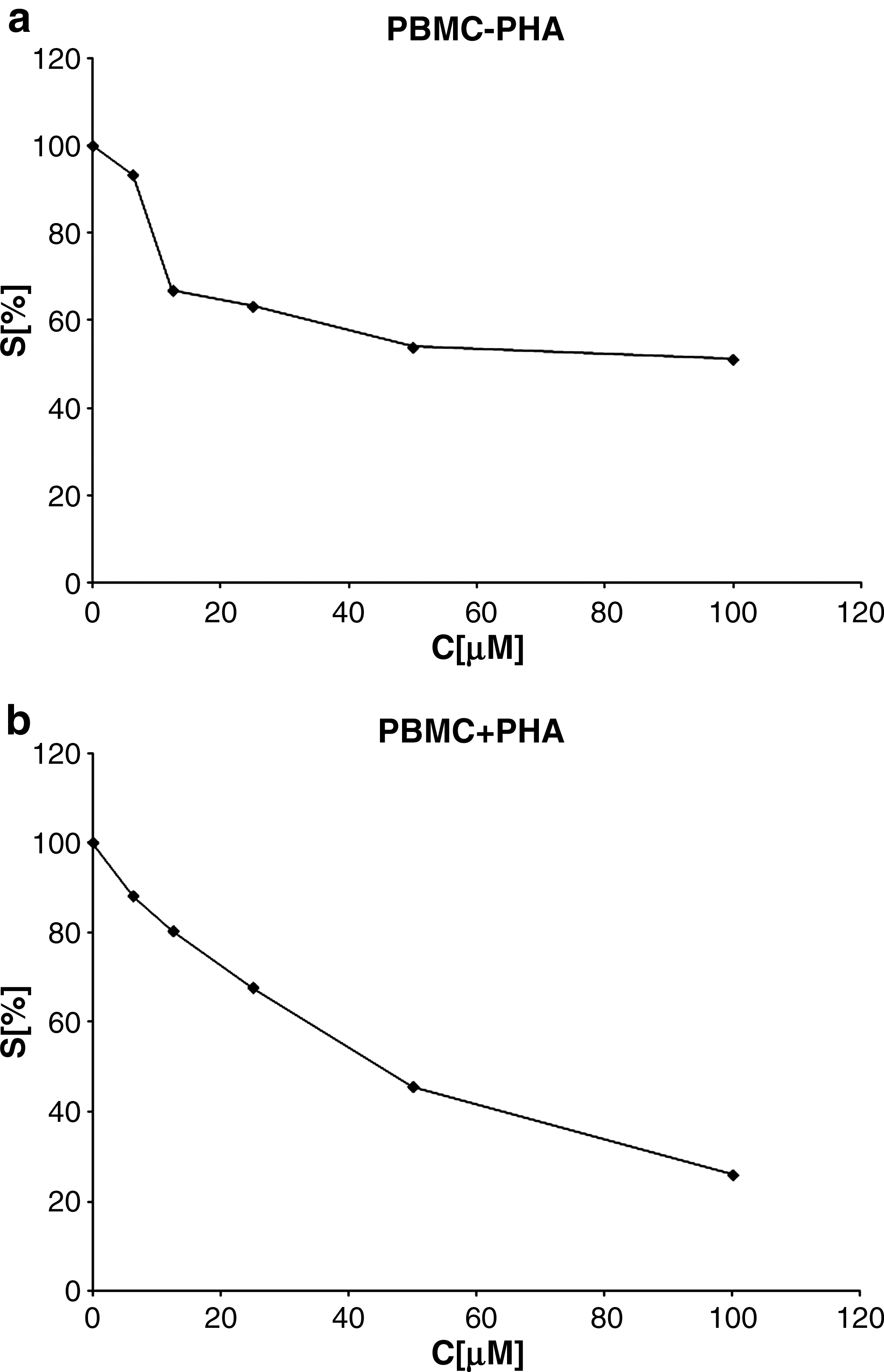
Survival of (
IC50 data are mean ± SD values of two independent experiments except for cisplatin (n = 1). Red and white wine extract values are percentages; resveratrol and cisplatin values are μM.
Considering the cytotoxic effect of red wine polyphenolic extract, it seems that this extract applied at concentrations up to 3% does not exhibit a significant cytotoxic effect against unstimulated human PBMCs. On the other hand, it displays a rather modest antiproliferative activity on stimulated PBMCs. However, white wine extract demonstrates a pronounced cytotoxic effect toward unstimulated PBMCs. In addition, its activity toward stimulated PBMCs is higher. Resveratrol is found to display a cytotoxic effect on stimulated PBMCs, but at the concentrations applied, this polyphenol has a very low cytotoxicity on unstimulated PBMCs.
Analysis of HeLa cell death
Morphological analysis of acridine orange/ethidium bromide-stained HeLa cells reveals that white wine polyphenolic extract, applied at a concentration double the 72-hour IC50 over 24 hours, induces apoptosis in these cells. The apoptotic effect of white wine extract is shown in Figure 8. Typical signs of programmed HeLa cell death could be observed, such as cell shrinkage, blebs, and nucleus condensation or fragmentation. Cells in late stages of apoptosis and secondary necrosis stained in orange-red could be noticed, as well. There were no signs of apoptosis or necrosis in HeLa cells treated with red wine polyphenolic extract for 24 and 48 hours (data not shown). These results clearly indicate that the cytotoxicity of white wine polyphenolic extract is due to the antiproliferative and apoptotic effect. The anticancer activity of red wine polyphenolic extract could be attributed to its antiproliferative effect.

Photomicrographs of acridine orange/ethidium bromide-stained (
Selectivity in the antitumor action
Selectivity coefficients in the antitumor action of red and white wine extracts and resveratrol are shown in Table 5. This parameter points to differences in cytotoxic activity of these agents toward malignant and healthy nontransformed cells. Results from this study indicate that the selectivity in the antitumor action of investigated agents is greater in the case when these substances perform its action on unstimulated PBMCs in comparison to stimulated PBMCs. It is important to observe that white wine polyphenolic extract has greater selectivity and cytotoxic activity than red wine polyphenolic extract. Resveratrol, a polyphenol that exerts strong cytotoxic activity, also exerts great selectivity in the antitumor action.
Discussion
Many recent studies have demonstrated that wine polyphenolic extracts have an important role in the reduction of malignant transformation. Wine polyphenols exhibit anticancer activity by inhibiting growth, inducing apoptosis, changing cell cycle regulation, and affecting target molecules of intracellular signal transduction pathways in malignant cells. Moreover, cytotoxicity of polyphenols may be due to their pro-oxidant properties. Phenolic compounds may exert both antioxidant and pro-oxidant action depending on their concentration and free radical source, 5,21 i.e., in general, their actions depend on the redox potential within the target cell. The pro-oxidant activity of polyphenols accelerates oxidative damage of DNA and induces apoptotic DNA fragmentation by producing reactive oxygen species.
Anticancer activity of wine polyphenols has been reported in numerous publications. 25 –28 For example, red wine polyphenols were shown to inhibit the proliferation of human colon carcinoma cells significantly by modulating activation of mitogen-activated protein kinases 15 (also see references cited by He et al. 15 ). In addition, anticancer action was also shown in breast cancer cells. 26,27 In a recent study, it was noticed that a hydrophobic polyphenol fraction isolated from Merlot wine exerted selective cytotoxicity against MCF-7 breast cancer cells, increased cytosolic calcium levels, and induced necrosis. 27 Furthermore, wine polyphenolic extracts have been demonstrated to exert antiproliferative and apoptotic effects in MOLT-4 human leukemia cells. 28 Regarding the molecular mechanisms of cytotoxic action, results of many studies indicate that wine polyphenols inhibit nuclear factor κB and phospholipase C signaling pathways, reduce cyclooxygenase-2 expression, and down-regulate cyclins, cyclin-dependent kinases, and the BCL2L1 anti-apoptosis gene. Therefore, wine compounds could suppress inflammation and carcinogenesis.
The results of the present study show that the white wine polyphenolic extract exhibits a significantly stronger cytotoxic activity toward all investigated cancer cell lines in comparison to the red wine polyphenolic extract. The observed effect is not related to their total phenolic content. This is in contrast to previous findings that the ability of red wine extract to decrease cancer cell survival is significantly higher than that of white wine extract. 25 The differences in the cytotoxic activity of red and white wine polyphenolic extracts could be attributed to different chemical composition. Although high contents of anthocyanins and gallic acid are found in the red wine extract, it exerts a weaker cytotoxic effect. Moreover, these phenols might be the active red wine components because the cytotoxic effect of these agents has been well documented. 29 –33 Additionally, phenolic acids have been reported to perform antioxidant and cytotoxic activity. Strong cytotoxicity of the white wine polyphenolic extract might be partially due to the activity of the caffeic acid. Cancer-suppressive action of the caffeic acid has been shown in several reports. 32,34 –36 Caffeic acid exhibits a cytotoxic effect against human breast cancer T47D, human cervix adenocarcinoma HeLa, human mammary gland adenocarcinoma MDA-MB-231, and lymphoblastic leukemia MOLT-3 cells. It is noteworthy that the concentration of the caffeic acid in white wines is two times higher than in red wines. The results from a recently published study have shown that the content of caffeic acid in white wines increases with storage time. 37 Combined additive, synergistic, or antagonistic action between the different phenolic compounds in each wine extract might be responsible for their antiproliferative effect.
It has been observed that red and white wine polyphenolic extracts exhibit significantly higher cytotoxic activity against HeLa and MDA-MB-453 cells in comparison to MDA-MB-361 cells. This selectivity in the anticancer effect toward a specific cancer cell line is related to the action of wine polyphenols on certain target molecules of signaling pathways that regulate cell proliferation and apoptosis. Furthermore, the cytotoxic action of wine polyphenolic extracts is considerably stronger against cancer cells than against PBMCs. Also, wine polyphenols are more active on stimulated PBMCs than on resting PBMCs, which may indicate that these agents possess the ability to inhibit PHA-stimulated proliferation of PBMCs. These findings point out that wine extracts rich in polyphenols can suppress immune functions, such as antigen stimulation.
Antiproliferative and apoptotic activities of resveratrol have been demonstrated in a variety of cancer cell lines, such as human epidermoid carcinoma A431 cells, breast cancer cell lines (MCF-7, MDA-MB-231, MDA-MB-435, and MDA-MB-468), gastric adenocarcinoma and colon carcinoma cell lines (HCT-116 and SW480), medulloblastoma cell lines (UW-228-2 and UW-228-3), prostate cancer cells (DU-145 and PC-3), pancreatic cancer cells, hepatoma HepG2 cells, human uterine cancer cells (HeLa, Hec-1A, and KLE), and various leukemic cell lines (HL-60 and U937). 16,17,38 –46 It is important to keep in mind that resveratrol exerts a selective cytotoxicity against specific cancer cell types. Action on HeLa, MDA-MB-361, and MDA-MB-453 cell lines supports this fact. The anticarcinogenic potential of resveratrol is closely associated with its antioxidant and pro-oxidant activities. It has been shown to inhibit cyclooxygenase, hydroperoxidase, protein kinase C, ribonucleotide reductase, DNA polymerase, nuclear factor κB, and cell cycle regulators. In general, red wines contain higher amounts of resveratrol than white wines. 47 –49 Surprisingly, the presence of resveratrol was not detected in the wine extracts investigated, what might be due to the very low levels. Considering the activity of resveratrol, it should be noticed that low pH causes cis-resveratrol isomerization to the trans isomer, an active and more stable form. Because each of the three hydroxyl groups can be a proton donor, resveratrol might increase the acidity of the nutrient medium. It was observed that resveratrol displays minimal cytotoxicity against unstimulated PBMCs at concentrations up to 100 μM. The cytotoxic effect is more pronounced against stimulated PBMCs. Some data suggest that trans-resveratrol inhibits the ability of PHA to stimulate proliferation of PBMCs. 50 There is evidence that resveratrol suppresses interferon-γ production in stimulated PBMCs, which seems to be connected to the inhibition of nuclear factor κB. 51
In conclusion, results from this in vitro study indicate that red and white wine polyphenolic extracts and resveratrol exert selective cytotoxic activity against HeLa, MDA-MB-361, and MDA-MB-453 cell lines. The activity of these agents against HeLa and MDA-MB-453 cell lines, which have higher proliferation rates, is more pronounced than against the MDA-MB-361 cell line, whose proliferation rate is lower. This suggests that wine polyphenols may regulate signal transduction pathways of cell proliferation. Antiproliferative activity of the white wine polyphenolic extract is significantly higher in comparison to activity of the red wine polyphenolic extract. It is also observed that a high concentration of white wine extract induces apoptosis and secondary necrosis in HeLa cells. It is noteworthy that wine polyphenolic extracts and resveratrol show a weaker cytotoxic effect on unstimulated normal PBMCs than on cancer cell lines. In addition, the agents investigated exert stronger cytotoxicity on stimulated PBMCs. Further studies should be conducted in order to investigate the cancer chemopreventive and therapeutic potential of the red and white wine polyphenolic extracts.
Footnotes
Acknowledgments
The authors are grateful to the Ministry of Science and Technological Development of the Republic of Serbia for financial support (Project Number 145006). Also, the authors would like to thank Tatjana Petrović for her excellent technical assistance.
Author Disclosure Statement
No competing financial interests exist.