
Abstract
Echinodorus macrophyllus leaf has been used in Brazilian folk medicine to treat inflammatory conditions and kidney dysfunctions. The present study evaluated the effects of leaf ethanolic extract from E. macrophyllus (EEEm) in acute and subchronic models of inflammation. The EEEm was found to cause significant and potent inhibition of carrageenan- and dextran-induced paw edema in rats and marked decreases in the exudate volume and leukocyte migration in rats with carrageenan-induced pleurisy, the vascular permeability increase induced by intraperitoneal acetic acid, and the croton oil-induced topical ear edema in mice. On the other hand, the EEEm was not active in the test model of cotton pellet-induced granuloma in rats. Phytochemical analysis with E. macrophyllus leaves revealed the presence of triterpenoids, steroids, flavones, flavonols, and xanthones. Two flavonoids were isolated from the ethyl acetate fraction and identified as isovitexin and vitexin. Our results support the traditional use of E. macrophyllus leaves in the treatment of acute inflammatory conditions.
Introduction
E
The main compounds that were already isolated from the leaves are echinophyllins A, B, C, and F, chapecoderins A and C, 2 –4 and echinodolides A and B. 5
Da Costa Lopes et al. 6 reported the absence of mutagenic, genotoxic, and cytotoxic effects in the aqueous extract of E. macrophyllus leaves, as well as the reduction of body weight and the presence of hepatic alterations in mice exposed to subchronic treatment with that extract.
Pinto et al. 7 reported that the immunosuppressive effect supports a potential therapeutic use of aerial parts of the E. macrophyllus plant to control exacerbated humoral and/or cellular immune responses, as in autoimmune rheumatic diseases.
There are very few scientific reports that support the traditional use of this plant in inflammatory conditions. Additionally, this is the first report that described the isolation of isovitexin and vitexin from E. macrophyllus. This study evaluated the topical and systemic anti-inflammatory effects of leaf ethanolic extract from E. macrophyllus (EEEm) so as to validate its traditional use in inflammatory conditions. A phytochemical examination was also carried out using dried leaves of the plant.
Materials and Methods
Animals
Seven to 10 Swiss mice (weighing 20–30 g) and albino male Wistar rats (weighing 180–250 g) were used in each group tested. The animals were kept in propylene cages at 22 ± 2°C with a 12-hour photoperiod and were allowed free access to food and water. Before the bioassays were set up, the animals were deprived of food for a period of 16–18 hours. The experimental protocols were approved by the Committee for Ethics in Animal Experimentation of the Federal University of Mato Grosso, Cuiabá, MT, Brazil, according to the Federal Government's legislation on animal care.
Plant material
E. macrophyllus leaves were collected from muddy areas located nearby rivers in Poxoreo County, MT, Brazil (GPS coordinates: 15°50′957″ S and 54°20′302″ W). The identification and authentication of the plant were carried out by the Plantarum Institute, Nova Odessa, SP, Brazil. A voucher specimen was deposited in the Herbarium of the Federal University of Mato Grosso under reference number 30486. Plant collection was authorized by the Brazilian Institute of Environment and Renewable Natural Resources.
Preparation of the ethanol extract
The leaves of E. macrophyllus (0.2 kg) was ground to a powder and extracted by sequential maceration with hexane, dichloromethane, ethyl acetate, and ethanol/water 75% (3:1, wt/vol) for 7 days at 25°C. The ethanol/water macerate was filtered, and the solvent was fully evaporated under reduced pressure with a rotary evaporator (model TE-210, Tecnal, Piracicaba, SP, Brazil) to obtain the dried ethanol extract (yield, 2.5 g) used for pharmacological assays.
Phytochemical analysis
Dried leaves of E. macrophyllus (650 g) were cut into small pieces and macerated in methanol for 7 days. The solvent was concentrated under reduced pressure to give the respective methanol extract (19 g).
In order to verify the presence of different chemical classes, the methanol extract was subjected to a standard screening test. 8 Conventional protocols for detecting the presence of alkaloids, flavonoids, saponins, tannins, xanthones, steroids, anthraquinones, triterpenes, etc., were used.
Part of this extract (15 g) was then successively partitioned with solvents of increasing polarity to give the following fractions and yields: n-hexane (2.7 g), dichloromethane (4.4 g), and ethyl acetate (5.2 g). The ethyl acetate fraction was chromatographed using a silica gel column eluted with a CHCl3-methanol gradient, furnishing a pure yellow amorphous powder, which was identified as isovitexin (145 mg) on the basis of spectral data (infrared and 1H and 13C nuclear magnetic resonance) compared with an authentic sample. From this fraction were detected by thin-layer chromatography several other compounds, particularly phenol compounds. One of them seems to be the flavonoid vitexin, first described for the genus Echinodorus.
Carrageenan- and dextran-induced paw edema
Edema was induced in rats by subplantar injection of 0.1 mL of 1% carrageenan or 1.5% dextran in normal saline in the left hind paw of the rats. 9 An equal volume of vehicle was injected in the contralateral paw. Volumes in both paws were measured in a plethysmometer (model 7150, Ugo Basile, Comerio, Varese, Italy), prior to (0 hour) and during the 4 hours after carrageenan injection or 2 hours after dextran injection. The difference in the volumes (in mL) was taken as the measure of the edema. The tested drugs—EEEm (1, 5, or 30 mg/kg), indomethacin (5 mg/kg), and cyproheptadine (5 mg/kg)—or vehicle (10 mL/kg distilled water) were given orally 1 hour before the subplantar injection of phlogistic agents.
Carrageenan-induced pleurisy
Pleurisy was produced in rats by injection of 0.1 mL of 2% carrageenan in normal saline in the thoracic cavity. 10 The EEEm (1, 5, or 30 mg/kg), dexamethasone (0.5 mg/kg), or vehicle was given orally 1 hour prior to the injection of the irritant. Six hours after the inflammatory challenge, the animals were killed by excess of ether, the thoracic cavity was opened, and the exudate was harvested by aspiration. Exudate volume was measured (in mL), and the mobilized leukocyte number in the exudate was quantified using improved Neubauer's counting chambers.
Acetic acid-induced peritoneal vascular permeability
Groups of mice were treated orally with vehicle (0.1 mL/10 g), EEEm (1, 5, or 30 mg/kg, p.o.), or indomethacin (5 mg/kg, p.o.). After 1 hour, each animal received 0.1 mL/10 g 2% Evans blue in normal saline, intravenously. After 10 minutes, 0.4 mL of 0.5% acetic acid in normal saline was given by the intraperitoneal route. Twenty minutes later, the mice were killed by an excess of ether, and their abdominal cavity was washed with distilled water (6–8 mL). The washings were pooled, the volume was made up to 10 mL with distilled water, and the absorbance of the solution was measured at 590 nm. The amount of dye leakage was calculated from the absorbance measurements. 11
Croton oil-induced dermatitis
The mice were treated topically, in each ear, with 20 μL of vehicle, EEEm (4, 20, or 100 μg), or dexamethasone (5 μg). After 30 minutes, each animal received, in the inner surface of the right ear, 20 μL of croton oil in acetone (5 mg/mL, 100 μg), and an equal amount of acetone was applied in the left ear. Six hours after the administration of the irritant, the animals were killed by cervical dislocation, and the ears were removed at the base and weighed. The inflammatory response was monitored by measuring differences in weight (in mg) between the right and left ears. 12
Cotton pellet-induced granuloma
Each rat received two subcutaneous implants of sterilized cotton pellets weighing 50 mg each in the dorsum (one on each side) under ether anesthesia. 13 At 24 hours after the incision, the animals were deprived of food for 1 hour and started oral treatment for 6 days with vehicle, EEEm (1, 5, or 30 mg/kg, p.o.), or dexamethasone (0.5 mg/kg, p.o.). After day 6, the animals were killed with high doses of ether, and the pellets surround by granuloma tissues were dissected out (two per animal), weighed, and dried at 60°C for 24 hours. The variation in the edema was calculated by the difference in humid weight of each tissue of cotton weight. The difference between the weight of each encapsulated dry tissue and implanted cotton was taken as the measure of the granuloma, considering the average of the duplicates.
Statistical analysis
Results were analyzed using one-way analysis of variance, followed by the Student-Newman-Keuls test, and expressed as mean ± SEM values. The difference between means of treated and control groups was considered significant at P < .05.
Results
Phytochemical screening revealed the presence of saponins, alkaloids, quinones, flavones, flavonols, xanthones, triterpenoids, and steroids.
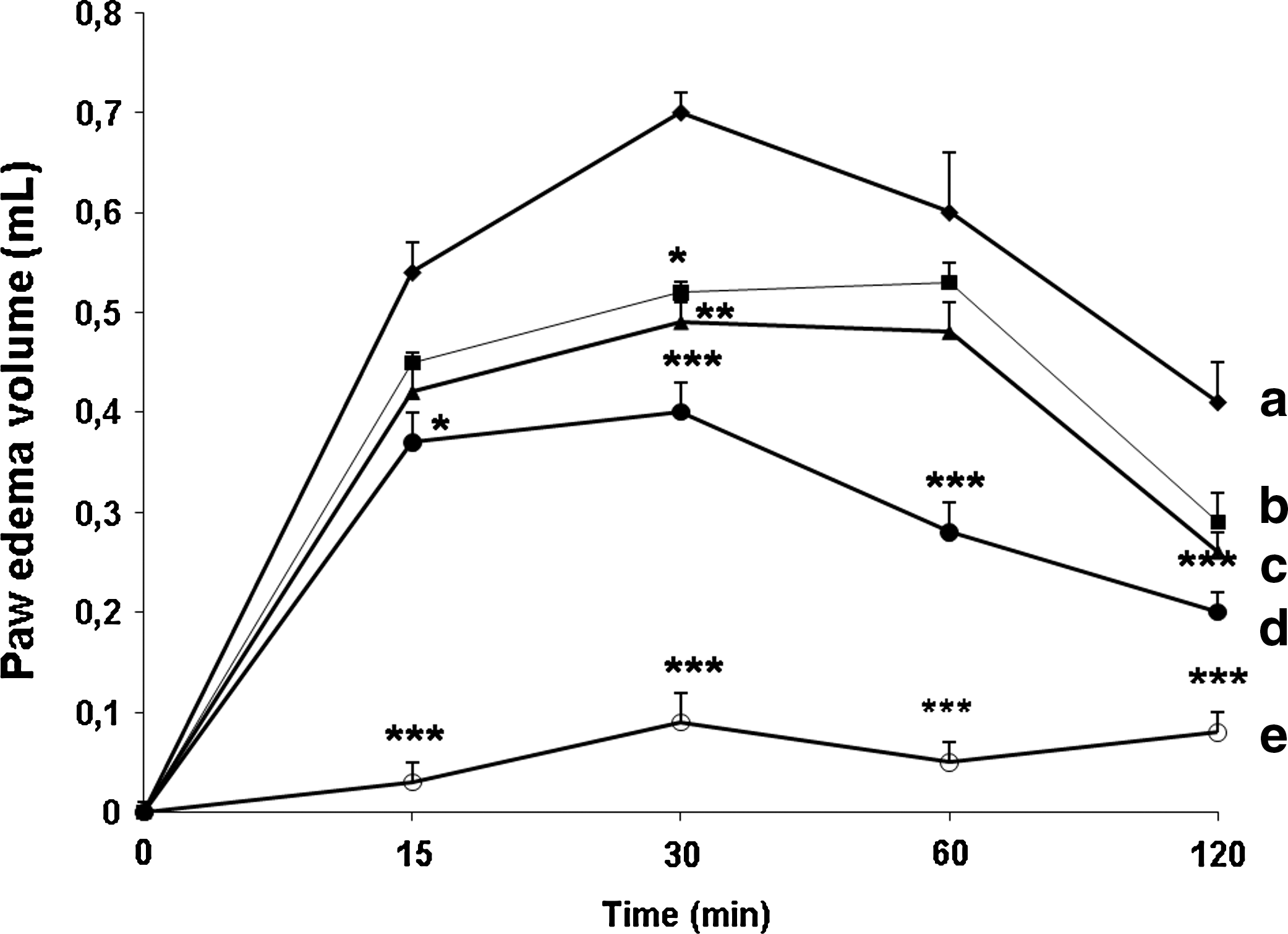
The results show that the EEEm (1, 5, or 30 mg/kg) caused significant dose-related reduction in swelling induced by subplantar injection of carrageenan to the 4th hour (25% [P < .001], 46% [P < .001], and 63% [P < .001], respectively) compared to the control (0.70 ± 0.3 mL). Indomethacin (5 mg/kg), the reference nonsteroidal anti-inflammatory agent, caused significant (P < .001) inhibition of paw edema by 82% compared to the vehicle-treated control (Fig. 1).

Effect of administration of (
The effects of the EEEm (1, 5, or 30 mg/kg) on dextran-induced paw edema are shown in Figure 2. At 30 minutes after subplantar dextran, the paw edema value in vehicle controls was 0.69 ± 0.21 mL. The EEEm (1, 5, or 30 mg/kg) and cyproheptadine (5 mg/kg) caused significant inhibition of paw edema by about 30% (P < .01), 23% (P < .05), 42% (P < .001), and 92% (P < .001), respectively, compared to vehicle-treated controls (Fig. 2).
Effect of administration of (
The injection of 0.4 mL of intraperitoneal 0.5% acetic acid caused, in the control group, an increase of vascular permeability, verified by the overflowing of 140 ± 15 μg of Evans blue dye to the peritoneal cavity. The orally administered EEEm (1, 5, or 30 mg/kg) reduced this rate to 90 ± 9 μg (P < .01), 110 ± 7 μg (P < .05), and 62 ± 5.5 μg (P < .001), respectively.
Indomethacin (5 mg/kg) produced significant (33 ± 5 μg, P < .001) inhibition of the vascular permeability increase induced by intraperitoneal acetic acid in mice (Fig. 3).

Effect of oral administration of vehicle, leaf ethanolic extract of E. macrophyllus (EEEm) in doses of 1, 5, and 30 mg/kg, or 5 mg/kg indomethacin (Indo) on acetic acid-induced vascular permeability in mice. Data are mean ± SEM (vertical bar) values from eight to 10 mice. Significantly different from the control group: *P < .05, **P < .01, ***P < .001.
Table 1 shows that the EEEm (30 mg/kg) and dexamethasone (0.5 mg/kg) were also effective in reducing the exudate volume (1.1 ± 0.11 mL [34%, P < .001] and 0.38 ± 0.05 mL [79%, P < .001], respectively) and the leukocyte migration (90 ± 3 × 10⁶ cells [40%, P < .05] and 40 ± 0.03 × 10⁶ cells [73%, P < .001], respectively) in the rat model of acute pleurisy induced by carrageenan compared with the control group (exudate volume, 1.80 ± 0.1 mL; leukocyte migration, 149 ± 14 × 106 cells).
Data are mean ± SEM values from eight to 10 rats.
Significantly different from the control group: *P < .05, ***P < .001.
The topical application of croton oil in the control group promoted the development of ear edema, evidenced by an increase in ear weight (91 ± 10 mg). The topically applied EEEm (4, 20, or 100 μg) and dexamethasone (5 μg) significantly reduced the cutaneous edema induced by croton oil (32% [P < .01], 39% [P < .01], 41% [P < .001], and 98% [P < .001], respectively) compared to the control (Fig. 4).
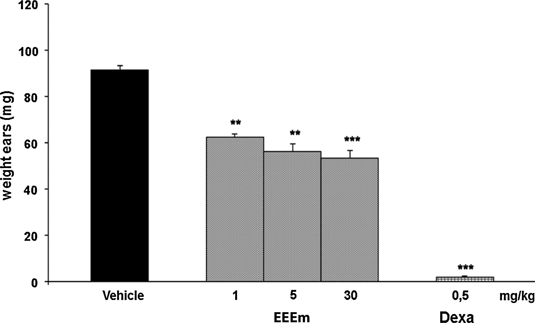
Effect of topical administration of vehicle, leaf EEEm in doses of 4, 20, and 100 μg, or 5 μg dexamethasone (Dexa) on croton oil-induced ear edema in mice. Data are mean ± SEM (vertical bar) values from eight to 10 mice. Significantly different from the control group: **P < .01, ***P < .001.
The pretreatment of rats with EEEm (1, 5, or 30 mg/kg) did not significantly inhibit the development of cotton pellet-induced granuloma in rats. On the other hand, dexamethasone (0.5 mg/kg) significantly inhibited the formation of edema (40% [P < .001]), as well as the development of granulomatous tissue (43% [P < .01]), compared with the control group (formation of edema, 0.90 ± 0.04 mg and development of granulomatous tissue, 0.14 ± 0.01 mg).
Discussion
In order to provide a scientific basis for the folklore use of E. macrophyllus in inflammatory conditions, its leaf extract (EEEm) was evaluated for anti-inflammatory-related effects using diverse experimental models of inflammation. Carrageenan-induced paw edema as an in vivo model of experimental inflammation has been frequently used to assess the anti-edematous effect of natural products. Its has been reported that various mediators are released by carrageenan in the rat paw; thus, while the initial phase (first 1.5 hours) may be due to the release of histamine and serotonin, the second phase (1.5–2.5 hours) is attributed to the release of kinins, followed by release of prostaglandins (PGs) in its final phase (1.5–5 hours), and is sensitive to most clinically effective anti-inflammatory drugs. 14,15 In the carrageenan test, EEEm (1, 5, or 30 mg/kg, p.o.) produced a significant anti-edema effect, which started 2 hours after the injection of the phlogistic agent and persisted until the 4th hour of testing, simulating the effect of reference compound indomethacin (5 mg/kg, p.o.). It is likely that this anti-edema response of EEEm might be a result of its ability to suppress PG biosynthesis from arachidonic acid through cyclooxygenase inhibition.
EEEm also caused significant inhibition of dextran-induced paw edema in mice, but its potency appeared much less than in the carrageenan test. The highest dose of the extract (30 mg/kg) was able to block the peak inflammatory edema that started at 30 minutes, and its action sustained all along the experiment (2 hours). However, cyproheptadine (5 mg/kg, p.o), the reference drug that has both anti-histaminic and antiserotonin properties, 16 was more potent in reducing the dextran-induced edema than EEEm. It is known that histamine and serotonin are the principal mediators of dextran-induced paw edema. 17,18 The fact that EEEm inhibits the dextran edema could imply that it inhibits either the degranulation of mastocytes and thus the release of mediators, histamine, and serotonin or their actions at the receptor level.
Pleurisy and carrageenan-induced paw edema are considered the most appropriate experimental models for the evaluation of anti-inflammatory substances. 17,19 In the pleurisy model, one can study the efficacy of anti-inflammatory agents that act differently, regulating the fluid and cellular components of pleural exudates. 12
It is believed that the overflow of plasma is a response to the local increases of vasoactive substances such as the PGs (PGE1 and PGE2), histamine, bradykinin, etc. 20 The migration of leukocytes, especially neutrophils, reaches its maximum between the period of 4 and 6 hours after the injection of carrageenan in the pleural cavity. 21 There is an increase in the production of PGs, especially PGE2, due to induction of the synthesis of cyclooxygenase-2 by the leukocytes of pleural exudates. 22,23 PGs, however, are not much involved in the chemotaxic response, even though they promote the migration of leukocytes to the inflamed area through an increase in blood flow. Then, the inhibition in the accumulation of neutrophils is independent of the effects on the cyclooxygenase activity 24 and might be related to the reduction of cellular activities such as cell proliferation and generation of chemotaxic factors (for example, leukotrienes). 25
The animals treated with dexamethasone (0.5 mg/kg, p.o) exhibited significant reduction, both in volume of exudates and in the accumulation of total leukocytes in the pleural cavity. The EEEm was effective in reducing exudation and cellular migration only at the highest dose (30 mg/kg, p.o.).
As with the carrageenan-induced paw edema, it might be assumed that the anti-exudation effect of EEEm in the pleurisy model is due to cyclooxygenase inhibition, whereas the reduction of cellular migration may be a result of lipoxygenase inhibition.
The most significant effect of EEEm on the exudation was clear when the increase in vascular permeability was induced by the intraperitoneal injection of 0.5% acetic acid in mice. In this model, EEEm significantly reduced the overflow of Evans blue to the peritoneal cavity in all doses tested (1, 5, and 30 mg/kg, p.o.), and its anti-exudation effect at the highest dose (30 mg/kg) was comparable to that of dexamethasone (0.5 mg/kg, p.o.).
The suggested mediators in the induction of croton oil-induced ear edema were the arachidonic acid metabolites (PGs and leukotrienes) and the pro-inflammatory cytokines. 26 It is characterized by increased vasodilatation, infiltration of polymorphonuclear leukocytes, and localized edema formation that reaches its peak at the 6th hour. This model is used to detect inhibitor activity of the cyclooxygenase and/or lipoxygenase in vivo, and it is very sensitive to steroidal anti-inflammatory agents and less sensitive to non-steroid anti-inflammatory agents. 12,27
In the present work, EEEm (4, 20, and 100 μg) produced a significant, dose-dependent anti-edema effect compared to the control group. Dexamethasone (5 μg) also promoted significant reduction of the edema in the ear treated with the irritating agent. This result strengthens the possibility that the anti-inflammatory effect of the extract in the carrageenan-induced paw edema test, pleurisy, and vascular permeability is the consequence of lipoxygenase inhibition. This fact is contrary to the hypothesis that the anti-inflammatory activity of EEEm is dependent, at least in part, on the inhibition of cyclooxygenase because drugs that behave as non-steroid anti-inflammatory agents are not very efficient in inhibiting croton oil-induced topic inflammation. 12,27
Following the encouraging results obtained in acute inflammatory models, we were prompted to verify the anti-inflammatory potential of E. macrophyllus in the cotton pellet-induced granuloma model, which represents subchronic inflammation.
The granuloma model has been widely used to evaluate anti-inflammatory drugs, especially steroid anti-inflammatory agents. 28 It consists of an acute transitory inflammatory response, followed by intense accumulation of activated macrophages, enveloped by a capsule of fibroblasts and conjunctive tissue (granuloma), oriented by products of lipoxygenase activity, especially leukotrienes (leukotriene B4), which explains its greater sensitivity to non-steroid anti-inflammatory agents. 29
The administration of EEEm (1, 5, and 30 mg/kg, p.o.) did not alter either the formation of exudate or the dry weight of granuloma compared to the control group. Dexamethasone (0.5 mg/kg, p.o.) showed a significant reduction both in the exudates and in the development of granulomatous tissue. Phytochemical analysis conducted with E. macrophyllus leaves revealed the presence of triterpenoids, steroids, flavones, flavonols, and xanthones as the main secondary metabolite classes, referred to in the literature as possessing anti-inflammatory activity. 30 –34 The ethyl acetate fraction furnished isovitexin and vitexin as the main flavone C-glucosides. Some pharmacological activities have also been attributed to vitexin and isovitexin, such as anti-inflammatory, 35 antimicrobial, 36 and antioxidant/free radical scavenging. 37,38
The results of the present study validate, in a preclinical point of view, the use of E. macrophyllus leaves in inflammatory processes.
Footnotes
Acknowledgments
The authors are grateful to the CAPES CNPq, Pantanal Research Center (CPP, Mato Grosso), and the FAPEMAT for financial support. We also thank the Plantarum Institute for providing botanical identification.
Author Disclosure Statement
No competing financial interests exist.