
Abstract
Cratoxylum formosum is an edible plant that is commonly consumed among the people in Northeast Thailand. This study aimed to investigate the gastroprotective effect of the ethanolic extract of C. formosum leaves (C. formosum ethanolic extract [CFE]). Gastric ulceration was induced in Wistar male rats by oral administration of acid/alcohol. Oral dosing with CFE at 250 and 500 mg/kg of body weight after the acid/alcohol induction significantly decreased the number of bleeding spots, area of bleeding, ulcer score, and ulcer index. Pretreatment with 500 mg/kg CFE significantly prevented the gastric damage. Histological studies of the acid/alcohol-induced animals indicated the gastric inflammation with lesion depth through the mucosal layer. Whereas the gastric lesion of the CFE-treated animals at both 250 and 500 mg/kg doses was decreased to be one-fourth of the mucosal layers, pretreatment with 500 mg/kg CFE prior to acid/alcohol induction completely protected against the mucosal damage. Biochemical analysis of gastric mucosa revealed a significant decrease of malondialdehyde in the CFE-treated group in a dose–response manner. These findings suggest that the gastroprotective activity of CFE could be mediated possibly through its antioxidant effect.
Introduction
G
Several plant extracts containing polyphenolics and other antioxidative constituents have been shown to have gastroprotective effects, such as grape seeds, 8 pear, 9 Phyllanthus emblica fruit, 10 Aloe vera, 11 and Centella asiatica. 12 –14
Cratoxylum formosum Dyer (Family Guttiferae) is an indigenous plant that is commonly found in Northeast Thailand. Fresh shoots and young leaves of this plant are traditionally consumed as a vegetable side dish or ingredient in the local style soup. The plant tastes sour and a little astringent because of the phenolic contents. The leaf extract showed strong antioxidant and antimutagenic activity. 15,16 The aqueous extract from the leaf of this plant has also demonstrated strong radical scavenging and vascular protective activities. 17 Phytochemical characterization revealed that this plant is rich in phenolic compounds. The main component was identified as chlorogenic acid, and the minor compounds were dicaffeoylquinic acid and ferulic acid derivatives. 18 Therefore, the present study was conducted to evaluate the gastroprotective effect of C. formosum ethanolic extract (CFE) using acid/EtOH-induced gastric lesions in rats as a model. The possible mechanism of CFE action via reactive oxygen species was also investigated by determination of malondialdehyde (MDA), a lipid peroxidation product.
Materials and Methods
Plant extract
C. formosum Dyer was collected in Khon Kaen Province, Thailand. The plant was identified, and a voucher specimen (KP-006) has been deposited as the herbarium at the Center for Research and Development of Herbal Health Products, Faculty of Pharmaceutical Sciences, Khon Kaen University, Khon Kaen, Thailand. CFE was prepared as described in the petty patent document. 19 In brief, the dried leaves were blended, pulverized, and then cold-extracted with 50% EtOH in a closed container for 1 week. The liquid part was filtered and centrifuged at 1,500 g (Kubota 5220, Kubota Corp., Tokyo, Japan) for 10 minutes; the supernatant was collected, dried using a rotary vacuum evaporator, and freeze-dried. The dried extract was obtained at a yield of 24.8% and kept in a tight and light-protected container at −20°C until used. The extract was suspended in distilled water, sonicated for 10 minutes, and then filtered (pore size, 0.45 μm) for intragastric administration to experimental animals.
Chemicals
All chemicals were purchased from local chemical distributors. β-NADPH, glutathione peroxidase, glutathione reductase, glutathione reduced form, dithiothreitol, hydrogen peroxide, thiobarbituric acid, 1,1′,3,3′-tetramethoxypropane, sodium dodecyl sulfate, EDTA, sodium azide, monobasic and dibasic sodium phosphate, and chlorogenic acid were products from Sigma (St. Louis, MO, USA). The other chemicals were products from various manufacturers as follows: methanol from Fisher Scientific (Loughborough, UK); sodium carbonate from Ajax Finechem (Seven Hills, NSW, Australia); glacial acetic acid from J.T. Baker (Philipsburg, NJ, USA); omeprazole from Berlin Pharm (Bangkok, Thailand); 2,2-diphenylpicrylhydrazine (DPPH) from Fluka Biochemika (Buchs, Switzerland); and Folin-Ciocalteu phenol reagent, EtOH, HCl, eosin, and hematoxylin from E. Merck (Darmstadt, Germany).
High-performance liquid chromatography analysis of chlorogenic acid content in CFE
Because chlorogenic acid was reported to be the main antioxidant component of C. formosum, CFE was subjected to high-performance liquid chromatography analysis using an Agilent (Palo Alto, CA, USA) Hypersil ODS column (particle size, 5 μm; 4.6 × 250 mm) with a mobile phase containing acetonitrile and 0.4% phosphoric acid (1:9 vol/vol). 20 The chromatogram was recorded at 327 nm with a flow rate of 1 mL/minute. Chlorogenic acid was used as a standard compound for the method validation.
Antioxidative activity and total phenolic content of the plant extract
Antioxidative activity of CFE was determined by the DPPH radical scavenging method. 21 The dried CFE was dissolved in methanol at various concentrations, and then 200 μL of the extract solution were incubated with 2,800 μL of 1 mM DPPH solution at room temperature for 15 minutes. The absorbance was measured at 515 nm, and then the DPPH radical scavenging activity was determined and expressed as the 50% effective dose. The Folin-Ciocalteu method as described by Makkar et al. 22 was used for determination of total phenolic content. The dried CFE was dissolved in 50% EtOH at various concentrations and then reacted with 2.5 mL of Folin-Ciocalteu reagent and 0.25 mL sodium bicarbonate solution (20%) for 40 minutes at room temperature. The absorbance was measured at 725 nm. The phenolic content was expressed as tannic acid equivalence and gallic acid equivalence in terms of mg/g of extract.
Animal treatment
Male Wistar rats (weighing 300–400 g) were purchased from the National Animal Center (Mahidol University, Bangkok). The animals were placed in suspended cages and housed in a climate-controlled room at 25°C with a 12-hour dark-light cycle. All the animals were acclimatized for 7 days before the experiment, fed standard food pellet (Chareonpokpan, Bangkok), and given drinking water ad libitum. The rats were food- and water-deprived for 24 hours prior to the experiments. The experimental protocols were approved by the Animal Ethics Committee of Khon Kaen University (AE009/50), in accordance with the national guidelines. The rats were randomly divided into seven groups (14 animals per group; seven animals for anatomical and histological studies and seven animals for biochemical analysis). An acid/alcohol solution composed of concentrated HCl:EtOH:distilled water (1.7:60:38.3 by volume) was used to provoke the inflammatory damage. 23 Group I (control), Group II, and Group III were given distilled water, CFE at 500 mg/kg of body weight, and acid/alcohol solution (1.5 mL/kg of body weight), respectively. Groups IV–VI were given acid/alcohol solution (1.5 mL/kg of body weight) 30 minutes prior to the administration of the standard drug omeprazole (20 mg/kg of body weight) and CFE at doses of 250 and 500 mg/kg of body weight, respectively. Group VII received the plant extract at 500 mg/kg of body weight 30 minutes prior to administration of acid/alcohol solution (1.5 mL/kg of body weight).
Assessment of acid/EtOH-induced gastric mucosal damage
After 6 hours of treatment, seven animals from each group were sacrificed by intraperitoneal injection of pentothal sodium (60 mg/kg of body weight), and the abdomens were opened. Then the pyloric sphincter and esophagogastric junction were ligated, and the stomach was rapidly removed. After injection of 1 mL of 10% formalin in 0.1 M phosphate buffer (pH 7), the stomach was further fixed in 10% formalin in 0.1 M phosphate buffer (pH 7) for 24 hours. The stomach was opened along the greater curvature, rinsed with ice-cold 0.9% NaCl, and extended on the paraffin board by fixing with needles. The mucosal lesion was measured by determination of the bleeding spot, bleeding area, and total mucosal area using an image analyzer (UTHSCSA image tool, version 3.00 [
Determination of the in vivo antioxidative effect of the plant extract
Another seven animals from each group were sacrificed; the stomach was rapidly removed as earlier described. Then the stomach was opened along the greater curvature and rinsed with ice-cold 0.9% NaCl. The gastric mucosal layer was scraped and homogenized in cold 0.1 M phosphate buffer solution, pH 7 (phosphate-buffered saline). After centrifugation at 6,500 g (Eppendorf Minispin, Eppendorf AG, Hamburg, Germany) at 4°C for 15 minutes, the supernatant was assayed for protein content 26 and used for the determination of glutathione peroxidase activity and MDA content.
Determination of glutathione peroxidase activity
To determine glutathione peroxidase activity, 50 μL of supernatant was added to 1 mL of a reaction mixture containing 0.35 mM β-NADPH, 25 units of glutathione reductase, 1 mM glutathione, 0.39 mM EDTA, and 0.98 mM sodium azide in 49 mM phosphate-buffered saline and 50 μL of 0.042% hydrogen peroxide solution. A blank was assayed in parallel by using 50 μL of 10 mM phosphate-buffered saline containing 1 mM dithiothreitol in place of the supernatant. The changes of absorbance at 340 nm were determined, and the specific activity was expressed as units/mg of protein as described by Wendel. 27
Determination of MDA
The method described by Ohkawa et al. 28 was used to determine MDA content. Supernatant (0.1 mL) was mixed with 1.9 mL of a mixture containing 0.1 mL of 8.1% sodium dodecyl sulfate, 0.75 mL of 20% acetic acid, 0.75 mL of 0.8% thiobarbituric acid, and 0.3 mL of distilled water and heated at 95°C for 60 minutes. After the mixture stood at room temperature for 10 minutes, 0.5 mL of distilled water and 2.5 mL of a mixture containing n-butanol and pyridine (15:1 vol/vol) were added and vigorously mixed, and then the whole mixture was centrifuged at 1,700 g (IEC clinical centrifuge, International Equipment Co., Needham, MA, USA) for 10 minutes. The upper layer was measured for the absorbance at 532 nm. The amount of MDA was expressed as nmol/mg of protein by using 1,1′,3,3′-tetramethoxypropane as the standard.
Statistical analysis
Data were analyzed by one-way analysis of variance followed by Fisher's Least Significant Difference test for multiple comparisons. The statistical significance was accepted at P < .05.
Results
Analysis of chlorogenic acid content in CFE by high-performance liquid chromatography
The high-performance liquid chromatogram demonstrated that chlorogenic acid was the major peak of CFE, which was eluted at 14.98 minutes (Fig. 1). By comparing the peak area with that of standard chlorogenic acid, CFE contained 67.3 ± 0.8 mg chlorogenic acid/g of dried extract.
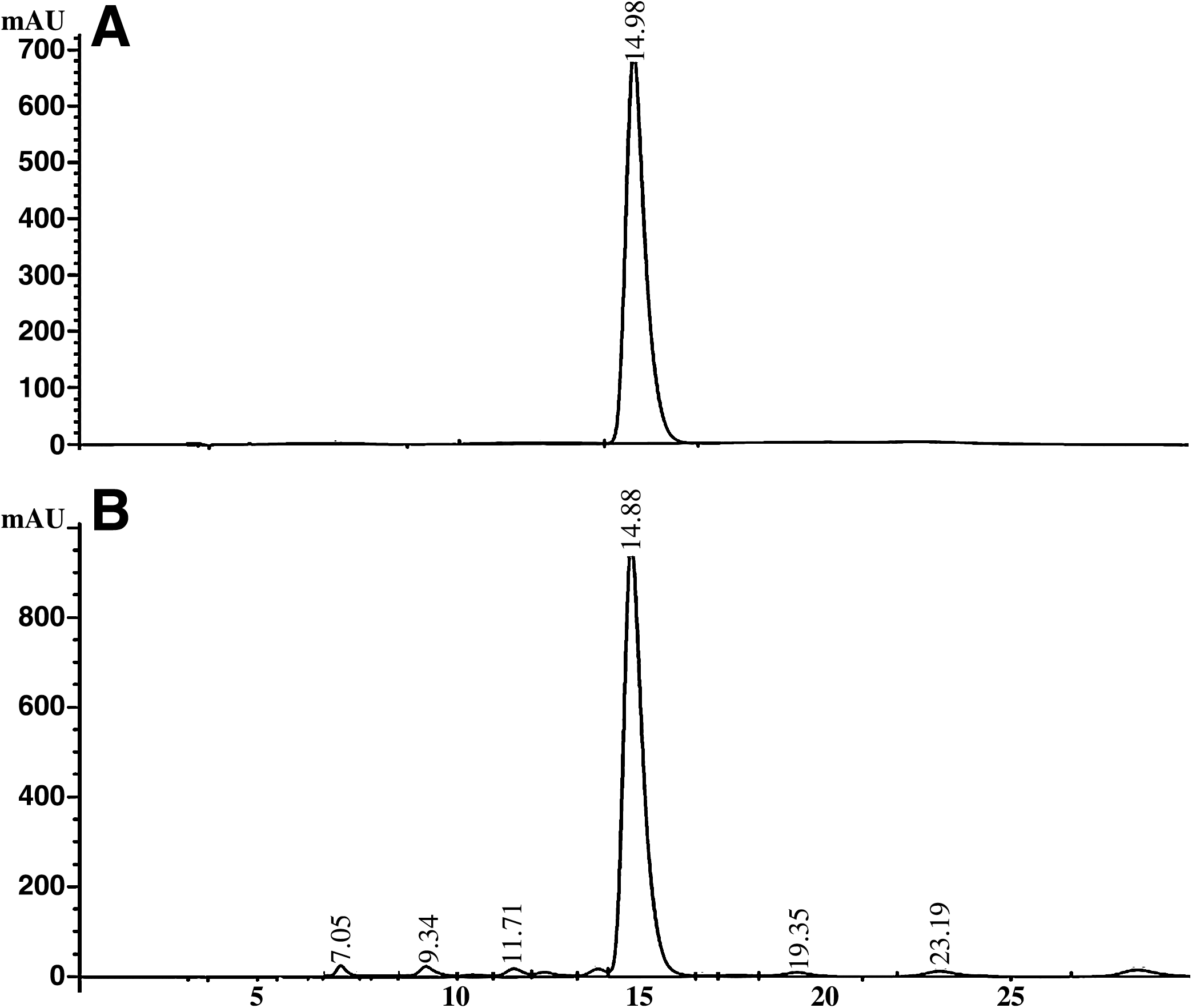
High-performance liquid chromatogram of (
Antioxidative activity and total phenolic content
CFE had strong antioxidative activity with a 50% effective concentration at 10.5 μg/mL, a value about 3 and 1.7 times of that of the standard antioxidants ascorbic acid and α-tocopherol, which were 3.6 and 6.1 μg/mL, respectively. Although the plant extract antioxidative activity is lower than that of the standard ascorbic acid and α-tocopherol, it contained high phenolic content, with a tannic acid equivalence of 307.5 ± 7.8 mg/g and gallic acid equivalence of 161.7 ± 2.7 mg/g.
Gastroprotective effect of CFE on HCl/EtOH-induced ulcer
Oral administration of CFE at doses of 250 and 500 mg/kg significantly inhibited gastric ulcers induced by HCl/EtOH (Table 1). Compared to the positive control, giving CFE after HCl/EtOH induction significantly reduced the number of bleeding spots, area of bleeding spots, ulcer index, and ulcer score in a dose-dependent manner. The ulcer score and ulcer index of the omeprazole-treated group were comparable to those of the 250 mg/kg CFE group. Moreover, giving CFE before HCl/EtOH induction completely prevented gastric ulcer formation as the ulcer score was equal to the negative control group. The plant extract itself did not cause any gastric ulceration. Histological studies confirmed the gastroprotective effect of CFE (Fig. 2). The stomachs of HCl/EtOH-treated animals had ulceration in the mucosa. The mucosal erosion showed injury closed to the submucosa layer (Fig. 3A). Ulcer induction by HCl/EtOH caused certain cell deaths as it was observed that some epithelial cells did not have a nucleus (necrotic cells). Furthermore, there were numerous red blood cells and polymorphonuclear cells, which represented tissue inflammation (Fig. 3B). These inflammation features were not seen in the negative control group (animals receiving distilled water). Giving CFE at doses of 250 and 500 mg/kg can prevent the bleeding and inflammation of the stomach. The tissue erosion observed was not more than one-fourth of the mucosal thickness. Moreover, pretreatment with CFE at a dose of 500 mg/kg inhibited edema and hemorrhage in the gastric mucosa, and results were better than with omeprazole treatment (see Fig. 2C, c, F, and f).
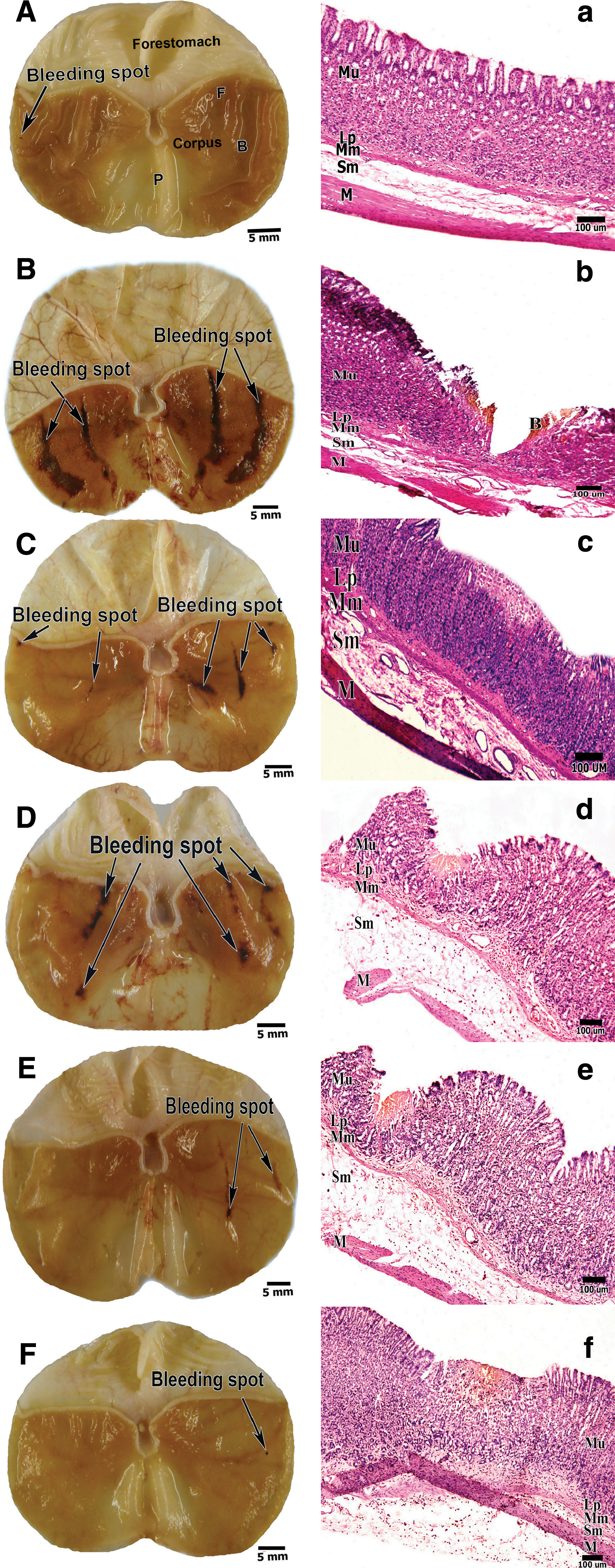
Stomach of rats receiving (
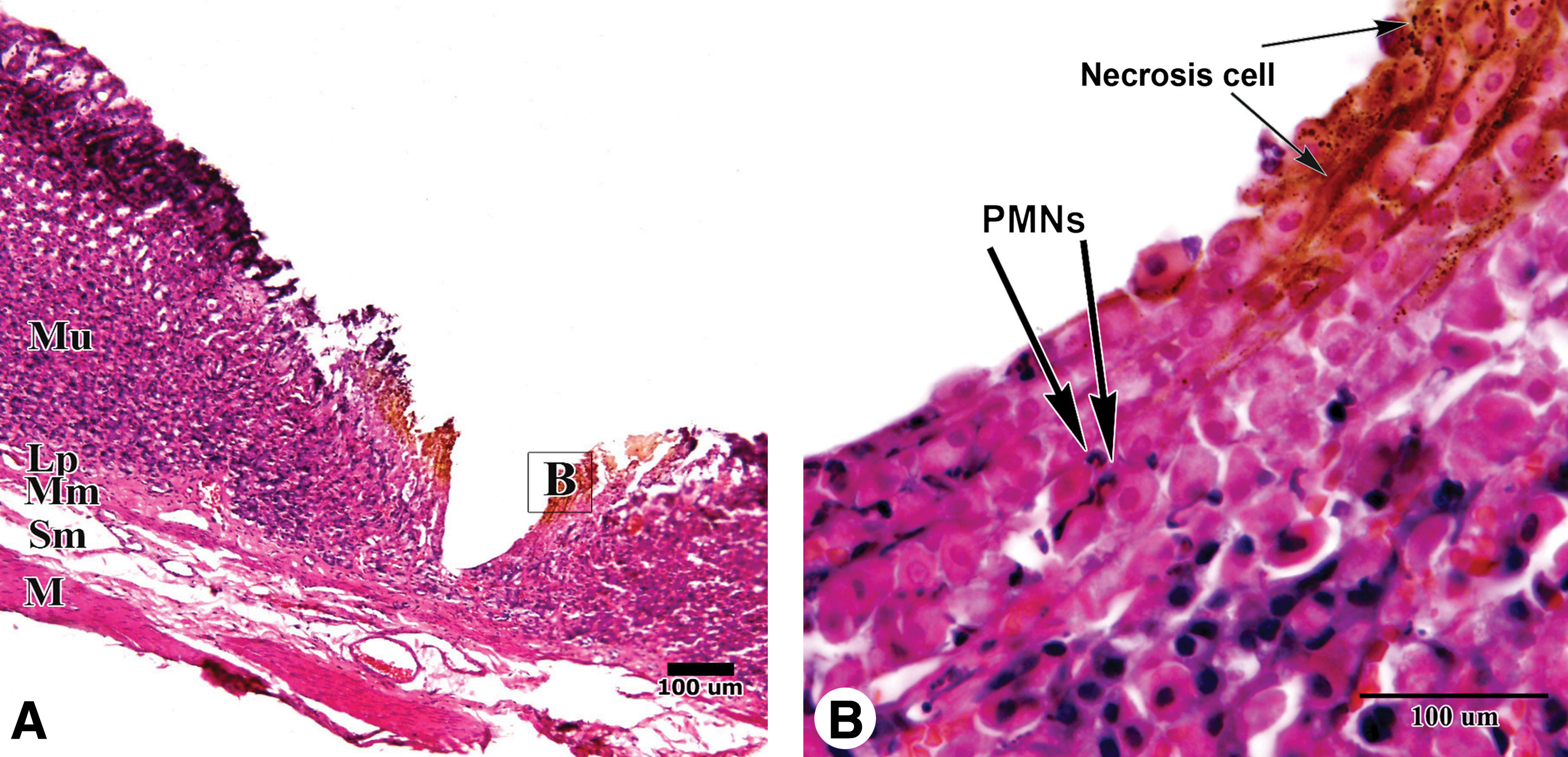
Histological features of stomachs from rats receiving HCl/EtOH. (
Data are mean ± SEM values (n = 7).
Significant differences compared to distilled water, C. formosum ethanolic extract (CFE), HCl/ethanol (EtOH), HCl/EtOH + omeprazole, HCl/EtOH + CFE 250 mg/kg, HCl/EtOH + CFE 500 mg/kg, and CFE 500 mg/kg + HCl/EtOH, respectively, at P < .05.
Effect of CFE on glutathione peroxidase activity and MDA levels
CFE or HCl/EtOH treatment did not affect mucosal glutathione peroxide activity, but giving CFE at the 250 mg/kg dose after HCl/EtOH induction of gastric damage significantly increased glutathione peroxidase activity. In contrast, gastric damage induced by HCl/EtOH showed an increase of MDA. However, giving CFE after the HCl/EtOH induction significantly decreased MDA levels in a dose–response relationship, to values that were better than that giving the standard drug, omeprazole (Table 2).
Data are mean ± SEM values (n = 7). Standard glutathione and 1,1′,3,3′-tetramethoxypropane gave the calibration equations of y = 0.0828x + 0.0001 (r 2 = 0.999) and y = 0.0477x + 0.013 (r 2 = 0.992), respectively.
Significant differences compared to distilled water, CFE, HCl/EtOH, HCl/EtOH + omeprazole, HCl/EtOH + CFE 250 mg/kg, HCl/EtOH + CFE 500 mg/kg, and CFE 500 mg/kg + HCl/EtOH, respectively, at P < .05.
GP, glutathione peroxidase; MDA, malondialdehyde.
Discussion
Several plant or fruit extracts with strong antioxidative activity have been reported to have a gastroprotective effect in rats and mice. 8 –14,29 –31 CFE was shown here to have antioxidative activity. Gastric injury is associated with inflammation is due to the toxicity of reactive oxygen species generated in the stomach. 32 Exposure of gastric mucosa to EtOH has been shown to affect cellular integrity and to be associated with oxidative stress. 33 This study provides evidence that CFE has protective and healing effects against HCl/EtOH-induced gastric ulceration. When CFE was given at a dose of 500 mg/kg prior to HCl/EtOH administration, the number and area of bleeding spots decreased to the control levels. Our results support the earlier report on wound healing by this plant. 18 Areas of bleeding spots in animals given omeprazole were not different from those in the 250 mg/kg and 500 mg/kg CFE-treated groups, suggesting the similar gastroprotective effect of omeprazole and CFE. Administration of HCl/EtOH alone or CFE at 250 mg/kg after HCl/EtOH, however, manifested in an increase of areas of bleeding spots and percentage of bleeding areas compared with the control or the group pretreated with 500 mg/kg CFE. It can be concluded that C. formosum has a gastroprotective effect more than a healing effect. Histological findings showed some epithelial cells without nuclei. Red blood cells and polymorphonuclear neutrophil cells migrated into the gastric mucosa, which represents tissue inflammation. The mechanism of ulceration might come from the production of reactive oxygen species from polymorphonuclear neutrophils that damage the mucosa. 7,9,30 The mechanism of HCl/EtOH induction of gastric mucosa damage was reported to be partially caused by reactive oxygen and nitrogen species. 34,35 The role of free radicals in the process of ulceration was confirmed according to the finding that mucosal MDA levels of animals that received omeprazole or CFE at 250 and 500 mg/kg after HCl/EtOH were less than that of the animals that received HCl/EtOH induction. Because CFE gave a gastroprotective effect similar to that of the proton pump inhibitor omeprazole, the other possible mechanism involved in HCl/EtOH induction of gastric damage should be also further investigated. It is also interesting to note that the 250 and 500 mg/kg doses of CFE are several times less than the plant 50% lethality dose, which was >3 g/kg in mice when given orally, 18 suggesting it is safe and potentially useful as a source of natural plant antioxidants. In term of chemical constituents, chlorogenic acid was found to be the major compound of CFE as previous reported. 18 However, Hamauzu et al. 9 reported that only the extract of Pyrus communis L. containing a mixture of procyanidins and chlorogenic acid had an anti-ulcer effect in rats, whereas chlorogenic acid alone did not have this effect. Therefore, the mixture of polyphenols showed significant gastroprotection.
In conclusion, we report the gastroprotective action of CFE in association with its antioxidative activity. This may come from the high total phenolic content of this plant. However, the relationship of its activity and its protective action needs to be further studied, which may provide new alternatives for clinical management of gastric ulcer disease.
Footnotes
Acknowledgments
This project was supported by the General Research Fund of Khon Kaen University and was approved by the Animal Ethic Committee, Faculty of Medicine, Khon Kaen University (AE 009/50). The authors would like to thank the staff of the Center for Research and Development of Herbal Health Products for preparing the plant extracts used in this study.
Author Disclosure Statement
No competing financial interests exist.