
Abstract
We previously reported that chicken collagen hydrolysate (CCH) has strong angiotensin I converting enzyme (ACE) inhibitory activity and antihypertensive effects on spontaneously hypertensive rats. Here, we investigated the chronic therapy effects of CCH on blood pressure and vascular relaxation in a cardiovascular damage model of Wistar-Kyoto rats induced by N-nitro-
Introduction
H
Chicken extract is widely used as a food throughout the world. Chicken broth contains abundant nitrogenous compounds such as free amino acids and peptides, as well as inosinic acid. 7 Collagen is a major component of the extracellular matrix and exists as a main structural protein for animals, including mammals, birds, and fish. The effects of collagen on suppressing increases in blood viscosity during shock, improving the microcirculation, and promoting recovery and stabilization of blood pressure have been reported. 8 However, the basic mechanism of these effects has not been fully clarified.
Previously, we have reported that chicken breast muscle hydrolysate exerts an antihypertensive effect in spontaneously hypertensive rats. 9 Collagen-derived angiotensin I converting enzyme (ACE) inhibitory peptides have been isolated as the active entity. 10,11 The antihypertensive effect of chicken collagen hydrolysate (CCH) has also been confirmed in mildly hypertensive subjects. We deduced that, in addition to its antihypertensive effect, the CCH had a protective effect on the blood vessels through the activation of endothelial progenitor cells. 12
ACE converts angiotensin I to angiotensin II, which is a strong vasopressor, and inactivates bradykinin, a vasodilator.
13
Studies in animals show that chronic administration of N-nitro-
Endothelial NO inhibits platelet aggregation, thrombogenesis, leukocyte adhesion, and proliferation of vascular smooth muscle cells. 18,19 Decrement of NO from the vascular beds results in increased leukocyte–endothelium interaction via up-regulation of endothelial cell adhesion molecules, which include intercellular adhesion molecule-1 (ICAM-1) and vascular cell adhesion molecule-1. 20 Under this condition, numerous leukocytes adhere to the vascular endothelium and migrate across it, thus aggravating endothelial dysfunction and tissue injury. 21 In contrast, systemic administration of NO donors to NO-deficient animals preserves endothelial function and attenuates pathological interactions between circulating leukocytes and the vascular endothelium.
CCH, with its antihypertensive effect, may be useful as a functional food in the treatment of cardiovascular diseases such as hypertension and atherosclerosis, but this issue has not yet been fully explored.
In this study, we investigated whether CCH can prevent hypertension and cardiovascular disorders, as well as the pathway of the putative protective effect of CCH on structural cardiovascular remodeling in a model of cardiovascular damage induced by
Materials and Methods
CCH preparation
The unfeathered portions of chicken legs (with claws) were solubilized by acid treatment and then digested with Aspergillus oryzae protease (Mitsubishi-Kagaku Food Co., Tokyo, Japan). Fractions with molecular masses less than 3,000 Da were collected with an ultrafiltration membrane (Millipore Co., Bedford, MA, USA) and were designated as CCH. After ultrafiltration, CCH solution was eluted on an activated carbon column for decolorization and then lyophilized.
Experimental animals
Eight-week-old male WKY rats were fed on a commercial chow (MF; Oriental Yeast, Tokyo) and water for 2 weeks ad libitum in an environmentally controlled room (23°C, 55% humidity). Then, 36 male WKY rats were randomly divided into three groups. The first group (control group) received untreated chow and drinking water. The second group (
Measurement of systolic blood pressure (SBP)
Tail SBP was measured every 2 weeks by the tail-cuff method with a plethysmographic tail apparatus (model 98A, Softron Co., Tokyo). Before the measurements, the rats were kept at 37°C for 10 minutes to make the pulsations of the tail artery detectable.
Vasorelaxation assay
The vasorelaxation assay was performed on eight or nine rats from each group at 8 weeks post-treatment. The rats were euthanized with diethyl ether, and the thoracic aorta was removed and placed in Krebs-Henseleit solution at 4°C.
22
The solution contained the following chemicals: 118.3 mM NaCl, 4.7 mM KCl, 2.0 mM CaCl2, 1.2 mM MgSO4, 25.0 mM NaHCO3, 1.2 mM KH2PO4, 0.026 mM calcium EDTA, and 11.1 mM glucose. The surrounding connective tissue and fat were carefully removed away from the thoracic aorta and cut into 2–3-mm-wide rings. Segments of thoracic aorta were mounted between two steel hooks in isolated tissue chambers containing Krebs-Henseleit solution (pH 7.4) at 37°C; the solution was continuously aerated with a mixture of 95% O2 and 5% CO2. An optimum resting tension of 1.0 g was applied to all aortic segments, and the tension was adjusted every 15 minutes during a 45-minute equilibration period before addition of the reagents. The isometric tension was recorded with an isometric force displacement transducer (model UL-10GR, Mineber, Tokyo) connected to an acquisition system (MSC-2, Labo Support Co., Ltd., Osaka, Japan). After the equilibration period, segments were initially exposed twice to 75 mM KCl to test their functionality and their maximum contractility. After a 60-minute washout period,
Determination of soluble ICAM-1 (sICAM-1) concentration
Plasma sICAM-1 levels were determined every 4 weeks by using commercially available enzyme-linked immunosorbent assay kits (R& D Systems Inc., Minneapolis, MN, USA). 23 Blood samples were collected from the tails of rats using a heparin-treated capillary tube and then centrifuged for 20 minutes at 2,000 g. Anti-sICAM-1 monoclonal antibody labeled with horseradish peroxidase (R& D Systems Inc.) and sICAM-1 standards and samples were added to the wells and incubated for 2 hours at room temperature. The wells were washed five times, and then the substrate solution was added. After 30 minutes of incubation at room temperature, the reaction was stopped by addition of 0.1% HCl. Absorbance was measured at a wavelength of 450 nm, with the correction wavelength set at 540 nm, on a model 3550 microplate reader (Bio-Rad, Tokyo).
Determination of serum NOx concentration
Blood samples were collected from the tails of rats anesthetized with diethyl ether and centrifuged at 10,000 g for 5 minutes. The supernatant was mixed with an equal amount of ethanol and then centrifuged at 10,000 g for 5 minutes. The supernatant collected was used for measurement of NO x , as previously described. 24 The sample was put into an automatic sample injector connected to an automatic detector–high-performance liquid chromatography system (model ENO-20, Eicom, Kyoto, Japan). NO2 − and NO3 − in the dialysate were separated on a reverse-phase separation column packed with polystyrene polymer (NO-PAK, 4.6–50 mm; Eicom), and NO3 − was reduced to NO2 − in a reduction column packed with copper-plated cadmium filings (NO-RED; Eicom). NO2 − was mixed with a Griess reagent to form a purple azo dye in the reaction coil. Contamination of NO2 − and NO3 − in Ringer's solution and the reliability of the reduction column were examined in each experiment.
Histopathological observations
Histopathological observation was performed by a single observer who was blinded to all treatment protocols. Five paraffin-embedded sections were prepared from each tissue, as previously described. 25 In brief, tissues isolated from each group were fixed with formalin, then embedded in paraffin, sectioned, and stained with hematoxylin and eosin or Masson's trichrome staining solution. Slices were examined under a light microscope for histopathological changes. Representative sections were photographed by an automatic photomicrographic system (Vanox, Olympus, Tokyo).
Statistical evaluations
All data were analyzed and expressed as mean ± SE values. Comparisons were performed by Tukey's test to detect differences between the groups. A probability value less than .05 was considered as statistically significant.
Results
Survival curves
At 8 weeks post-treatment, the survival rate of
Survival curves of the control group,
Effect on antihypertensive measures
The effects of oral administration of

Changes of SBP in the control group,
Effect on vasorelaxant measures
Figure 3 shows the relaxation caused by administration of CCH for 8 weeks, as determined by screening of the rat thoracic aortic segments. Treatment with acetylcholine chloride caused concentration-dependent relaxation of the thoracic aortic preparations from all groups after the preparations had been precontracted with L-norepinephrine bitartrate. The acetylcholine chloride induced a relaxant response in the thoracic aorta from the
Relaxation of rat thoracic aortas. Rat thoracic aorta was precontracted with
Effects on plasma sICAM-1 concentration
Plasma sICAM-1 levels did not differ significantly between 4 and 8 weeks post-treatment with
Concentration of sICAM-1 in rat plasma. Blood samples were collected from the tail vein into heparinized capillary tubes and then centrifuged at 12,000 g for 5 minutes at 4°C. Plasma was collected and stored at −80°C until analysis. Data are mean ± SE values (n = 8–9 rats). *P < .05 versus
Changes in serum NOx concentration
Changes in NO x were measured in the sera of the rats. After 8 weeks, serum NO x concentrations did not differ significantly among the groups (data not shown). However, we found in an additional experiment that the NO x concentration in the serum of rats that ingested CCH was significantly greater than that of control rats that ingested water at 1 hour post-treatment, whereas at 2 hours post-treatment they were nearly same (Fig. 5). This result indicated that CCH treatment could increase the NO concentration transiently.
Transient change of NO x concentration in serum from a rat treated with a single dose of CCH (2 g/kg of body weight). Blood samples were collected from the tail vein of the rat, stored at 4°C for 10 minutes, and then centrifuged at 10,000 g for 5 minutes at 4°C. Serum was collected and stored at −80°C until analysis. Data are mean ± SE values (n = 6 rats). *P < .05 versus no CCH treatment.
Histopathology of heart, kidney, and thoracic aorta
The size of focal lesions and the extent of invasion by inflammatory cells were evaluated by histopathological methods. The results revealed that the focal fibrosis appeared in sections of rat heart from the

Histopathological photomicrographs of sections from the control group,
Discussion
This study demonstrated that treatment with CCH partially restored survival rates and attenuated the increase in inflammatory and proliferative changes induced in the heart, kidney, and thoracic aorta by
We previously reported that CCH reduced blood pressure in spontaneously hypertensive rats by inhibiting ACE activity.
9,11
Several reports have shown that local expression of ACE plays a key role in the pathogenesis of the coronary vascular and myocardial remodeling induced in rats by long-term administration of
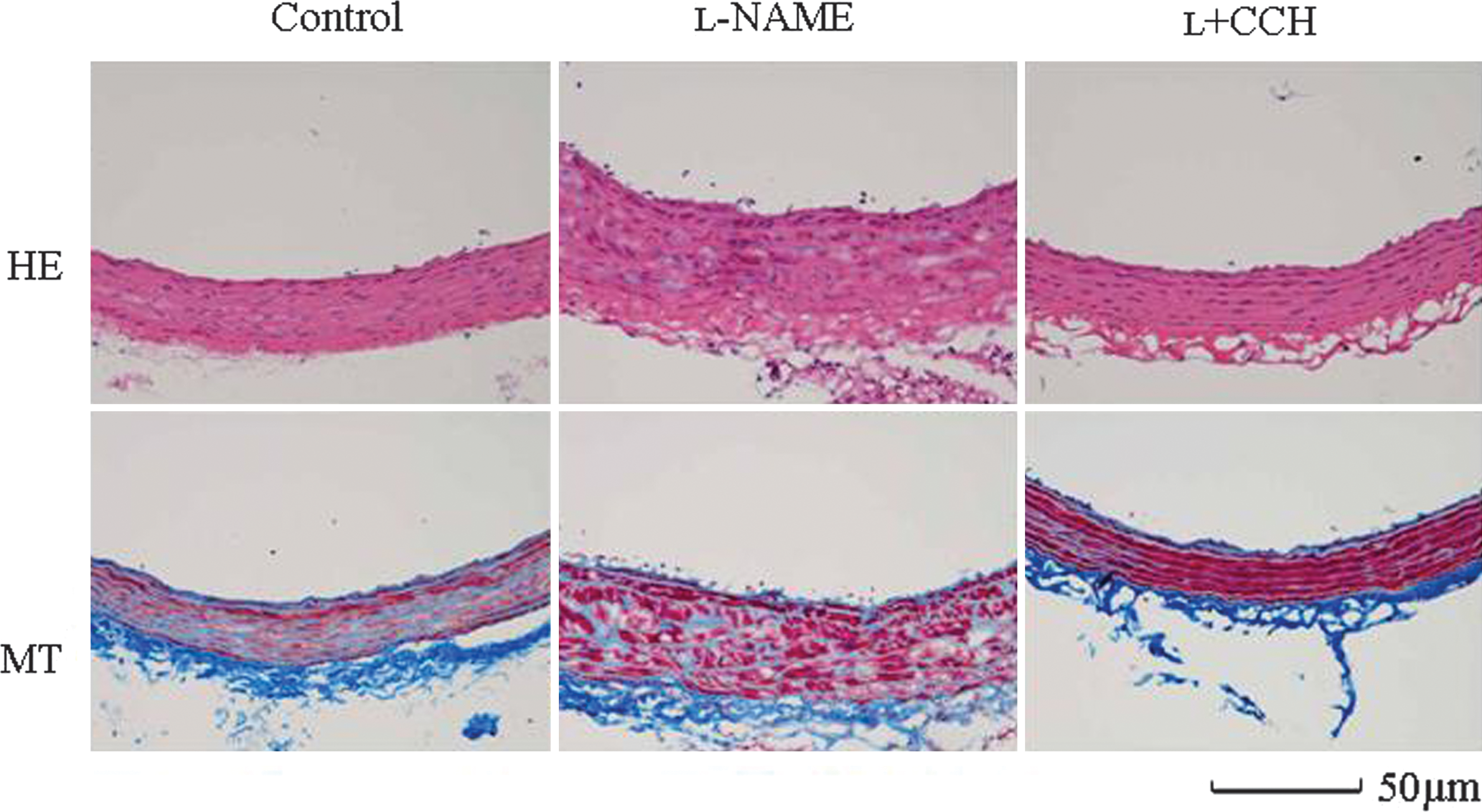
Histopathological photomicrographs of thoracic aorta sections from the control group,
To investigate the antihypertensive mechanism of CCH and the pathway of prevention of cardiovascular remodeling in the heart, kidneys, and thoracic aorta, we determined the changes of NO
x
concentration in sera of the L + CCH group rats. The serum concentration of NO
x
did not change in rats that ingested CCH for 8 weeks. However, an increase in tissue ACE activity precedes the development of vascular and myocardial remodeling induced by long-term inhibition of NO synthesis.
15
Therefore, we performed an additional experiment in which we gave a single dose of CCH to rats and measured the change in serum NO
x
concentration. The NO level increased at 1 hour post-treatment of CCH and then returned to the basal level (Fig. 5). This result indicated that the serum NO level was transiently increased by treatment with CCH, suggesting that CCH treatment could ameliorate the
Various studies have reported that several peptides derived from food materials reduce vascular resistance and have blood pressure-lowering effects in rats. 32 We isolated nine specific peptides from human blood after oral intake of CCH, and we found that these peptides possessed ACE inhibitory activities, 33 although the mechanisms by which CCH inhibits remodeling activity have not been completely proven in vivo. More studies are needed to reveal the active substances in CCH and to clarify the antihypertensive and vasodilatory mechanisms of these substances.
Footnotes
Acknowledgment
This study was supported by a 2006 Grant-in-Aid for scientific research on “Study of the high value of foods and materials from stockbreeding that prevent lifestyle-related diseases,” from the Bio-oriented Technology Research Advanced Institution of Japan.
Author Disclosure Statement
All authors are employees of Nippon Meat Packers Inc. No competing financial interests exist.