
Abstract
Vineatrol®30 (developed and produced jointly by Breko GmbH [Bremen, Germany] and Actichem [Montauban, France]) is a grapevine-shoot extract that contains resveratrol as well as considerable amounts of resveratrol oligomers. In the present study it is shown that Vineatrol30 at a noncytotoxic concentration of 2.3 μg/mL significantly reduced the number of malignantly transformed foci induced by a sequential treatment of BALB/c-3T3 cells with 3-methylcholanthrene and 12-O-tetradecanoylphorbol 13-acetate in the so-called BALB/c-3T3 cell transformation assay. At a higher concentration Vineatrol30 drastically decreased the relative plating efficiency of the cells. Furthermore, the results suggest that the resveratrol oligomers present in Vineatrol30, independently from resveratrol itself, were indeed able to inhibit the formation of malignantly transformed BALB/c-3T3 foci.
Introduction
Several different in vitro cell transformation assays for the detection of carcinogenic compounds have been established. The advantages, limitations, and recommendations for future use of three of them—the Syrian hamster embryo cell transformation assay, the C3H10T1/2 cell transformation assay, and the BALB/c-3T3 cell transformation assay—have recently been reviewed in detail by the Organization for Economic Co-operation and Development. 13 As pointed out in this detailed review the transformation process in BALB/c-3T3 cells parallels the induction and progression of tumors in vivo. 13 A two-stage protocol for the BALB/c-3T3 cell transformation assay was described by Sakai and Sato 14 and optimized over the years by Umeda and colleagues at the Hatano Research Institute, Hatano City, Japan. 15 –17 In the meantime the BALB/c-3T3 cell transformation assay has successfully been used to analyze the transforming capacity of pesticides, metal compounds, quartz, and 3-methylcholanthrene (MCA). 18 –23
In the present study the BALB/c-3T3 cell transformation assay was used to determine whether Vineatrol30 is able to inhibit the malignant transformation of cells. The individual polyphenols present in complex polyphenol mixtures such as Vineatrol30 may act synergistically and therefore elicit a stronger health-promoting effect than resveratrol alone. This in fact was demonstrated when comparing the antiproliferative effects of Vineatrol30 and resveratrol in chronic B lymphocytic leukemia cell cultures. 11 Hence, in the present study we also compared the capacity of Vineatrol30 with that of resveratrol to inhibit the malignant transformation of BALB/c-3T3 cells.
Materials and Methods
Chemicals
The grapevine-shoot extract Vineatrol30 was kindly provided by the joint manufacturers, Breko GmbH and Actichem. Vineatrol30 includes 15.2% resveratrol, 13.2% ɛ-viniferin, 4.4% ampelopsin A, 2.8% hopeaphenol, 2.1% iso-trans-ɛ-viniferin, 1.9% vitisin A, 1.9% vitisin B, 1.8% piceatannol, and 1.6% miyabenol C. Resveratrol, MCA, and 12-O-tetradecanoylphorbol 13-acetate (TPA) were purchased from Sigma-Aldrich (Steinheim, Germany), methanol from Carl Roth (Karlsruhe, Germany), and Giemsa staining solution (0.76% Giemsa stain in a 1:1 methanol/glycerin mixture) from AppliChem (Darmstadt, Germany). Stock solutions of resveratrol, MCA, TPA, and Vineatrol30 were prepared in dimethyl sulfoxide (DMSO), aliquoted, and stored at –20°C.
Cell culture
The BALB/c-3T3 clone A31-1-1 from the laboratory of Makoto Umeda (Hatano Research Institute) was kindly provided by Dr. A. Poth (Harlan Cytotest Cell Research, Roßdorf, Germany). Cells were grown in minimum essential medium from Invitrogen (Karlsruhe) supplemented with 10% fetal bovine serum (Moregate, Bulimba, Australia), 100 μg/mL streptomycin, and 100 U/mL penicillin (Biochrom, Berlin, Germany) at 37°C under a humidified 5% CO2 atmosphere. Cells were passaged after reaching 80% confluence, and a working stock (i.e., the one cell passage used for the complete study) was frozen in liquid nitrogen in a medium consisting of 80% minimum essential medium, 10% fetal bovine serum, and 10% DMSO.
BALB/c-3T3 cell transformation assay
The ability of Vineatrol30 and resveratrol to inhibit the malignant transformation of BALB/c-3T3 cells was tested by performing the two-stage cell transformation assay according to Sakai and Sato. 14 A scheme of the treatment protocol used in the present study is shown in Figure 1. Exponentially growing BALB/c-3T3 cells were plated at a density of 104 cells per 60-mm dish in 4 mL of cell culture medium; eight dishes per resveratrol/Vineatrol30 concentration were used. Twenty-four hours later the cell culture medium was replaced by a cell culture medium containing 0.5 μg of MCA/mL. Thereafter the medium containing MCA was replaced by MCA-free cell culture medium. Three days later (i.e., on day 8 after beginning the assay) the cell culture medium was replaced by medium containing 0.3 μg of TPA/mL, and the cells were cultured up to day 22 in TPA-containing medium. After the TPA-containing cell culture medium was replaced, the cells were cultured for another 3 weeks in normal (MCA- and TPA-free) cell culture medium. Resveratrol or Vineatrol30 was present at the given concentrations in the cell culture medium during the whole 6-week time period (Fig. 1). At the end of week 6 cells were fixed in methanol and stained with Giemsa solution. The cell culture medium was changed every 3–4 days, and whenever a medium change was performed the medium included a fresh aliquot of the corresponding test compound(s). In control experiments MCA, TPA, or both were replaced by DMSO.
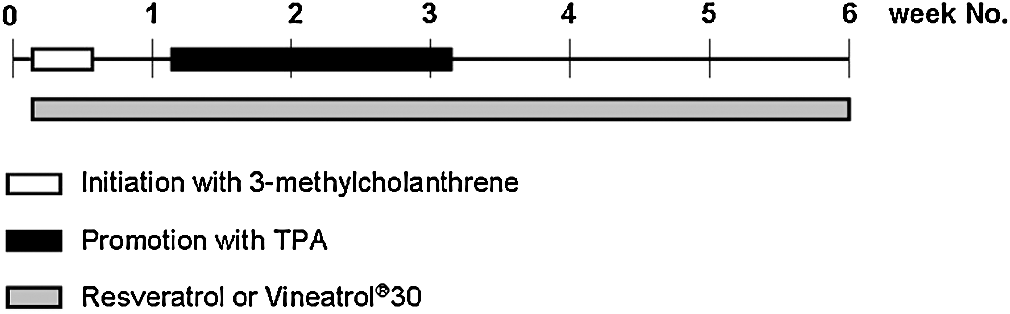
Scheme of the BALB/c-3T3 cell transformation assay as performed in the present study. Cells were initiated by treating them between day 2 and 4 with 0.5 μg of 3-methylcholanthrene/mL. The tumor promoter 12-O-tetradecanoylphorbol 13-acetate (0.3 μg/mL) was included in the cell culture medium between day 8 and day 22. Resveratrol or Vineatrol30 was present in certain experimental groups during the whole 6-week time period. At the end of the 6-week time period foci were fixed in methanol, stained with Giemsa solution, and counted.
The final concentration of DMSO in the cell culture medium was 0.1% (vol/vol), and DMSO was also present in control cultures that were not incubated with resveratrol or Vineatrol30 to maintain the same level of solvent.
Three types of foci (type I, II, and III foci) were identified and scored according to Reznikoff et al. 25 Type I foci are considered not to be fully transformed, whereas type II and type III foci are fully transformed. Type I foci are composed of tightly packed cells. Type II foci are composed of strongly basophilic cells that show massive piling up, but neither criss-crossing at the edge of the foci nor invasion of the surrounding cell monolayer. Type III foci are composed of strongly basophilic cells that show massive piling up and invade the surrounding cell monolayer. Furthermore, in the case of type III foci criss-crossing is evident at the edge of them. Foci of less than 3 mm in diameter were not scored.
Plating efficiency assay
The plating efficiency was determined in parallel to the cell transformation assay. Two hundred cells per 60-mm dish were seeded and treated for 11 days with the corresponding test compounds as in the case of the cell transformation assay (Fig. 1). All treatments were performed in quadruplicate. After 11 days dishes were fixed in methanol and stained with Giemsa solution. All the cell colonies were counted, and the relative plating efficiency was calculated according to the following equation:
Statistical analysis
The statistical analysis of the results was carried out by performing an analysis of variance followed by Dunnett's multiple comparison test 26 with the software SPSS version 11 for Windows (SPSS, Inc., Chicago, IL, USA).
Results
Incubation of BALB/c-3T3 cells with MCA or TPA alone only led to a modest increase in the number of type I, II, and III foci compared to DMSO-treated cells (Table 1). In contrast, the sequential treatment of BALB/c-3T3 cells with the tumor initiator MCA and the tumor promoter TPA resulted in a strong increase in the number of type I, II, and III foci per dish compared with DMSO-, MCA-, or TPA-treated cells (Table 1). Hence, the malignant cell transformation assay described by the research group of Umeda at the Hatano Research Institute could successfully be established in our laboratory.
The values for treatment groups 1–4 and 6 represent the mean ± SD values of the number of type I, II, and type III foci formed in three to five independent experiments. The values for treatment groups 5, 7, and 9 represent the means of two independent experiments.
Significantly different from dimethyl sulfoxide (DMSO)-treated cells (P < .05).
Significantly different from 3-methylcholanthrene (MCA)- and 12-O-tetradecanoylphorbol 13-acetate (TPA)-treated cells (P < .05).
Significantly different from MCA + TPA-treated cells (P < .05).
Res, resveratrol.
In a second step the effect of resveratrol on the malignant transformation of BALB/c-3T3 cells was studied. Resveratrol at a concentration of 1.1 μg/mL of cell culture medium was not able to reduce the number of foci per dish formed by MCA + TPA (Fig. 2), whereas addition of 2.3 μg of resveratrol/mL to the cell culture medium of cells being treated with MCA + TPA led to a statistically significant reduction in the total number of foci counted per dish compared to the MCA + TPA group (Fig. 2). Furthermore, 2.3 μg of resveratrol/mL significantly decreased the number of type I, II, and III foci per dish compared with cells being incubated with only MCA and TPA (Table 1). In order to be able to interpret the results obtained and as recommended by the IARC/NCI/EPA Working Group, 27 cytotoxicity was examined by determining the relative plating efficiency. MCA itself led to a strong cytotoxic effect, which was not further enhanced by addition of TPA (Table 2). The two concentrations of resveratrol tested in the present study resulted in a strong cytotoxic effect on BALB/c-3T3 cells as evidenced by the extremely strong decrease in the relative plating efficiency observed in the two MCA + TPA + resveratrol groups compared to the MCA + TPA group (Table 2).
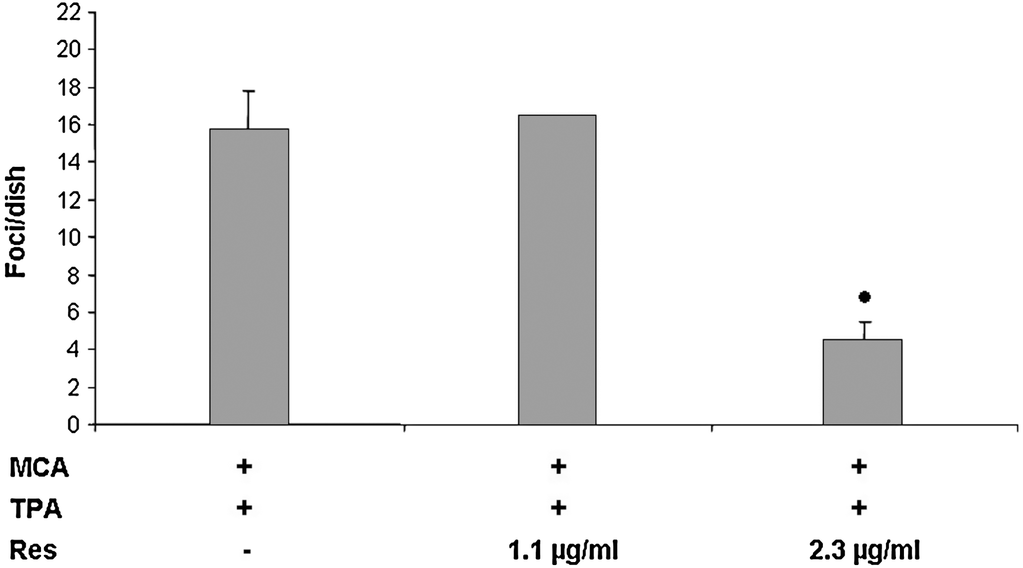
Effect of Res on the malignant transformation of BALB/c-3T3 cells. BALB/c-3T3 cells were treated with MCA and TPA as described in Figure 1. Res was present during the whole 6-week time period. The total number of foci per dish is shown. The results are expressed as the mean of two independent experiments in the case of the incubation of the cells with 1.1 μg of Res/mL and as the mean ± SD values of three independent experiments in the case of the incubation of the cells with 2.3 μg of Res/mL or without Res. Eight dishes per experiment and experimental group were assayed in parallel. The black dot indicates a value significantly different from the solvent control value (analysis of variance followed by a two-sided Dunnett's test, P < .01).
Data are means of two independent experiments.
Incubation of MCA + TPA-treated BALB/c-3T3 cells with Vineatrol30 decreased the total number of foci per dish formed; this decrease became statistically significant at a Vineatrol30 concentration of 2.3 μg/mL (Fig. 3). At this concentration Vineatrol30 reduced the number of type I, II, and III foci per dish having been formed by treatment of the cells with MCA + TPA (compared to cells having been treated with MCA + TPA alone), but only the reduction in the number of type I and II foci per dish reached a statistically significant level (Table 1). The relative plating efficiency of the cells that had been incubated with MCA, TPA, and 0.2 or 2.3 μg of Vineatrol30/mL was similar to that of cells that had been treated with MCA + TPA alone, whereas a drastic decrease in the relative plating efficiency was observed with 13.7 μg of Vineatrol30/mL (Table 2).
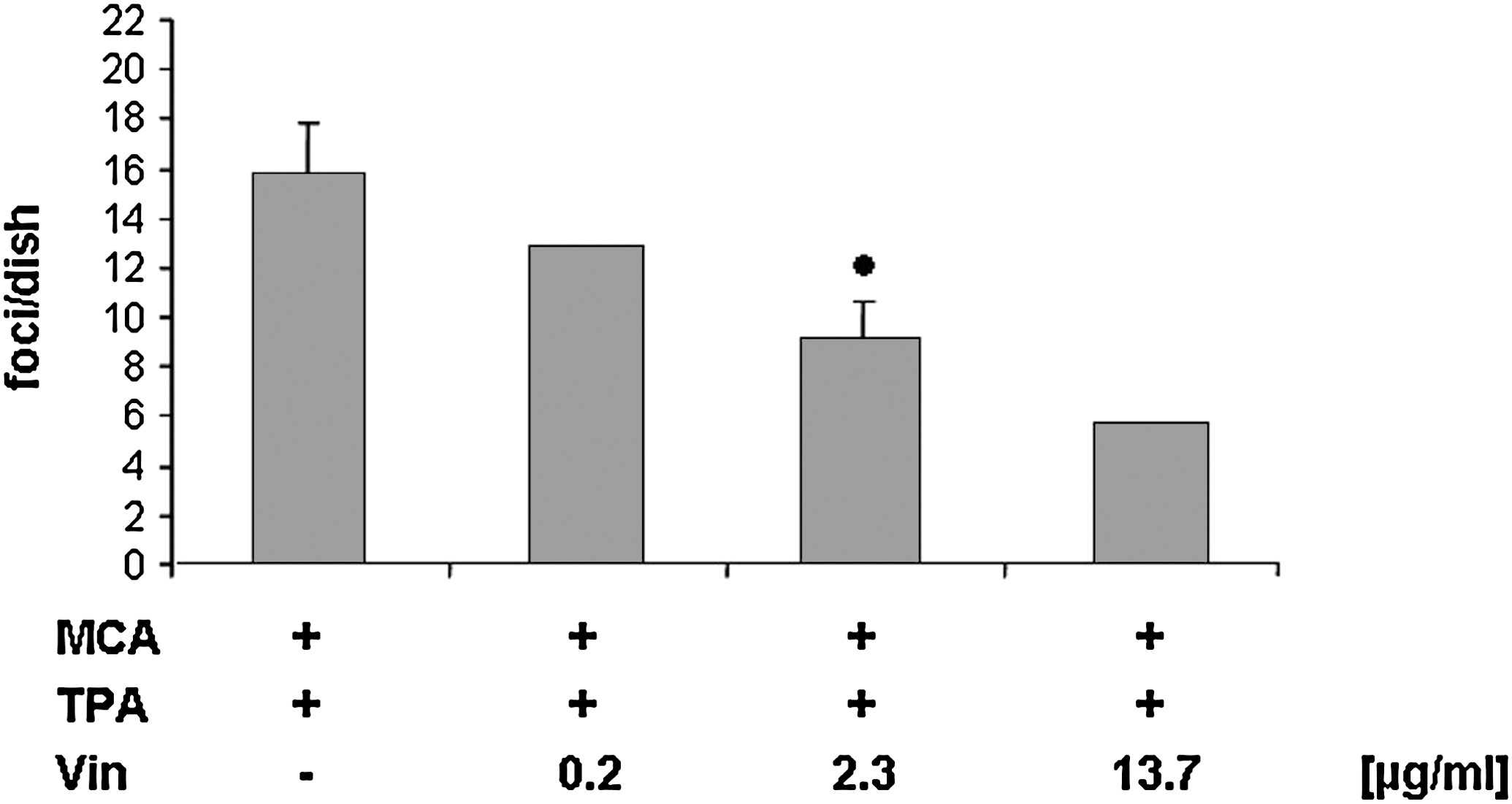
Effect of Vineatrol30 (Vin) on the malignant transformation of BALB/c-3T3 cells. BALB/c-3T3 cells were treated with MCA and TPA as described in Figure 1. Vin was present during the whole 6-week time period. The total number of foci per dish is shown. The results are expressed as the mean of two independent experiments in the case of the incubation of the cells with 0.2 and 13.7 μg of Vin/mL or without Vin and as the mean ± SD values of three independent experiments in the case of the incubation of the cells with 2.3 μg of Vin/mL. Eight dishes per experiment and experimental group were assayed in parallel. The black dot indicates a value significantly different from the solvent control value (analysis of variance followed by a two-sided Dunnett's test, P < .01).
Discussion
The BALB/c-3T3 cell transformation assay has been used over the years to test the tumor-initiating and tumor-promoting capacity of chemicals. In a limited number of studies the two-stage cell transformation assay has also been shown to be suited for the identification of compounds being able to suppress the initiation and/or promotion steps of tumor development. 28,29 In the present study it is shown for the first time that the grapevine-shoot extract Vineatrol 30 inhibits the malignant transformation of BALB/c-3T3 cells treated with MCA and TPA. Furthermore, Vineatrol30 led to a decrease in the number of foci that are considered to be “not fully transformed” (type I foci) as well as in the number of foci considered to be “fully transformed” (type II and III) according to Reznikoff et al., 25 whereas in the case of type III foci the decrease did not reach a statistically significant level. The lowest concentration of Vineatrol30 leading to malignant cell transformation inhibition (2.3 μg/mL) did not reduce the plating efficiency of the cells to a greater degree than the sequential treatment of the cells with MCA and TPA alone. However, at a concentration of 13.7 μg of Vineatrol30/mL the plating efficiency of the BALB/c-3T3 cells was dramatically reduced. These results suggest that the ability of Vineatrol30 to inhibit the chemically induced malignant transformation of BALB/c-3T3 cells relies on more than one mechanism of cell toxicity. In agreement with this suggestion is the observation made by Marel et al. 12 that low concentrations of Vineatrol (i.e., 1.1 μg/mL) were able to inhibit the proliferation of the human carcinoma cell line SW480 by elongating the S phase of their cell cycle, which in turn prevented the cells from entering the G2/M phase; after addition of higher concentrations of Vineatrol (i.e., >4.6 μg/mL) the SW480 cells died off by necrosis or apoptosis. Whether these mechanisms of cell toxicity also apply to the chemically transformed BALB/c-3T3 cells treated with low and high concentrations of Vineatrol30 remains to be demonstrated.
In terms of anticarcinogenic mechanisms one should bear in mind that Vineatrol30 also possesses antioxidative capacity. In a very recent study it has been shown that Vineatrol30 acts as a free radical scavenger, inhibits lipid peroxidation, and enhances human glutathione peroxidase 1 as well as human superoxide dismutase 1 gene promoter activities at noncytotoxic concentrations. 30 Oxidative DNA damage has been suggested to be involved in the initiation as well as in the promotion step of carcinogenesis. 31 Based on their antioxidative properties one would expect polyphenols to be able to inhibit the initiation and/or promotion step of carcinogenesis. In this context recent studies document that this is in fact the case. For example, quercetin is able to inhibit lipid peroxidation, to enhance the concentration of reduced glutathione, and to increase glutathione peroxidase, superoxide dismutase, and catalase activities in the liver of rats treated with diethylnitrosamine and in parallel to inhibit the development of preneoplastic foci in the livers of these animals. 32 Furthermore, the anti-tumor-promoting effects of polyphenols present in grape extracts 33 and black tea 34 have been documented.
The individual polyphenols present in complex polyphenol mixtures such as Vineatrol30 may act synergistically and therefore elicit a stronger decrease in the number of malignantly transformed foci formed than resveratrol alone. In the present study 1.1 μg of resveratrol/mL did not lead to a decrease in the number of foci per dish, whereas 2.3 μg of resveratrol/mL reduced the number of foci per dish by about 72%. When wanting to compare the effect of resveratrol with that of Vineatrol30 on the formation of MCA + TPA-treated foci, one has to bear in mind that Vineatrol30 includes 15.2% resveratrol. Vineatrol30 at a concentration of 2.3 μg/mL (resveratrol content, 0.35 μg/mL) decreased the number of malignantly transformed foci by 42%, whereas 13.7 μg of Vineatrol30/mL (resveratrol content, 2 μg/mL) reduced the number of foci by about 64%. Thus, it seems that polyphenols other than resveratrol present in Vineatrol30 are indeed able to inhibit the formation of foci when resveratrol is present in low amounts (i.e., when adding 2.3 μg of Vineatrol30/mL to the cell cultures), whereas the inhibiting effect of a high concentration of Vineatrol30 is mainly due to resveratrol present in the extract.
In a recent review by Yadav et al. 35 the biological and medicinal properties of grapes were described. In this context it was pointed out that the anticarcinogenic activity of grapes might be due to a variety of bioactive constituents present in them, such as resveratrol, procyanidins, epigallocatechin 3-gallate, and anthocyanins. 35 Because Vineatrol30 is an extract that includes resveratrol, resveratrol oligomers, and up to now unidentified polyphenols, at present one cannot ascribe the inhibiting effect of Vineatrol30 on the chemically induced malignant transformation of cells to a particular component. It very well could be that besides resveratrol and reveratrol oligomers, further compounds, such as those mentioned by Yadav et al., 35 might mediate the effects described in this study.
Taken together, the in vitro studies performed up to now have shown that Vineatrol30 exerts a growth-inhibiting effect on human cancer cells 11,12,36 and inhibits the chemically induced malignant transformation of BALB/c-3T3 cells at a noncytotoxic concentration (this study). If one takes into account that Vineatrol30 also shows a potent antioxidative activity, 30 this extract might be a promising candidate for cancer chemoprevention, in particular to avoid cancer development in the gastrointestinal tract.
Footnotes
Acknowledgment
This study was supported by grant 0315373C from the Federal Ministry for Education and Research in Germany.
Author Disclosure Statement
No competing financial interests exist.