
Abstract
Laurus nobilis L. (Family Lauraceae) is an evergreen tree widely distributed in the Mediterranean area and Europe. It is used in folk medicine of different countries as a stomachic and carminative as well as in treatment of gastric diseases. Extracts obtained with different methods (methanol and chloroform) from laurel leaves were evaluated for their gastroprotective activities in the rat. The antioxidant capacity of the different extracts has been also measured in vitro. In order to confirm the activities investigated, histological observations were performed. The gastric damage was significantly reduced by all extracts administered. The more effective protection was produced by chloroformic and methanolic crude extracts. The results obtained after oral administration of L. nobilis leaf extracts are in good agreement with their antioxidant capacity, confirming the relationship between pharmacological efficacy and antiradical activity. Histological evidences confirm the results evaluated with the animal procedures.
Introduction
L
The aim of the present study was to verify the folkloric claims for this plant in gastric ailments. As previously reported 10 the phytochemical content of Laurus leaves suggests a significant antioxidant activity. The protection of a chloroformic and a methanolic extract of laurel leaves against gastric damage was the object of our investigations in rats. At the same time the antioxidant activity of the extracts of L. nobilis was evaluated with three different chemical in vitro methods based on the free radical scavenging properties of antioxidants: the Briggs–Rauscher (BR) method under acidic conditions, the Trolox equivalent antioxidant capacity (TEAC) assay under physiological conditions (pH 7.4), and the 2,2-diphenyl-1-picrylhydrazyl (DPPH) (in methanol) assay. The total phenolic content of the extracts was also measured by using the Folin–Ciocalteu (FC) reagent, even if more selective, precise methods are now available such as 1 H-nuclear magnetic resonance or micromethods. Indeed, the FC method was very recently recommended for quantifying the reducing capacity of antioxidants in matrices. 11,12 The chemical in vitro evaluation of the antioxidant capacity is important because free radicals are developed in gastric mucosal lesions. 13 Finally, histological observations of the gastric mucosa were performed to confirm the investigated activity and to compare the efficacy of the three different extracts.
Materials and Methods
Plant material and extraction
Leaves of L. nobilis L. (Family Lauraceae) were collected in November 2003 at S. Basilio (province of Cagliari, Sardinia). The plant was identified by Dr. Maria Cecilia Loi, Department of Botanical Sciences, University of Cagliari, Cagliari, Italy, where a voucher specimen was deposited (voucher number LN031103SB).
Air-dried powdered leaves of L. nobilis (200 g) were exhaustively extracted in a Soxhlet apparatus (model B-811, Buchi, Flawil, Switzerland) with methanol. The solvent was removed under reduced pressure, and a methanolic crude extract (ME) was obtained. Yield in weight of residue, relative to the weight of dry material extracted, was 28.6%. Air-dried powdered leaves of L. nobilis (350 g) were defatted with petroleum ether and then exhaustively extracted in a Soxhlet apparatus (Buchi model B-811) with chloroform and methanol, to obtain two extracts. The solvents were removed under vacuum. Yields of the extracts were as follows: chloroformic (CLS), 6.51%; and ME, 17.8%.
Animals
Male Sprague–Dawley rats (Harlan-Italy, S. Pietro al Natisone, Italy) weighing 180–200 g, housed under standard environmental conditions (temperature 25 ± 1°C, 70% relative humidity, free access to commercial food pellets and tap water) on a 12-hour light/12-hour dark cycle, were used for the different experimental procedures. They were divided into groups of three and kept in plastic cages. The animals were fasted overnight before the experiments. Procedures and animal comfort were controlled by the University of Bologna Veterinary Service and Ethic Committee.
Extracts and drugs
L. nobilis leaves extracts were diluted with water and orally administered at the dose of 200 mg of extract/kg. Misoprostol (Biolab São Paulo, São Paulo, Brazil) was used as a gastric cytoprotector and orally administered (100 μg/kg).
Acute gastric ulcer induced by ethanol
Groups of six rats were pretreated with L. nobilis extracts (200 mg/kg): CLS and ME extracts. Misoprostol (100 μg/kg) as the reference drug and saline were administered to the control group by the intragastric route. After 30 minutes gastric damage was induced by oral administration of a necrotizing agent (1 mL of ethyl alcohol, 96%). After 1 hour animals were sacrificed, and their stomachs were removed, cut along the greater curvature, and examined to establish a lesion index for each one (Table 1). 14
Antioxidant activity
Chemicals
Malonic acid (reagent grade, >99%; Merck, Darmstadt, Germany), manganese (II) sulfate monohydrate (reagent grade, >99%; Merck), NaIO3 (reagent grade, >99.5%; Merck), Na2CO3 anhydrous (reagent grade, >99.9%; Merck), resorcinol (1,3-benzenediol, reagent grade, ≥99%; Merck), gallic acid (3,4,5-trihydroxybenzoic acid, reagent grade, ≥98%; Fluka, Buchs, Switzerland), resorcinol (benzene-1,3-diol, reagent grade, ≥98%; Fluka), K2S2O8 (reagent grade, ≥99%; Fluka), 2,2′-azino-bis-(3-ethylbenzthiazoline-6-sulfonic acid) (ABTS) (catalog number 11557, Fluka), Trolox (6-hydroxy-2,5,8-tetrametylchroman-2-carboxylic acid) (catalog number 39,192-3, Aldrich, Milwaukee, WI, USA), and DPPH (reagent grade, ≥99%; Sigma-Aldrich, St. Louis, MO, USA) were used without further purification. FC reagent (catalog number 47641, Fluka), HClO4, H2O2, and other chemicals were of analytical grade. All stock solutions were prepared with double distilled, deionized water. Perchloric acid was analyzed by titration versus a standard 0.1 M NaOH solution (from Merck). H2O2 was standardized daily by manganometric analysis. All measures were performed in triplicate.
Assay based on the BR reaction
The chemical in vitro BR method 15 is based on the inhibitory effects by reactive oxygen species scavengers on the oscillations of the BR reaction.
The BR system 16 consists of H2O2, acidic iodate, malonic acid, and Mn(II) as catalyst and works at a pH of about 2, similar to that of human gastric juice. The reaction method is based on the generation of free radicals in the reaction mixture. The generated hydroperoxyl radicals (HOO•) are among the main intermediates of the BR system. The mechanism of the action of antioxidants against HOO• radicals in the BR system has been described in detail elsewhere. 15,17 In brief, when antioxidant scavengers of free radicals are added to an active oscillating BR mixture there is an immediate quenching of the oscillations, an inhibition time that linearly depends on the concentration of the antioxidant added, and a subsequent regeneration of the oscillations. Relative antioxidant activities with respect to a substance chosen as a standard are determined on the basis of the inhibition times. Resorcinol (1,3-benzenediol) was chosen as the standard. One milliliter of suitably diluted samples of extracts was added to 30 mL of an active BR mixture (maintained at 25.0 ± 0.1°C) after the third oscillation. The oscillatory behavior was followed potentiometrically by recording the potential of the mixture using a coupled bright Pt electrode − reference electrode. Electrodes were connected to a multimeter controlled by an IBM-compatible PC. More details about the experimental procedure and relative antioxidant activity calculation have been reported elsewhere. 18 The relative antioxidant activity is expressed as μg/mL resorcinol equivalents.
Based on the TEAC assay
We used the protocol suggested by Re et al. 19 The green ABTS•+ radical cation was prepared by mixing ABTS stock solution (7 mM in water) with 2.45 mM potassium persulfate. The mixture was kept in the dark for 12–24 hours until the reaction is complete and absorbance is stable. For the measurements, the ABTS•+ solution was diluted with phosphate-buffered saline (pH 7.4) or ethanol to an absorbance of 0.800 ± 0.020 at 734 nm. A sample of an extract was dissolved in ethanol. This solution was suitably diluted. For the photometric assay 3.0 mL of diluted ABTS•+ solution and 30 μL of samples of extracts suitably diluted were mixed in a photometric cuvette (1.00 cm optical path length) for 45 seconds, and the absorbance was measured after exactly 6 minutes at 734 nm (temperature = 30.0 ± 0.1°C). A blank with distilled water was measured in the same way. The difference between the absorbances of the blank and the sample gave ΔE 6 (E 6, blank – E 6, sample = ΔE 6), the value used for further calculations of the Trolox equivalents (TEAC) in mg/L or mM. A 0.25 mg/mL stock solution of Trolox was prepared and diluted to an amount ranging from 0.05 mg/mL to 0.1875 mg/mL. Absorbance was measured by using a Shimadzu (Kyoto, Japan) UV-1601 spectrophotometer controlled by an IBM-compatible PC. The relative antioxidant activity is expressed as mM Trolox equivalents.
Based on the DPPH assay
In the DPPH assay antioxidants reduce the free radical DPPH, which has an absorption maximum at 515 nm. 20 The radical solution was prepared by dissolving 2.4 mg of DPPH• in 100 mL of methanol. For the photometric assay 1.95 mL of DPPH• solution and 50 μL of antioxidant solution were mixed. At first, the absorbance of the disposable cuvette with 1.95 mL of DPPH• was measured as the blank, and then the antioxidant solution was added and mixed. The absorbance of the reaction mixture was measured after 2, 3, 4, 5, and 10 minutes and then in intervals of 5 minutes until ΔE = 0.003 minute−1. The antioxidative activity was calculated by determining the decrease in absorbance (in percentage) at different concentrations and expressed as the IC50, i.e., the concentration of the sample required to reduce absorbance 50%.
Determination of total phenolics using the FC reagent
This test is based on the oxidation of phenolic groups by phosphomolybdic and phosphotungstic acids (FC reagent). After oxidation the absorbance of a green–blue complex can be measured at 765 nm. We used the procedure for the total volume (20 mL) of the reacting mixture. 21 Two milliliters of a suitably diluted sample solution of L. nobilis extract was mixed with 10 mL of FC reagent (diluted 1:10 with distilled water) in a 20-mL volumetric flask and allowed to stand at 24°C for exactly 5 minutes. Then 8 mL of a sodium carbonate solution containing 0.6 g of anhydrous Na2CO3 was added to the mixture. After exactly 2 hours at 24°C, absorbance was measured at 765 nm. The blank (2 mL of distilled water) was treated in the same way. Gallic acid was used as the standard, and the total phenolic content is expressed as gallic acid equivalents (GAE) in mg/L.
Statistical analysis
All results are from at least three replicates and reported as mean ± SEM values. Differences were determined by one-way analysis of variance followed by Dunnett's post test. Values of P < .05 were regarded as significant.
Histological evaluation
Specimens from different tissue targets were dissected, immediately fixed in 10% formaldehyde with 1% carbodiimide for 48 hours, embedded in paraffin, and sectioned (6 mm). Specimens from the acid-producing regions of the stomach wall were dissected. First the sections were stained with hematoxylin–eosin to show the structure of the gastric mucosa. The sections were then stained with the periodic acid Schiff technique in order to examine mucus production by the surface mucoid cells and the mucous neck cells of the main gastric glands.
Results
Pretreatment with extracts of L. nobilis caused a significant protection from gastric damage produced by ethanol administration. The detailed evaluation of ulcer scores (Fig. 1) shows significantly reduced values for CLS (1 ± 0.2) and ME (1 ± 0.1) in comparison with controls (2.7 ± 0.4) after oral pretreatment with 200 mg/kg.
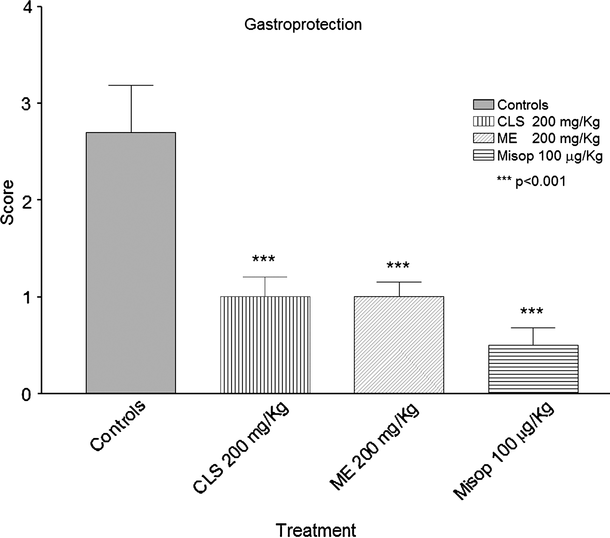
Comparison of gastric damage scores between stomachs of rats pretreated or not with L. nobilis extracts (200 mg/kg). The scores were calculated according to Robert et al.14 CLS, chloroform extract; ME, methanolic crude extract; Misop, misoprostol. ***P < .001.
Specimens from animals treated with the necrotizing agent revealed severe gastric epithelium damage, characterized by hyperemia of the mucosa with erosions (Fig. 2A and B). In contrast, specimens from animals pretreated with L. nobilis leaf extracts showed a different aspect of the gastric mucosa, with less severe damage, as can be seen in Figure 2C and E (stained with hematoxylin–eosin) and D and F (stained with periodic acid Schiff). In Figure 3 specimens of saline-solution treated animals (Fig. 3A and B) and of animals pretreated with misoprostol are reported as negative and positive controls (Fig. 3C and D).
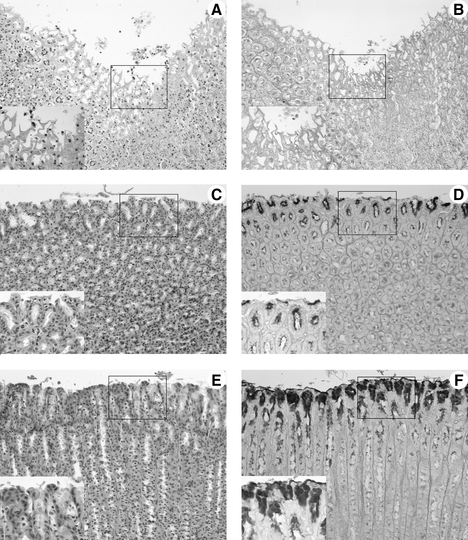
Gastric damage in gastric mucosa from (

Gastric mucosa from (
The in vitro antioxidant power was evaluated with different experimental methods, as previously described, and the measurements of this activity and of the total phenolic content (expressed in mg/L GAE), reported in Table 2, are in good agreement. Even if the FC reagent method suffers from several interfering substances so the GAE values can overestimate the total phenolic content, 12 it is the recommended method for measuring the total reducing capacity. 22 The good agreement between GAE values and antioxidant power could then be an indication that the total reducing capacity of the extracts is due only to their polyphenols content.
Data are mean ± SEM values.
ARC, antioxidant relative activity; DPPH, 2,2-diphenyl-1-picrylhydrazyl; GAE, gallic acid equivalents; IC50, concentration of the sample required to reduce absorbance 50%; RE, resorcinol equivalents; TEAC, Trolox equivalent antioxidant capacity.
Discussion
The in vivo results justify the traditional use of Laurus for the treatment of gastric ailments, and the gastroprotection observed in our experimental conditions may be explained by the presence of active principles possessing with good antioxidant capacity.
In fact, the in vitro results confirm this good capacity with some difference within methods of extraction. The order of tested extracts activity, evaluated with BR, TEAC, and DPPH methods, indicates that the methanolic leaf extract is more effective than the chloroformic one. This order, as far as that of the total phenolic content, is identical to that observed in the attribution of the scores in the in vivo test. Data reported in Table 2 indicate that the active compounds present in L. nobilis leaves are mainly hydrophilic (probably glycosylated compounds such as flavonoids and derivatives). This may justify the traditional use of Laurus leaf infusion that is an extraction in aqueous medium. In fact, a previous phytochemical study 10 suggested the presence in the infusion of a good pattern of antioxidant compounds.
The crucial role of reactive oxygen species in producing tissue damage is well established. The inflammatory response in the gastric mucosa, as well as in other tissues, is caused by an oxidative stress; for this reason antioxidant compounds can counteract, at least in part, the inflammatory response. We report here the data obtained for extracts of other plants with analogous relation between antioxidant and pharmacological activities. Wulfenia carinthiaca Jacq. showed a good anti-inflammatory activity correlated with good scavenger properties. 23 A similar correlation was observed for dichloromethane and methanolic extracts of Leontopodium alpinum Cass. 24 Finally, a commercial hydroalcoholic extract of artichoke (Cynara scolymus) leaves showed a significant antioxidant power strictly correlated with a significant protection of liver tissue from oxidative stress damage. 25
Hence it is possible to conclude that the antioxidant properties of the leaf extracts of L. nobilis (Table 2) can explain the gastric mucosal protection.
The mechanism responsible for gastric damage involves the increase of reactive oxygen species. It has been ascertained that reactive oxygen species play a role in the development of pathogenesis in acute experimental gastric lesions induced by stress, ethanol, and nonsteroidal anti-inflammatory drugs.
Thus the gastroprotective effect of L. nobilis leaf extracts here reported may be explained, at least in part, by their antioxidant capacity. The antioxidant properties of flavonoids and polyphenols have been related to anti-ulcer activity because free radicals are developed in gastric mucosal lesions. Moreover, flavonoids have shown cytoprotective activity in different animal models, as well as in parietal cell cultures. 13,26 Our in vivo results, confirmed by histological evidences and in good agreement with chemical results, seem to support a hypothesized mechanism of action proposed by other authors. 27,28
Footnotes
Acknowledgments
This work was supported by a research grant from the University of Bologna. We thank Dr. M.C. Loi (University of Cagliari, Cagliari, Italy) for the kind gift of the plants of L. nobilis.
Author Disclosure Statement
No competing financial interests exist.