
Abstract
The methanolic extracts of the leaves of Lippia species (L. pseudo-thea, L. hermannioides, L. alba, L. rubella, and L. sidoides) were tested for their antibacterial, antifungal, and antioxidant activity. Cytotoxicity was determined by using brine shrimp lethality bioassay. Phytochemical screening was also performed. The extracts showed a minimum inhibitory concentration (MIC) ranging from 78 to 5000 μg/mL for antibacterial activity against at least 2 species of bacteria, although none was active against Escherichia coli. Antifungal activity was found only for L. pseudo-thea (MIC, 625 μg/mL for Candida albicans) and L. sidoides (MIC, 625 μg/mL for both C. albicans and C. neoformans). The bioautography showed that flavonoids and coumarins are responsible for the antioxidant activity of the extracts and that the antimicrobial properties are due to flavonoids and terpenoids. The cytotoxic activity was stronger for L rubella extract. To our knowledge, this is the first report of the biological and chemical constituents of L. pseudo-thea, L. hermannioides, and L. rubella.
Introduction
T
The aim of this study was to evaluate the antioxidant and antimicrobial activities of the methanolic extracts of the leaves obtained from 5 Lippia species popularly used in traditional medicine. Cytotoxicity was determined by using brine shrimp lethality bioassay, and phytochemical screening was performed. The total amount of phenolics and flavonoids was determined. To our knowledge, this is the first report of the biological and chemical constituents of L. pseudo-thea, L. hermannioides, and L. rubella.
Materials and Methods
Plants
The Lippia species were collected in Juiz de Fora, Minas Gerais, Brazil, in July 2005. Voucher specimens were deposited at the Herbarium Leopoldo Krieger (CESJ) of Federal University of Juiz de Fora. The species used in this study were L. pseudo-thea Schauer [45171], L. hermannioides Cham. [46088], L. alba Mill. (N.E.) [46177], L. rubella Moldenke [46178], and L. sidoides Cham. [46180].
Preparation of the extract
The leaves (50 g each) were powdered and macerated with methanol (3 × 200 mL) for 5 days at room temperature. After evaporation of the solvent under reduced pressure at 45°C, the respective methanolic extracts were obtained. All the extracts were kept in tightly stoppered bottles under refrigeration until used for the biological testing and phytochemical screening.
Phytochemical screening
A portion of each extract that was subjected to the biological screening was used to identify the major secondary metabolites according to protocols described elsewhere. 8 Briefly, the extract was suspended in methanol (1 mg/mL) and submitted to the following identification reactions: The characterization for tannins was performed by gelatin, iron salt, and lead acetate reactions. Triterpenoids and steroids were investigated by using a Liebermann-Burchard reagent, and alkaloid analysis was done by precipitation reactions with the reagents of Dragendorff, Bouchardat, Mayer, and Bertrand. For flavonoids, the reactions of Shinoda and aluminum chloride were used. Antraquinones were characterized with a Borntraeger reaction. The presence of saponins was assessed according to the formation of foam, and coumarins were characterized by using KOH.
Amount of total phenolic compounds and flavonoids
The amount of phenolic compounds in the plant samples was determined by the Folin-Denis method. 9 Tannic acid was used as the standard for the calibration curve. One mL of the samples resuspended in ethanol (0.5 mg/mL) was mixed thoroughly with the Folin-Denis reagent (1 mL) and Na2CO3 2% in 0.1 N NaOH (8 mL). After a 60-minute incubation period at 30°C, the absorbance was read against a blank at 730 nm. All determinations were performed in triplicate. Total content of the phenolic compounds was expressed in mg/g plant extracts, in tannic acid equivalents.
The amount of flavonoids was determined as described elsewhere, with slight modifications. 10 Rutin was used as the standard for the calibration curve. One mL of plant samples was resuspended in methanol (0.5 mg/mL) and mixed with 2% aluminum trichloride in ethanol (1 mL). After the mixture was diluted with ethanol to 25 mL and allowed to stand for 40 minutes at 20°C, its absorbance was measured at 415 nm. The blank was prepared from the plant samples (1 mL) and 1 drop of acetic acid diluted to 25 mL in ethanol. All determinations were performed in triplicate. Total amount of flavonoids was expressed in mg/g plant extracts, in rutin equivalents.
Antioxidant activity
1,1-Diphenyl-2-picrylhydrazyl (DPPH) radical scavenging
The free radical–scavenging activity of sample solutions in methanol was determined on the basis of their ability to react with stable 1,1-diphenyl-2-picrylhydrazyl (DPPH) free radical (Sigma). 11 The plant samples at various concentrations (7.8–250 μg/mL) were added to a 152-μM solution of DPPH in methanol. After incubation at 37°C for 30 minutes, the absorbance of each solution was determined at 517 nm. The antioxidant activity of the samples was expressed as IC50 (inhibitory concentration), which was defined as the concentration (in μg/mL) of sample required to inhibit the formation of DPPH radicals by 50%. As positive controls, α-tocopherol and rutin were used.
Reducing power
The reducing power was determined according to a described procedure. 12 Ten mg of each sample was mixed with potassium phosphate buffer (0.2 M; pH, 6.6) (2.5 mL) and potassium ferricyanide (10 g/L) (2.5 mL). The mixture was incubated at 50°C for 20 minutes. A 2.5-mL aliquot of 10% trichloroacetic acid was added to the mixture, which was then centrifuged at 3000 rpm for 10 minutes. The upper layer of the solution (2.5 mL) was mixed with distilled water (2.5 mL) and 0.1% FeCl3 (0.5 mL), and the absorbance was measured at 700 nm. Ascorbic acid was used as reference material. All tests were performed in triplicate. Increase in absorbance of the reaction indicated the reducing power of the samples. A higher absorbance indicated a higher reducing power. EC50 (effective concentration) values (μg/mL) were calculated and indicate the effective concentration at which the absorbance was 0.5 for reducing power.
Antimicrobial activity
Microbial strains
The samples were evaluated against a panel of microorganisms, including the bacterial strains Staphylococcus aureus (American Type Culture Collection [ATCC] 6538), Pseudomonas aeruginosa (ATCC 15442), Salmonella typhimurium (ATCC 13311), Shigella sonnei (ATCC 11060), Klebsiella pneumoniae (ATCC 13866), Escherichia coli (ATCC 10536), and Bacillus cereus (ATCC 11778) and the yeasts Candida albicans (ATCC 18804) and Cryptococcus neoformans (ATCC 32608). Bacterial strains were cultured overnight at 37°C in Mueller Hinton agar. Yeasts were cultured for 48 hours at 30°C in Sabouraud dextrose agar.
Serial dilution assay for determination of the minimal inhibitory concentration (MIC)
The MIC of each extract was determined by using the broth microdilution techniques as described elsewhere for bacteria and yeasts. 13,14 MIC values were determined in RPMI 1640 (Sigma) buffered to a pH of 7.0 with 4-morpholinepropanesulfonic acid (Sigma) for yeasts and in Mueller Hinton broth (MHB) for bacteria. Yeasts were cultured at 30°C for 48 hours in Sabouraud dextrose agar, and bacteria were cultured overnight at 37°C for 24 hours in Mueller Hinton agar. Sample stock solutions were diluted from 500 to 2.5 μg/mL (final volume, 80 μL), with a final dimethyl sulfide (DMSO) concentration of 1% or less. Then, RPMI or MHB (100 μL) was added to microplates. Finally, 20 μL of 106 colony-forming units/mL (values of 0.08–0.10 at 625 nm, according to McFarland turbidity standards) of standardized yeast and bacterial suspensions were placed in microplates, and the test was performed in a volume of 200 μL. Plates were incubated at 30°C for 48 hours for yeasts and at 37°C for 24 hours for bacteria. The same tests were performed simultaneously for growth control (RPMI plus yeast and MHB plus bacteria) and sterility control (RPMI or MHB plus extract). The MIC values were calculated as the highest dilution showing complete inhibition of the tested strain. For bacteria, extracts were considered active if they gave an MIC of 500 μg/mL or less against the strains tested. 15,16 For antifungal evaluation, extracts with an MIC of 800 μg/mL or less were considered active. 17
Quantitative evaluation of antimicrobial activity
The antimicrobial activity of plant extracts may be expressed in different ways according to the technique used. The agar diffusion method is commonly used as a preliminary test in the screening of plants for antimicrobial activity. The microdilution method yields MIC values, the minimum concentration at which inhibition is observed (μg/mL). In our study, methods other than numeric values were used to express antimicrobial efficiency. In addition to recording results in terms of MIC (mg/mL), we used total activity values, as well as percentage activity values. The latter demonstrate the total antimicrobial potency of particular extracts and microbial susceptibility index (MSI), which is used to compare the relative susceptibility among the microbial strains: 18
Total activity was calculated as follows:
These values would indicate the largest volume to which biologically active compounds in 1 g of plant material can be diluted and still inhibit microbial growth.
Percentage activity (%) was calculated as follows:
The percentage activity demonstrates the total antimicrobial potency of particular extracts. It shows the number of microbes found susceptible to one particular extract.
The microbial susceptible index (MSI) was calculated as follows:
MSI is used to compare the relative susceptibility among the microbial strains. MSI values ranges from 0 (resistant to all samples) to 100 (susceptible to all samples).
Thin-layer chromatography analysis
Silica gel G60 F254 aluminium-backed plates (7 cm × 2 cm) were used. The methanolic extracts of Lippia species (10 mg/mL; 20 μL) were applied and the layers developed with MeCl2: MeOH (99:1). All thin-layer chromatography (TLC) plates were run in duplicate, and 1 set was used as the reference chromatogram. The separated components were visualized under visible and ultraviolet light (254 and 366 nm) or using spray reagent such Dragendorff (for alkaloids), sulfuric vanillin, Lierbermann-Burchard (for terpenoids), 10% KOH solution (for coumarins), and natural product-polyethylene glycol (for flavonoids).
TLC antimicrobial bioautography
The bioautography was performed only for the most active Lippia extracts (L. hermannioides, L. pseudo-thea, and L. sidoides) against B. cereus, C. albicans, and C. neoformans strains. Chromatograms were placed on sterile square Petri dishes with covers and exposed to ultraviolet light for 30 minutes. After this, overlay media (Mueller Hinton agar and Sabouraud agar, 10 mL, for bacteria and fungi, respectively), to which was added 1 mL of the detecting strain at a final concentration of 107 cells/mL, was distributed over the developed TLC plates. After solidification of the media, the TLC plates were incubated for 24 hours at 37°C for B. cereus, 48 hours at 35°C for C. albicans, and 72 hours at 35°C for C. neoformans. The bioautograms were sprayed with a solution of p-iodonitrotetrazolium violet, 2 mg/mL (wt/vol) for observation of the inhibition zones. Inhibition zones were compared with retention factors of the related spots on the reference plates. The experiment was performed in duplicate.
TLC antioxidant bioautography
The TLC plates of the most active antioxidant extracts (L. alba, L. rubella, and L. sidoides) were developed as described earlier and were allowed to air-dry for 30 minutes, followed by spray with a 2.54-mM DPPH. methanol solution for derivatization. Spots with the DPPH. scavenging activity were observed as white yellow bands on a purple background. Inhibition zones were compared with the retention factors of the related spots on the reference plates. The experiment was performed in duplicate.
Cytotoxicity assay
A brine shrimp lethality bioassay was carried out to investigate the cytotoxicity of the extracts. 19 Brine shrimp (Artemia salina Leach) eggs were hatched in a beaker filled with seawater under constant aeration. After 48 hours, the nauplii were collected by pipette and were counted macroscopically in the stem of the pipette against a lighted background. Solutions of the extracts were made in seawater containing 1% DMSO, at varying concentrations (10–1000 μg/mL) and incubated in triplicate vials with 10 brine shrimp larvae. After 24 hours of incubation, the nauplii were examined against a lighted background with a magnifying glass, and the number of survivors in each vial was counted and noted. Both positive (thymol) and negative (sea water containing 1% DMSO) control assays were carried out to verify the susceptibility of A. salina under assay conditions employed. LC50 was determined from 24-hour counts. The general toxicity activity was considered weak when LC50 was above 250 μg/mL.
Statistical analysis
The IC50 and EC50 values for DPPH radical-scavenging activity and reducing power, respectively, were calculated by linear regression analysis and expressed as mean ± standard error; the LC50 for cytotoxic activity was calculated by the probit analysis. 20 Statistically significant differences between the treatments and the control were evaluated by using the analysis of variance test using the GraphPad Prism 4 statistical software. A difference in the mean values of P < .05 was considered to be statistically significant.
Results
The leaves of 5 Lippia species used in folk medicine were evaluated for their antioxidant (DPPH radical-scavenging and reducing power) and antimicrobial (7 strains of bacteria and 2 yeasts) activity. Cytotoxicity was also evaluated by the brine shrimp lethality bioassay.
Table 1 summarizes the results of the yield and phytochemical screening of the extracts. Alkaloids, tannins, and flavonoids were found in all extracts.
Table 2 shows that all extracts exhibited antioxidant activity. The DPPH radical-scavenging assay showed that L. sidoides had the best antioxidant activity (IC50 of 5 μg/mL), followed by L. rubella and L. alba (IC50 of 7 and 9 μg/mL, respectively). This finding indicates that these samples have good potential as free radical scavengers. In addition, those extracts presented significant reduction power (EC50 of 107, 109, and 110 μg/mL, respectively) when compared with the control ascorbic acid (EC50 of 105 μg/mL). The bioautography showed through ultraviolet light and the characteristics colors presented with natural product-polyethylene glycol and 10% KOH solution that the compounds responsible for antioxidant activity could be flavonoids and coumarins (Fig. 1).
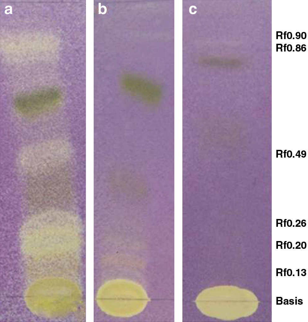
Thin-layer chromatography (TLC) plates stained with 2.54 mM 1,1-diphenyl-2-picrylhydrazyl (DPPH.) methanol solution and visualized under visible light. Twenty μL of the methanolic extract (10 mg/mL) of (
Values are expressed as mean ± standard error (n = 3); P > .05.
The numbers in parentheses are the 95% confidence limits.
Positive controls.
The content of phenolic compounds in L. sidoides extract was the highest, followed by the extracts of L. rubella, L. alba, L. hermannioides, and L. pseudo-thea. The amount of flavonoids presented the same profile (Table 2).
Table 3 shows the antimicrobial activity of the extracts. MIC values ranging from 5000 to 78 μg/mL were obtained. Extracts of L. pseudo-thea, L. hermannioides, and L. sidoides demonstrated strong activity against B. cereus (MIC of 156, 78, and 78 μg/mL, respectively) (Table 3).
– indicates no activity.
Chloramphenicol and amphotericin B were used as positive reference standard antibiotics for bacteria and fungi, respectively.
Total activity values revealed that L. hermannioides, L. sidoides, and L. pseudo-thea extracts had the highest antimicrobial activity against B. cereus (total activity, 1282, 1026 and 962 mL/g, respectively). Total activity, percentage activity, and MSI were also calculated. MSI values were useful in evaluating the susceptibility of the different strains of microbes toward the plant extracts investigated. B. cereus and P. aeruginosa were the test organisms found to be the most susceptible to the samples investigated (100% susceptibility). On the other hand, E. coli was resistant to all strains tested. Among the samples evaluated, the extract of L. sidoides was the most efficient (78% activity) against the different microorganisms used.
The bioautography showed that the compounds responsible for the antimicrobial activity of the extracts were flavonoids and terpenoids (Fig. 2). The flavonoids of L. sidoides with retention factors of 0.20 and 0.26 presented both antimicrobial for B. cereus and antioxidant properties. Although this species presented a moderate anti-Cryptococcus activity (Table 3), the active compounds were not detected by the bioautography assay.
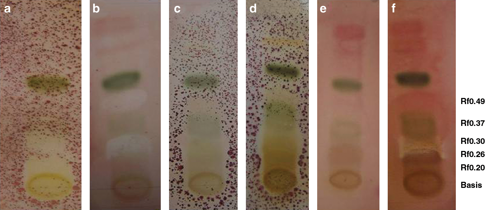
Thin-layer chromatography (TLC) plates stained with p-iodonitrotetrazolium violet (2 mg/mL) for observation of the inhibition zones. Twenty μL of the methanolic extract (10 mg/mL) of (
Only the extract of L rubella exhibited cytotoxic activity against A. salina (Table 2).
Discussion
The DPPH assay showed that the IC50 values of the extracts of L. sidoides, L. rubella, and L. alba were close to the IC50 of rutin (IC50 of 3 μg/mL), a well-known free radical scavenger and antioxidant used to decrease the risk for heart disease. 21 This finding demonstrates the strong ability of these extracts to act as a donor for hydrogen atoms. Like DPPH radical-scavenging activity, the reducing power increased in a dose-dependent manner. Oxygen radicals are the most responsible for the development of many diseases, including carcinogenesis, atherosclerosis, and heart diseases. 22 Although synthetic antioxidants have often been used against free radicals by scavenging reactive oxygen or ending radical chain reactions, recent health concerns have drawn much attention to the use of natural antioxidative compounds. 23
The biological activities of the polyphenols in different systems are believed to be due to their redox properties, which can play an important role in absorbing and neutralizing free radicals, quenching singlet and triplet oxygen, or decomposing peroxides. 24 Therefore, relationships between antioxidant activity (DPPH radical-scavenging activity and reducing power) and the content of phenolic compounds were evaluated.
It is extremely important to point out that there is a positive correlation between DPPH radical-scavenging activity potential and amount of phenolic compounds of the extracts (Table 2). In addition, antioxidant activity has been reported to be concomitant with the development of reducing power. 25 The reducing power of a compound may serve as an important indicator of its potential antioxidant activity. 26 The large amount of total phenols and flavonoids in the extracts of those species might explain their high scavenging ability and reducing power.
The extract of L. sidoides showed a strong activity against both fungal tested (MIC of 625 μg/mL for all). The antimicrobial activity of L. sidoides has been referred by substances found in the essential oil. 4 Although most of the MIC values found for the extracts were above the breakpoint (MIC ≤ 500 μg/mL for bacteria and 800 μg/mL for fungal), they show the potential clinical use and interest of these extracts, which are crude extracts of uncertain composition and have components that can have synergistic or antagonistic effects. 27 Plant extracts may be more beneficial than isolated constituents because other compounds present in the extracts can change the properties of bioactive individual component. 28 Thus, total activity, percentage activity, and MSI were calculated. The results presented reinforce the importance of Lippia extracts against B. cereus. While most Bacillus species are regarded as having little pathogenic potential, B. cereus has been known to act as a primary invader or secondary infectious agent in many diseases and has been implicated in some cases of food poisoning. 29
The brine shrimp lethality assay is based on the ability to kill laboratory-cultured A. salina nauplii brine shrimp and is considered one of the most useful tools for the preliminary assessment of general toxicity. 30 LC50 values less than 250 μg/mL are considered significant for crude extracts. 31 The toxicity against A. salina was correlated with the toxicity against human solid tumor cell lines, and the results suggested that this method can be used as a preliminary analysis of cytotoxicity of novel substances. 30
In conclusion, this research indicates that some Lippia species may be a good candidate for pharmaceutical plant-based products. Moreover, to our knowledge this is the first report of the antimicrobial and antioxidant activity of L. pseudo-thea, L. hermannioides, and L. rubella.
Footnotes
Acknowledgments
The authors are grateful to Fundação de Amparo a Pesquisa do Estado de Minas Gerais (FAPEMIG) and Universidade Federal de Juiz de Fora (UFJF)/ Brazil for financial support and to Dr. Fatima Regina Salimena for the botanical identification of species.
Author Disclosure Statement
No competing financial interests exist.