
Abstract
English walnuts have been shown to decrease cardiovascular disease risk; however, black walnuts do not appear to have not been studied for their cardioprotective effects. The purpose of this study was to determine the effects of English versus black walnut consumption on blood lipids, body weight, fatty-acid composition of red blood cell (RBC) membranes, and endothelial function. Consumption of 30 g of English walnuts per day for 30 days, by 36 human participants, improved blood lipids; the effects of black walnuts were dependent on the participant's sex. Addition of either nut to the diet did not result in weight gain. The fatty-acid composition of RBC membranes was favorably affected by walnut consumption. RBC polyunsaturated fatty acids increased after consumption of either type of nut; however, eicosapentaenoic acid increased significantly more after English walnut consumption. Endothelial function of 6 unmedicated humans with hypercholesterolemia was maintained after consumption of English walnuts with a meal high in high saturated fats; however, consumption of black walnuts with the same meal did not maintain endothelial function. Overall, these results support the recommendation that consumption of 1 oz of English walnuts per day may decrease cardiovascular risk, but more research on black walnut consumption is necessary before an appropriate recommendation can be made.
Introduction
N
In addition to the nutrients that directly improve lipoprotein levels, nuts also contain nutrients that have other cardioprotective qualities. Some nuts are high in antioxidant compounds, including large amounts of tocopherols, tannins, ellagic acid, and other polyphenols. 5,6 In fact, a study revealed that English walnuts had a higher amount of total antioxidants than 121 of 122 whole foods tested. 7 Nuts are also high in arginine, a precursor of nitric oxide, a vasodilator that may also downregulate adhesion molecule expression, LDL uptake, and smooth muscle proliferation, all of which improve endothelial function. 8 Finally, nuts also contain folate, which may lower the levels of homocysteine, a marker of inflammation. 9
The relationship of English walnuts and cardiovascular disease is the most studied among all nuts. Numerous English walnut–feeding trials have found significant reductions in LDL and total cholesterol, along with significant increases in high-density lipoprotein (HDL), with 1–2 oz of walnuts consumed daily for 4–8 weeks. 10,11 The participants in these studies have been male and female (both pre- and postmenopausal), young, old, healthy, overweight, and dyslipidemic. The studies have been conducted in many different locations. Some studies had a specific diet for the participants to eat, whereas others had participants eat walnuts in addition to their current diet. Regardless of the amount of nuts, time of study, participant characteristics, or diet conditions, almost all studies of English walnut consumption have found significant improvements in the blood lipid profile. 10,11
The composition of red blood cell (RBC) membranes reflects changes in fatty-acid intake in the diet and has also been correlated to cardiovascular disease risk or risk factors. 12 –16 To date, only 1 known study has looked at the effects of English walnut consumption on fatty-acid composition of RBC membranes. In a crossover study, Rajaram et al. 17 fed participants 1 of 3 diets for 4 weeks: a diet that contained 1.5 oz of walnuts per day, 6 days a week; a diet that contained 2 fatty-fish meals per week; or a control diet that did not contain foods high in omega-3 fatty acids. The walnut diet resulted in the greatest amount of PUFA, linoleic acid, and alpha-linolenic acid in the membrane. The fish diet resulted in the greatest amount of eicosapentaenoic acid (EPA) and docosahexaenoic acid (DHA). The EPA in the walnut diet was also slightly increased from the control diet, but there was no difference in DHA. The small increase in EPA in the walnut diet may be due to the slow conversion of linolenic acid to EPA. 18
Many cardiovascular disease factors, such as smoking, obesity, diabetes, and high levels of LDL, lead to atherosclerosis by impairing the ability to maintain proper vascular tone and leading to endothelial dysfunction. Researchers have recently found that endothelial dysfunction is reversible and affected by diet, which has led to numerous studies on the effects of consumption of specific nutrients. 9,19
Walnuts are high in antioxidants, alpha-linolenic acid, arginine, and folate, making these nuts an ideal food with which to study the effects of dietary intervention on endothelial function. 20 Two known studies have correlated walnut consumption and endothelial function. Ros et al. 21 had participants consume a diet in which either walnuts or olive oil replaced 32% of the energy from monounsaturated fat for 4 weeks. They found that the walnut diet improved occlusion-mediated vasodilation measured by ultrasonography of the brachial artery. Cortés et al. 22 looked at the effects of addition of 40 g walnuts or olive oil to a meal high in saturated fat. A high-saturated-fat meal was used because it had been shown to decrease vasodilation in response to occlusion, particularly in participants who were hypercholesterolemic. 9 Cortés et al. 22 found that in normal healthy participants, there was no significant difference in either group; however, in hypercholesterolemic participants, the walnut meal increased vasodilation while the olive oil meal decreased vasodilation. These findings suggest that English walnuts have additional cardiovascular benefits beyond lipoprotein improvements.
All the previous studies assessed English walnuts; to our knowledge, black walnuts have not been studied with regard to their cardiovascular or other health benefits. According to limited U.S. Department of Agriculture food and nutrition database information, 23 black and English walnuts have a different fatty-acid, antioxidant, and amino acid content than their English walnut relatives. It is also thought that black walnuts may contain a significant amount of antioxidant chemicals that may be further protective to the cardiovascular system on the basis of their rich color and flavor.
These nuts contain less linoleic (18:2) and alpha-linolenic (18:3) acid than English walnuts, which may result in a smaller effect on membrane composition. 23
The purpose of the current study was to determine how black walnuts influence blood lipid levels, incorporation of PUFA into RBC membranes, and endothelial function in comparison with English walnuts.
Materials and Methods
Approval and nut donation
All human participant procedures were approved by the University of Wisconsin-La Crosse Institutional Review Board for the Protection of Human Subjects. Black walnuts were obtained through donations from Hammons Products Co. (Stockton, Missouri, USA), Michael Winfrey (Ettrick, Wisconsin, USA), and Harry Lundstrom (Unionville, Virginia, USA). Thirty-gram portions of black walnut meats were weighed and placed in individual zipper-top plastic bags. Thirty-gram portions of unsalted English walnuts, purchased in bulk from the People's Food Coop (La Crosse, Wisconsin, USA), were also placed in individual bags.
Walnut consumption crossover study
Thirty-six participants were recruited primarily through print advertisements. Upon completion of written informed consent, participants' heights and weights were measured. Medication and food-frequency questionnaires were also completed at this time. Participants were randomly assigned to black or English walnuts and were given and instructed to eat one 30-g bag of assigned nuts each day for 28–30 days. Participants were advised to eat the nuts daily in addition to their usual diet and record any deviations, but they were not asked to eat nuts at a specific time or in a specific context. A crossover portion of the study was done after a 12-week washout period. Twenty-nine participants switched to the other type of walnut and consumed one 30-g bag of the other nut each day for 28–30 days. All participants reported that nuts were eaten as directed and that no other changes in the usual diet or medications occurred during nut consumption.
Venous blood was drawn by trained phlebotomists into Vacutainer clot activator/gel tubes (BD Diagnostics, Franklin Lakes, New Jersey, USA) from arm or hand veins in 12-hour fasted participants before and after 28–30 days of nut consumption. Body weight was also measured again at this time. Blood lipids in serum samples were analyzed with spectrophotometry (Cobas 501; Roche Diagnostics, Indianapolis, Indiana, USA) by technicians blinded to participant condition at Gundersen Lutheran Medical Center Laboratory. Additional plasma and RBCs were stored at −80°C until fatty-acid composition was analyzed.
Fatty-acid analysis
Fatty-acid composition of RBC membranes was analyzed with the fatty-acid methyl ester (FAME) procedure and Agilent 6850 Series II gas chromatograph (Agilent Technologies, La Jolla, California, USA). Samples were prepared by using solid and liquid reagents from Sigma Aldrich (St. Louis, Missouri, USA) and gas reagents from Airgas (Waterloo, Iowa, USA) unless otherwise specified. RBCs were combined with 0.9% cold saline, CH3Cl: MeOH (1:2), and 2-diheptadecanoyl-sn-glycero-3-phosphorylcholine (internal control; Matreya LLC #1400, Pleasant Gap, Pennsylvania, USA), vortexed, and incubated on ice for 20 minutes in the dark. Saturated NaCl and chloroform were added and samples were vortexed, incubated on ice in the dark for 10 minutes, and centrifuged for phase separation. The chloroform layer was transferred and evaporated to dryness under nitrogen gas with the N-Evap system (Organomation Associates Inc., Berlin, Massachusetts, USA). A second extraction was performed as described earlier. Fatty acids were saponified in 0.5 N NaOH/MeOH, capped tightly, vortexed, and incubated for 15 minutes at 86°C. Once cooled, BF3 in methanol was added and the tubes were capped tightly, vortexed, and incubated at 86°C for 15 minutes for methylation. Once cool, 0.7 N HCl in MeOH was added for neutralization. Hexane and saturated NaCl were added and samples were vortexed, incubated at room temperature, and centrifuged for phase separation. The hexane layer was transferred and evaporated to dryness under nitrogen gas. A second hexane and saturated NaCl extraction was performed as described earlier, and the hexane layer was transferred to the tubes already drying under nitrogen gas. Once dry, hexane was added to resuspend the fatty-acid methyl esters. The FAME samples were vortexed, transferred to V-vials (National Scientific Company #C4012-10, Rockwood, Tennessee, USA), capped, and frozen until injected into the gas chromatograph.
All samples were run on the gas chromatograph within 36 hours of preparation. One microliter was autoinjected into the gas chromatograph. The inlet was split with H2 gas at a ratio of 40.0:1. The split flow was 73.6 mL/min and the total flow was 83.0 mL/min. Inlet temperature and pressure were held at 270°C and 23 PSI, respectively. A DB-23 column (60 m×250 μm×0.15 μm, Agilent Technologies, La Jolla, California, USA) was held at a constant pressure of 23 PSI. The column flow was 1.8 mL/min and the average velocity was 45 cm/sec. The oven temperature was held at 130°C for 1 minute. The temperature was ramped 6.50°C/min to 170°C and then 2.50°C/min to 215°C, which was held for 12 minutes. To clean off the column, the oven was ramped 40.00°C/min to 230°C and held for 3 minutes. The detector was held at 280°C with an H2 flow of 40.0 mL/min, air flow of 450 mL/min, and make up gas flow (N2) of 40.0 mL/min. The data were analyzed at 50 Hz and reported in pA. For quality assurance and quality control, 80% of samples were run in duplicate, 10% were selected at random and run in procedural triplicate, and 10% were selected at random and run in analytical triplicate. Hexane blanks were run before and after each set of samples. A set of fatty-acid standards (Nu-Chek Prep Inc. #461 or 68B, Elysian, Minnesota, USA) were run with each set of samples.
Statistics for blood lipids and RBC fatty acids
The SPSS version 16.0 linear model (SPSS Inc., Chicago, Illinois, USA) with multivariate repeated-measures procedure was used with 2 within-subject factors with 2 levels (before and after nut consumption and type of walnut [English and black walnut]) and 3 between-subject factors (sex, order, and consumption of fatty-acid supplement) for the dependent variables (13 individual fatty acids, total saturated fatty acids [SFAs], total unsaturated fatty acids, total monounsaturated fatty acids, total polyunsaturated fatty acids, and omega-3 index). Significant findings (P<.05) with multivariate analysis were also analyzed by univariate analysis. Data from participants who did not complete the crossover study were not included in the multivariate analysis.
Hyperemia methods
Serum was collected from 6 unmedicated hypercholesterolemic (LDL>130 mg/dL) participants for blood lipid screening to verify hypercholesterolemia, as described previously. Serum collection occurred at least 1 week before hyperemia measurements, and arm selection for sampling was left up to the phlebotomist. On 2 separate days at least 1 week apart, hypercholesterolemic participants were instructed to fast overnight and eat a breakfast of cereal, skim or 1% fat milk, and their usual beverage (with no cream) the morning of each study appointment. Each participant arrived at the University of Wisconsin-La Crosse Student Health Center at approximately noon for measurement of flow-mediated vasodilation of the participant's right brachial artery, approximately as described by Corretti et al. 24 Brachial artery diameter and blood flow rate were measured before blood pressure cuff occlusion and 60–90 seconds after release of cuff pressure after 4.5 minutes of occlusion. Occlusion pressure applied was 50 mm Hg above participant systolic blood pressure measured earlier the same day.
All ultrasound measurements were conducted by a family practice clinical investigator, Dr. Brian Allen, who was blinded to nut condition, with a General Electric LOGIQ Ultrasound Recorder (GE Healthcare Technologies, Waukesha, Wisconsin, USA). Four sites in each longitudinal section recording were measured and averaged. The percentage difference between before and after occlusion brachial artery diameters was calculated.
After the vasodilation test, participants ate a meal containing a sandwich with 75 g salami and 50 g cheddar cheese on 100 g white bread smeared with 2 teaspoons of butter along with 125 g 10% fat yogurt and 40 g black or English walnuts. This high-saturated-fat meal was used because previous research has shown that it decreases flow-mediated vasodilation, especially in hypercholesterolemic patients. 9,22 Participants were sent home or back to work and advised to fast and limit physical activity. They returned at 4 p.m., at which time another brachial artery vasodilation test was performed. Changes in vasodilation (before and after occlusion diameters) of and blood flow in the brachial artery pre- to post-nut (black vs. English) and high-saturated-fat meal consumption were analyzed by paired t-test (SPSS software, version 16.0); a P value less than .05 was considered to represent a statistically significant difference.
Results
Blood lipids
Of the 36 participants who began the study, 29 completed the entire crossover portion of the study and were used for analyses. There were no significant differences in age, number of participants receiving cholesterol-lowering medications, or fatty-acid supplements by nut type (Table 1) or nut type by gender (Table 2). As expected, women had significantly higher average HDL levels (P=.027) and significantly lower average body weight (P=.001) than men.
Unless otherwise noted, data are mean±standard error.
Number of participants using fish oil and/or fatty-acid supplements.
HDL, high-density lipoprotein; LDL, low-density lipoprotein; TC, total cholesterol.
Unless otherwise noted, data are means±standard errors.
Significantly different by sex (P<.05).
BF, females who consumed black walnuts; BM, males who consumed black walnuts; EF, females who consumed English walnuts; EM, males who consumed English walnuts; HDL, high-density lipoprotein; LDL, low-density lipoprotein; TC, total cholesterol.
After nut consumption, there was no significant difference in body weight (Table 1). Consumption of nuts resulted in reductions in total cholesterol and LDL and increased HDL, differences that were not statistically significant when assessed with multivariate analysis. No main effects of nut type or sex or nut-by-sex interactions were found with multivariate analysis. However, analysis of variance of pre- to post-change scores from all 65 nut trials (in 29 participants who crossed over and 7 who did not) revealed a significant nut-by-sex interaction in total cholesterol (P<.05), HDL (P<.02), and LDL (P<.05). Total cholesterol, HDL, and LDL levels increased in women who consumed black walnuts and decreased in men who consumed black walnuts (Fig. 1). English walnut consumption, regardless of sex, resulted in a decrease in total cholesterol and LDL levels. HDL levels increased in men who consumed English walnuts. Men who consumed black walnuts had a larger decrease in LDL levels than men who consumed English walnuts. There were no significant changes in triglycerides or total cholesterol:HDL ratio.
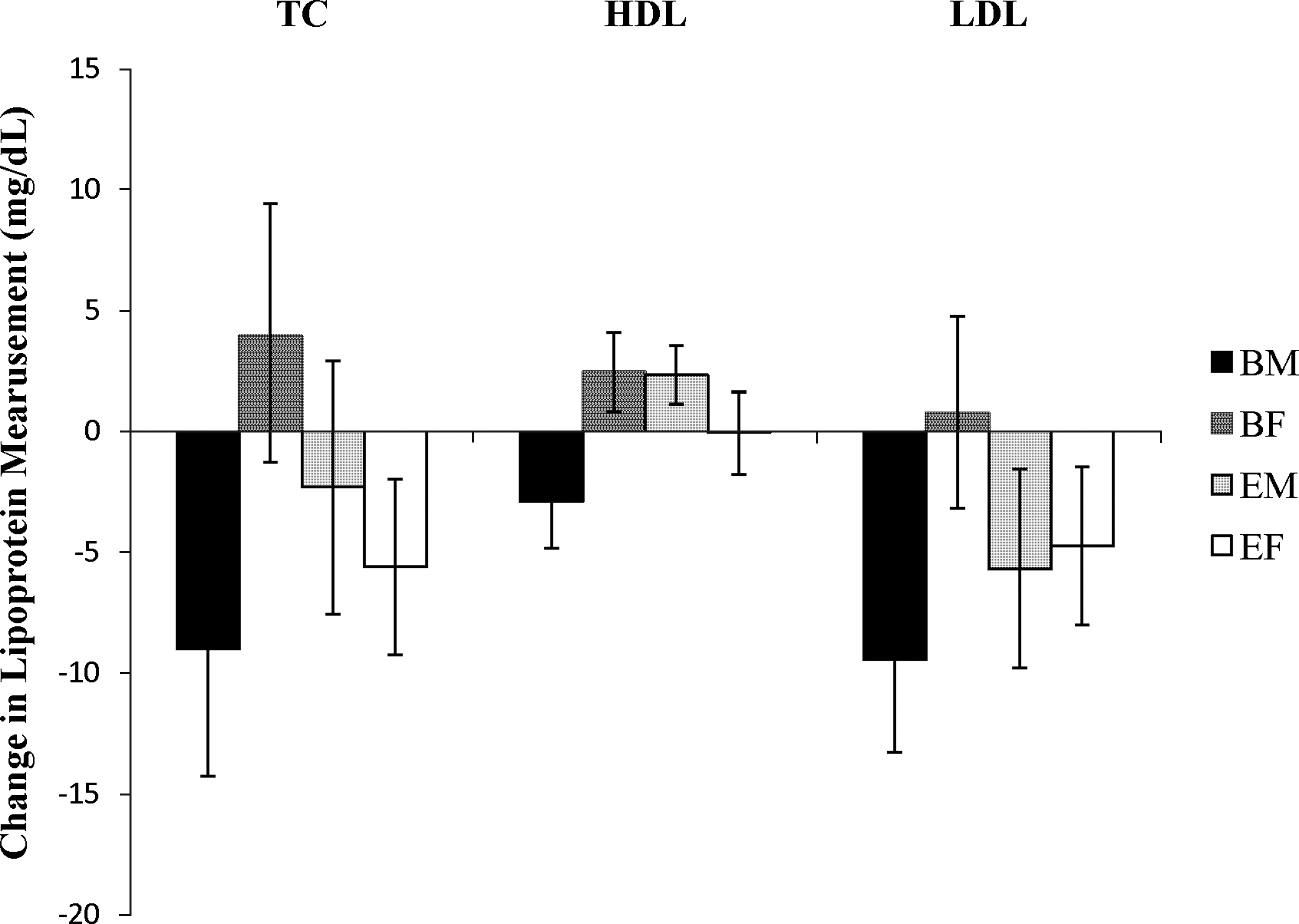
Change in total cholesterol, high-density lipoprotein (HDL), and low-density lipoprotein (LDL) after 28–30 days of walnut consumption. BF, females who consumed black walnuts; BM, males who consumed black walnuts; EF, females who consumed English walnuts; EM, males who consumed English walnuts. Values are expressed as the mean±standard error of mean (SEM).
RBC membrane composition
The RBCs from 29 participants who completed the entire crossover portion of the study were used for analysis of membrane fatty acids. There were no significant differences in any SFA (Table 3), MUFA (Table 4), or PUFA (Table 5) before nut consumption. Individuals who consumed a fatty-acid supplement (fish oil, DHA, EPA, or essential fatty acids) before and during the study had significantly different RBC membrane fatty-acid composition compared with those who were not taking supplements (P=.028). Significantly lower 18:1 n-9 (P=0.017), 20:4 (P=.029), and 22:4 (P=.001) and lower 16:1 (P=.084) were found in RBC membranes of participants who were consuming fatty-acid supplements. Significantly higher 20:5 (P<.001), 22:5 (P=.003), and 22:6 (P=.005) were also observed in participants who consumed fatty-acid supplements.
Significantly different pre- to post-consumption (P<.05).
SE, standard error.
Significantly different by nut type (P<.05).
Significantly different pre- to post-consumption (P<.05).
Almost significantly different by nut type (P<.1).
MUFA, monounsaturated fatty acids; SE, standard error.
Significantly different pre- to post-consumption (P<.05).
Significantly different by nut type (P<.05).
PUFA, polyunsaturated fatty acids; SE, standard error.
Regardless of nut type, a significant (P=.015) change in membrane fatty-acid composition with consumption was observed (Fig. 2). Significant decreases in 16:0 (P=.006), 18:1 n-9 (P=.002), 18:1 n-7 (P=.041), and 20:3 (P<.001) were observed after consumption of either type of walnut. Linoleic acid significantly increased (18:2; P<.001) after nut consumption, regardless of nut type. A significant decrease in MUFA (P=.002) and a significant increase in PUFA (P=.044) was also observed after nut consumption regardless of nut type (Fig. 3). No significant difference was observed in total SFA with nut consumption (P=.310).

Changes in percentage composition of red blood cell membrane fatty acids pre- and post-nut consumption (English or black walnut) (pooled data). Error bars represent SEM.

Changes in percentage composition of red blood cell membranes after 28–30 days of English or black walnut consumption (pooled data). MUFA, monounsaturated fatty acids; PUFA, polyunsaturated fatty acids; SFA, saturated fatty acids. Error bars represent SEM.
Nut type significantly (P=.015) affected the fatty-acid composition of RBC membranes over time, as shown in Figure 4. English walnut consumption resulted in a significant decrease in 16:1 (P=.041). A decrease in 18:1 n-9 that almost reached significance (P=.083) was observed with walnut consumption, and the decrease in 18:1 n-9 was greater with English than with black walnut consumption. EPA decreased with black walnut consumption but increased with English walnut consumption (P=.001). Membrane composition of all other individual fatty acids analyzed was not significantly affected by nut type. The degree to which MUFA decreased over time was affected by nut type (P=.033). MUFA decreased significantly more after English walnut consumption than after black walnut consumption. Changes in relative amounts of total SFA (P=.304) and total PUFA (P=.956) were not significantly affected by nut type.
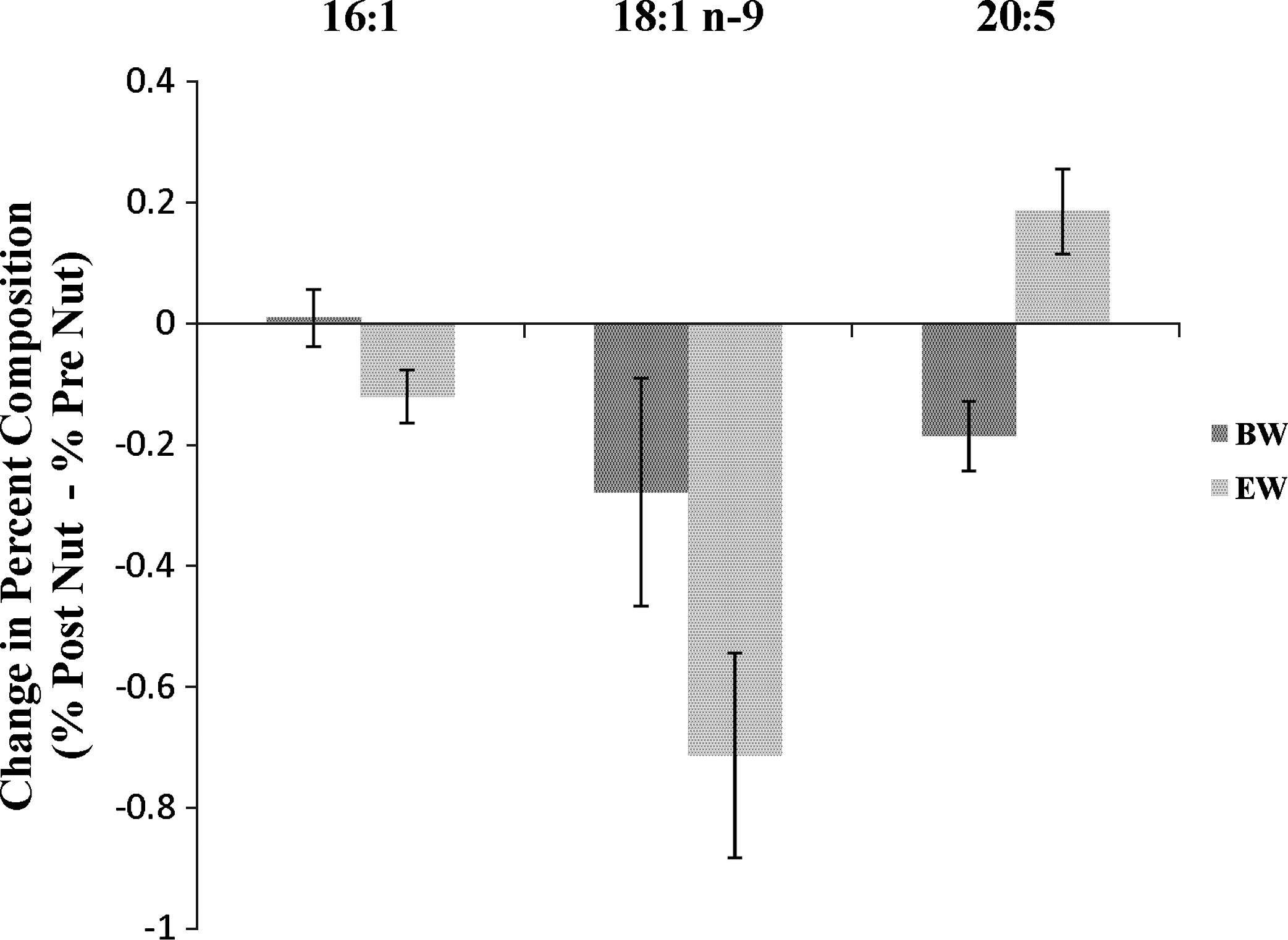
Changes in fatty-acid composition of red blood cell membranes pre- to post-consumption of English and black walnuts. BW, black walnuts; EW, English walnuts. Error bars represent SEM.
Endothelial function
In the 6 participants completing the acute endothelial function experiment, there were no significant initial differences between percentage vasodilation in response to occlusion or flow rate (Table 6). The effects of the high-fat meal on vasodilation in response to occlusion significantly differed by nut type (P<.02). After consumption of the high-saturated-fat meal containing black walnuts, endothelial function significantly decreased (P<.02). After consumption of the same high-fat meal containing English walnuts, there was no significant change in percentage dilation. No significant differences were observed in flow rates before or after dilation either before or after the meal.
Values are expressed as mean±standard error.
Significantly different by nut type (P<.05).
BMI, body mass index; LDL, low-density lipoprotein.
Discussion
Blood lipids
In this study, English walnut consumption resulted in decreased total cholesterol, increased HDL, and decreased LDL; however, none of these changes were significant according to multivariate analysis. Although this trend supported previous observations, the amount of change in these variables was smaller than in previous studies of English walnut consumption. 11,25
Many factors may have led to reduced lipoproteins in this study versus others. The dose of nuts (30 g) and duration of feeding (28–30 days) in this study were at the low end of those used elsewhere. 10,11 Many previous studies also controlled diet; however, others have not, and diet was not controlled in the present study. 10,11 In addition, participants in our 30-day nut consumption study were not denied participation if they were taking fatty-acid supplements or cholesterol-lowering medications (Tables 1 and 2). Instead, participants were instructed to continue consuming medications, supplements, and foods as they had been previously and add the walnuts to usual intake. Although these factors may have resulted in smaller changes in lipoprotein levels for those already taking such corrective measures, our effects may be more representative of a real-life scenario in which a person adds a handful of nuts per day to improve blood lipids, while keeping medication and eating habits relatively constant.
Black walnuts have not been previously studied for their effects on blood lipids. The nutritional profiles of English and black walnuts vary 23 and nuts within each type may vary by cultivar. In preliminary studies, we found no differences in fatty-acid profiles of black walnuts from Wisconsin versus Virginia (data not shown). It may be thought that black walnuts contain more phytochemicals than English walnuts because of their richer color and flavor. Although repeated-measures multivariate analysis did not reveal any significant changes due to consumption of either nut, analysis of variance of change scores revealed interesting sex-by-nut interactions. Total cholesterol, HDL, and LDL levels decreased in men and increased in women with consumption of black walnuts. This significant sex difference suggests that there may be different substances present in the black versus English walnuts that affect liver production or peripheral tissue clearance of lipoproteins. Many compounds in black walnuts need to be tested further in order to determine and further define a potential sex effect. Phytoestrogens, including daidzein, genistein, and lignan, have been found in walnuts and, through their interaction with estrogen receptors, may be able to affect the lipid profile. 26,27 Wilson et al. 28 has found a large number of polyphenolic compounds in both black and English walnuts that merit further investigation for effects on blood lipid metabolism.
Consumption of either type of nut did not result in a significant change in body weight, supporting previous English walnut findings and suggesting that black walnut consumption may also result in lipoprotein changes independent of changes in body weight. 29, 30 Perhaps nuts high in fat and calories do not increase body weight because they have a satiating effect and lead to reduced intake of other high-calorie foods, although this has not yet been established. 30 Regardless of the mechanism, consumption of both English and black walnuts without instruction to decrease or replace any other food resulted in changes in blood lipids without affecting body weight.
Limitations of this study included reliance on participants' accurate reporting and maintenance of typical diets and medications, as well as shorter duration of nut consumption compared with previous studies. In addition, the diversity of the participant pool with regard to age, sex, and medication/supplements taken for dyslipidemia probably added to variability and possibly reduced detection of significant effects. Overall, addition of 1 oz of walnuts daily to the diet may improve blood lipid profiles without weight gain, but sex influences on the direction and degree of the effect require further study.
RBC membranes
In the present study, after consumption of 30 g of English or black walnuts daily, there was a significant increase in 18:2, the most abundant fatty acid in walnuts. 23 The significant increase in 18:2 was coincident with significant decreases in 16:0, 18:1 n-9, 18:1 n-7, and 20:3. Overall, this intervention significantly increased total PUFA and significantly decreased total MUFA, which was also observed by Rajaram et al. 17 Of interest, neither this study nor the one by Rajaram et al. 17 observed a significant change in total SFA after walnut consumption. This finding might reflect RBC membrane fluidity requirements.
The changes in fatty acid composition after English walnut consumption supports previous findings that consumption of this nut resulted in a slight increase in EPA because of a slow conversion of alpha-linolenic acid to EPA. 18 DHA did not increase in this or previous studies because of an extremely low conversion rate from alpha-linolenic acid to DHA. 18 The differences in membrane EPA composition observed with English versus black walnut consumption may also result in differences in cardiovascular effects because the increased EPA with English walnut consumption can be used as substrate for EPA. 31 Thus, consumption of English walnuts may result in production of more heart-healthy eicosanoids than is seen with black walnut consumption; however, because EPA only slightly increased with English walnut consumption, it is unclear whether this small change will result in significant changes in eicosanoid production in cells other than RBC.
The long amount of time between blood draws and sample analysis, in which samples were stored at −80°C and were subjected to thawing and refreezing because of freezer malfunction, was a limitation because it may have led to sample degradation. Because of this degradation, alpha-linolenic acid (18:3) content of walnuts was not reliably detectable due to the small amounts found in RBC membranes. It can be assumed that 18:3 increased after walnut consumption because it is the second most abundant fatty acid in English walnuts; 23 however, further research needs to be done using fresher samples to investigate this claim.
Endothelial function
In this study, endothelial function was maintained in unmedicated hypercholesterolemic participants after consumption of English walnuts and a meal high in saturated fat. Vasodilation was shown to increase after English walnut consumption in previous studies by Cortés et al. 22 and Ros et al.; 21 however, we did not observed this increase. In the present study, no significant change was observed after a high-saturated-fat meal that included English walnuts. This finding is still significant because a high-saturated-fat meal, without English walnuts, has been shown to decrease vasodilation. 9,22 Thus, consumption of English walnuts by hypercholesterolemic individuals may help maintain proper endothelial function.
In this study, consumption of black walnuts in addition to a high-saturated-fat meal significantly decreased vasodilation in response to occlusion. Because vasodilation in response to occlusion is decreased after consumption of a high-saturated-fat meal, it is assumed that black walnuts do not favorably affect endothelial function. 9
The different effects on endothelial function may be attributed to the numerous nutritional differences between the nuts. English walnuts contain more alpha linolenic acid, arginine, and folate than black walnuts. 23 A collaborative study with Wilson et al. 27 also found that English walnuts have nearly 10 times as much antioxidant capacity as black walnuts. These nutritional differences suggest that English walnut consumption should result in more improvements in endothelial function than black walnut consumption and explains the differences observed in the present study. 20
Limitations
Limitations of this study included a low participant number because of strict qualifications (being under the age of 60, being hypercholesterolemic, not receiving cholesterol medication, and not being hypertensive). Another limitation was that the ultrasound technique to measure brachial artery function is highly user-specific; however, possible error was reduced by having the same trained individual performing all of the measurements. The average before-meal percentage vasodilation in the black walnut trial was greater than that reported in the literature for even normal individuals, but was within the normal range for the English walnut trial. Order of the meals was randomly assigned, the ultrasound practitioner was blinded to the nut treatment, and these were the same individuals on different days; this calls into question whether there is significant variation with method or within individuals. There was also no control group with which to observe the effects of a high-saturated-fat meal without walnuts; however, all participants completed both treatments and previous research has shown a decrease in flow-mediated vasodilation in response to occlusion and with essentially the same high-fat meal used in this study and with hypercholesterolemic participants. 9,22 Finally, this study was limited because participants were allowed to go home or to work between meal consumption and the post-meal measurements. Although they were instructed to avoid physical activity and they all reported they followed instructions, we did not monitor participants' activities between measurements.
Conclusion
This study reinforces studies showing that a single high-saturated-fat meal may impair vascular function. In addition, when English walnuts were consumed with a high-saturated-fat meal, endothelial function was maintained. Black walnuts did not improve endothelial function, which can be explained by nutritional differences between the nuts.
In summary, further research needs to address the effects of both short- and long-term consumption of English or black walnuts to determine whether changes observed in lipid levels, RBC membrane fatty-acid content, and endothelial function are actually correlated with fewer cardiovascular incidents. This study does support that consumption of 1 oz of either English walnuts per day may reduce the risk for cardiovascular disease through improvement of blood lipids and cell membrane fatty-acid profile. Moreover, consuming 1.5 oz of English walnuts may preserve endothelial function when it is challenged with a high-saturated-fat meal. In contrast, while black walnuts improved RBC membrane fatty-acid composition, their effects on lipoprotein levels and endothelial function require further study with attention to individual nutrients and sex differences.
Footnotes
Acknowledgments
We wish to acknowledge Drs. Jay Whelan and Sophia Huang of the University of Tennessee Nutrition Department for their assistance with fatty-acid gas chromatography procedures. Thanks to Grace Smith, A.J. Christnovich, Andrea Pierce, Pamela Jensen, and Mary Klos for volunteering to be phlebotomists for our study. Thanks to Harry Lundstrom, Unionville, Virginia, USA; Michael Winfrey, Blair, Wisconsin, USA; and Hammons Products Co., Stockton, Missouri, USA, for their generous contributions of black walnuts. Thanks to the University of Wisconsin-La Crosse Statistical Consulting Center staff for their assistance with statistical analyses. This research was supported by University of Wisconsin-La Crosse undergraduate and faculty research grants.
Author Disclosure Statement
No competing financial interests exist.