
Abstract
Vitamin D and certain natural compounds have been shown to regulate both lipid metabolism and bone formation. Treatments that prevent or reverse age-related increase in bone marrow adiposity could both increase new bone formation and inhibit bone destruction. We tested the hypothesis that dietary supplementation with combinations of vitamin D and phytochemicals inhibits bone loss and decreases adiposity to a greater extent than control or vitamin D–alone diets. Aged ovariectomized female rats (12 months old, n=50, initial body weight=240 g) were given control (AIN-93M diet), vitamin D (2,400 IU/kg), or vitamin D plus resveratrol (16, 80, or 400 mg/kg of diet [low, medium, and high dose, respectively]), quercetin (80, 400, or 2,000 mg/kg of diet), and genistein (64, 256, or 1,040 mg/kg of diet) for 8 weeks. The high-dose treatment (vitamin D+400 mg/kg resveratrol+2,000 mg/kg quercetin+1,040 mg/kg genistein) reduced body weight gain (P<.05) and the fat pad weights (P<.05). This treatment also increased the serum concentration of insulin-like growth factor-1 (P<.05) and the bone mineral content of the femur. Micro-computed tomography and histomorphometric analyses indicated that the high-dose treatment prevented loss of trabecular bone (P<.05) and reduced marrow adipocytes (P<.001) and osteoclasts (P<.05) compared with the control and vitamin D alone (P<.05). We conclude that aged ovariectomized female rats supplemented with vitamin D combined with genistein, quercetin, and resveratrol had improved bone mineral density and reduced body weight gain and a significant decrease in bone marrow adipocytes. The synergistic effects of a combination of phytochemicals with vitamin D may be effective in reducing bone loss and weight gain after menopause.
Introduction
O
It is well known that mesenchymal stem cells (MSCs) in bone marrow can differentiate to form a variety of cell types, including osteoblasts and adipocytes. As a person ages, adipocytes tend to accumulate in certain regions of the body such as bone marrow, liver, and muscle. Studies have demonstrated that women with osteoporosis have higher numbers of marrow adipocytes than women with healthy bone quality, and bone formation rate is inversely correlated with the number of adipocytes in bone marrow. 4,5 Therefore, treatments that direct MSCs towards the osteoblast lineage rather than adipocyte lineage should prevent the accumulation of adipocytes within bone marrow, decreasing the risk of osteoporosis and fractures in the aging population. 6
Studies have shown that estrogen and vitamin D can be effective in the management of postmenopausal osteoporosis. Likewise, the isoflavone genistein (4,5,7-trihydroxyisoflavone), which shares a structural similarity with estrogen and has a high affinity for estrogen receptor-β, has been shown to improve bone parameters in several studies. 7,8 For example, Morabito et al. 9 have shown that supplementation with genistein (54 mg/day) significantly increased bone mineral density (BMD) in the femur and lumbar spine in postmenopausal women without exerting adverse effects on uterus and breast. Genistein has also been shown to decrease food intake, body weight, and fat pad weight and induce apoptosis of adipose tissue in ovariectomized mice. 10
Quercetin (3,5,7,3′,4′-pentahydroxyflavone) is a flavonol present in a wide variety of plants and is especially predominantly found in onions. 11 It is interesting that onions have been observed to exert a particularly strong effect in regulating bone metabolism in ovariectomized rats. 12 Quercetin itself has been shown to have inhibitory effects on osteoclasts, 13 –16 to promote the differentiation of human MSCs to osteoblasts and to inhibit their differentiation to adipocytes, 17,18 and to reduce bone resorption in ovariectomized rats 19 and mice. 20 Quercetin has also been shown to inhibit glucose uptake in isolated rat adipocytes 21 and to increase lipolysis, an effect that was synergistic with epinephrine. 22 Recently, quercetin was shown to improve dyslipidemia, hypertension, and hyperinsulinemia in obese Zucker rats. 23
Resveratrol (trans-3,5,4′-trihydroxystilbene), a naturally occurring phytoalexin found in red wines, is also believed to stimulate proliferation and differentiation of osteoblasts and inhibit activity of osteoclasts. 24 It is interesting that, in the last 5 years, the data from lower organisms have provoked intense study, indicating that resveratrol has ability to retard the aging process by stimulating sirtuin enzymatic activity (Sir2). 25,26 Furthermore, activation of the mammalian Sir2 homolog SIRT1 by resveratrol has been demonstrated and appears to mimic some aspects of caloric restriction in multiple organisms, indicating that resveratrol can prevent or delay the progression of a wide variety of illnesses. 27 Like genistein, resveratrol has been shown to stimulate proliferation and differentiation of osteoblasts and to inhibit activity of osteoclasts because of the ability to bind estrogen receptor-β, which has been shown to be expressed in higher levels in bone marrow. 24,28 Furthermore, resveratrol (0.7 mg/kg) was shown to increase BMD and to inhibit bone loss in ovariectomized rats, suggesting that it could play a role in protecting against bone loss induced by estrogen deficiency. 29 Although the mechanisms of action for phytoestrogens remain elusive, the presence of estrogen receptors in bone and the wide-ranging biological properties of phytoestrogens indicate that they likely play a role in bone remodeling.
In addition to regulating the concentration of plasma calcium, vitamin D can act directly on stem cells to alter the developmental path towards osteogenesis to increase bone formation and alter adipogenesis. 30 It is interesting that accumulating evidence indicates that the vitamin D receptor (VDR) exists in a variety of cell types, in addition to intestine, bone, kidney, and parathyroid gland, resulting in non-calcemic actions of VDR ligands. A growing body of evidence suggests that vitamin D has direct effects on adipocytes. For example, the expression of adipocyte-specific transcription factors like CCAAT/enhancer-binding protein β and peroxisome proliferator-activated receptor γ was markedly suppressed by 1,25-dihydroxyvitamin D3 [1,25(OH)2D3] in mouse epididymal fat tissue cultures, and in addition to inhibiting adipogenesis, 1,25(OH)2D3 also induced apoptosis in 3T3-L1 preadipocytes. 30,31 Furthermore, osteogenic differentiation of adipose tissue-derived MSCs and bone marrow-derived MSCs can be induced by 1,25(OH)2D3, which acts directly on stem cells to alter the developmental path towards osteogenesis. 32,33 A significantly negative association between serum 25(OH)D3 concentration and body fat mass or body mass index indicates the potential role of vitamin D in modulation of adiposity. 34,35
Numerous studies have investigated the effects of individual natural compounds to determine underlying mechanisms of action on bone and adiposity. However, relatively few studies have investigated the effects of vitamin D combined with natural compounds, although flavonoids occur naturally as combinations in low concentration. Previously, we found that the combination of genistein and 1,25(OH)2D3 cause significant increases in the expression of VDR in maturing 3T3-L1 adipocytes and results in an enhanced inhibition of lipid accumulation and induction of apoptosis. 36 We have also shown that the combination of genistein, resveratrol, and quercetin acted synergistically to decrease adipogenesis and promote apoptosis in human and mouse adipocytes. 37 We have proposed that the molecular mechanisms involved in the synergistic enhancement of activity with a combination of specific natural compounds and vitamin D in vitro may lead to a new strategy for preventing the increase in bone loss and adiposity that occurs with the onset of menopause. 37 –40 Thus, based on the results of our studies and those of others, we carried out this study to examine the effects of vitamin D in combination with genistein, quercetin, and resveratrol on body weight gain and bone mass in ovariectomized female rats.
Materials and Methods
Animals and design
Fischer 344 rats (12 months old, n=55) were obtained from the National Institute on Aging (Bethesda, MD, USA). Upon arrival the rats were housed in individual cages with food and water available ad libitum. They were adapted to a semipurified phytoestrogen-free casein-based diet with 4% safflower oil replacing the soybean oil for 5–7 days (Mod AIN-93M® 5SQV, TestDiet, Richmond, IN, USA), after which they were weighed and randomly assigned to one of five treatment groups, such that mean initial body weight was similar across groups (n=10) (Tables 1 and 2). The doses that are used in this study are based on the available published in vivo studies and the proposition, based on our in vitro work, that these compounds act synergistically when combined. Kato et al. 41 demonstrated that 1,25(OH)2D3 at 0.1 μg/kg of body weight given to rachitic rats decreased alkaline phosphatase activity by 40%. Other investigators reported that a vitamin D3 analog reduced diabetes incidence in rats that received 0.2 μg/kg of body weight. 42 A dose of genistein at 1,500 mg/kg of feed, when administered to ovariectomized female mice, caused weight loss and adipose tissue apoptosis. 10,43 Quercetin at 2.5% of the diet increased bone density of both the lumbar spine and femur in ovariectomized female mice. 20 Lagouge et al. 44 showed that dietary treatment of mice with resveratrol at either 200 or 400 mg/kg/day delivered in either chow or high fat diets significantly increased their resistance to high fat diet-induced obesity. The doses that are used in this study are based on the available published in vivo studies and the proposition, based on our in vitro work, that these compounds act synergistically when combined.
Treatments were given by average initial body weight of rats. Diets were prepared by TestDiet. Compounds (98–99% pure) were purchased from Spectrum (Gardena, CA, USA).
Data are mean±SEM values. Treatment groups are described in Table 1.
Means without a common letter are significantly different, P<.05.
BAT, brown adipose tissue; I, inguinal; RP, retroperitoneal.
Two rats from each treatment group were ovariectomized each day over a 5-day period. All rats were allowed to recover from surgery for 3 days before the experiment was begun. Compounds were thoroughly mixed with the diets and provided for the next 8 weeks. Food containing the test compounds was prepared weekly in the AIN-93M diet containing 2.4 IU/g vitamin D (Mod AIN-93M5SQU) and stored in plastic bags within air-tight containers at 4°C. Food intake was monitored on a daily basis, and body weights were measured weekly. At the end of the 8-week treatment, rats were fasted for 2 hours, weighed, and then killed by CO2 asphyxiation followed by decapitation.
Tissue collection
After rats were killed, trunk blood was collected into a tube containing a dipeptidyl peptidase-4 protease inhibitor (10 μL/mL of blood) (Millipore, Billerica, MA, USA), and the glucose concentration was determined by using the FreeStyle blood glucose monitoring system (Abbott, Alameda, CA, USA). The remaining blood was allowed to clot on ice and then centrifuged (2,000 g) for 20 minutes to obtain the serum. The serum samples were stored at −80°C for measurements. Inguinal and retroperitoneal fat pads were removed bilaterally, weighed, and fixed in 4% paraformaldehyde. Intrascapular brown adipose tissue and gastrocnemius and soleus muscles were removed, weighed, and frozen immediately in liquid nitrogen. The right hindlimb was fixed for 24 hours in 10% neutral buffered formalin and then stored in 70% ethanol prior to densitometry. Uteri were removed and sectioned so that the length from the junction and each horn was of equal distance. They were then weighed and stored in 10% neutral buffer formalin. All surgical and experimental procedures proposed in this study were conducted in accordance with the guidelines of the National Institutes of Health and were approved by the Animal Care and Use Committee for The University of Georgia prior to initiating the study.
Serum concentration of biomarkers
Serum concentrations were determined using the Luminex100™ instrumentation and following the manufacturer's instructions for the single-plex and multiplex kits (Millipore). Insulin, leptin, glucagon, and glucagon-like peptide-1 were determined using the rat endo multiplex kit (RENDO-85K-04). Rat bone single-plex kits were used to determine the serum concentrations of receptor activator for nuclear factor κB ligand (RBN-31K-1RANKL), osteoprotegerin (RBN-31K-1OPG), osteocalcin (RBN-31K-1OC), and insulin-like growth factor-1 (IGF-1) (RMIGF187K).
Bone densitometry
Muscle, skin, and major tendons were removed from the right femur before testing. The whole-bone mineral content and BMD of the right femur were determined by using dual-energy X-ray absorptiometry densitometry (PIXImus system, GE Lunar Corp., Waukesha, WI, USA).
Micro-computed tomography analysis
The trabecular bone microstructures of the right distal femur were measured with a high-resolution micro-computed tomography scanner (Skyscan 1072, Skyscan, Aartselaar, Belgium). Samples (n=4) were randomly selected from the control, vitamin D, and High treatment groups for analysis. The X-ray source was set at 60 kV and 165 μA, with a pixel size at 26 μm. The image slides were reconstructed using NRecon software (Skyscan). After images were reconstructed, the region for analysis was 1 mm below the growth plate and extended toward midshift for 3 mm; approximately 267 slides established the volume of interest. Trabecular bone was separated from cortical bone by free drawing the regions of interest using computed tomography analyzer software (Skyscan). A fixed threshold value of 250 was used to analyze the three-dimensional parameters and to obtain a three-dimensional image of the original trabecular bone. The tissue volume (TV) (in mm3), bone volume (BV) (in mm3), and bone surface (BS) (in mm2) were directly measured from the original three-dimensional images. The trabecular BV (BV/TV) (%) and BS density (BS/TV) were normalized to compare samples of different sizes. The parameters of trabecular microstructures were trabecular thickness (Tb.Th) (in μm), trabecular number (Tb.N) (in mm−1), and trabecular separation (Tb.Sp) (in mm). The structure model index is a parameter to quantify the characteristic in terms of the amount of plates and rods. The degree of anisotropy defines the magnitude of the preferred orientation of the trabeculae.
Bone histomorphometry
Muscle, skin, and major tendons were removed from the right tibia, which was fixed for 24 hours in 10% neutral buffered formalin and then stored in 70% ethanol. Subsequently, the bones were decalcified in 4% EDTA for 3 weeks, changing the buffer daily and keeping the samples refrigerated. The specimen was checked with a 26-gauge needle to determine the end point of decalcification. Once the needle could penetrate the bone with ease, the tibia was cut across the proximal third of the shaft. It was then embedded in paraffin followed by dehydration, cleaning, and infiltration. Transverse cross sections were cut 5 μm thick. Some sections were stained with hematoxylin and eosin for measuring the number of adipocytes. The number of adipocytes was counted over a 0.10-mm2 area. Other sections were stained for tartrate-resistant acid phosphatase (TRAP) (kit number 386, Sigma, St. Louis, MO, USA) as a marker of active osteoclasts. For TRAP staining, slides were deparaffinized, decalcified, and incubated for 1.5 hours (at 37°C) in the dark in a solution of 44 mL of water, 2 mL of acetate solution, 2 mL of naphthol AS-BI phosphoric solution, 2 mL of tartrate solution, and contents of one capsule of Fast Garnet GBC salt to develop bright-red TRAP localization. Osteoclasts were counted on the endosteal surface and expressed as the number of osteoclasts per bone perimeter.
Statistical analysis
STATISTICA software (version 7.0; Tulsa, OK, USA) was used for all statistical analysis. Significance of treatment effects was determined by one- or two-way analysis of variance (treatment×time). Significance of differences among treatment means was determined by Fisher's Least Significant Difference test. Significance was established at P<.05.
Results
Food intake and body weight
During 8 weeks of treatment, average daily food intake was not significantly affected by treatments (control, 14.39±0.2 g; vitamin D, 14.96±0.3 g; High, 14.22±0.3 g). Based on an approximate average daily food intake of 15 g, the calculated doses of phytochemicals per kg of body weight actually consumed per day were as follows: for low-dose treatment (Low), 4 mg/kg of body weight genistein+5 mg/kg of body weight quercetin+1 mg/kg of body weight resveratrol; for medium-dose treatment (Medium), 16 mg/kg of body weight genistein+25 mg/kg of body weight quercetin+5 mg/kg of body weight reservatrol; and for high-dose treatment (High), 65 mg/kg of body weight genistein+125 mg/kg of body weight of quercetin+25 mg/kg of body weight resveratrol. There was a trend for the mean final body weight for the High group to be lower than that of the control (P=.06) and vitamin D groups (P=.08) (Table 2). However, total weight gain was significantly reduced in the High group after 8 weeks of treatment (control, 80±5 g; vitamin D, 81.5±5 g; High: 62.0±3 g; P<.05) (Fig. 1).

Treatment effect on total body weight gain. Total body weight gain is given after 8 weeks of treatment in ovariectomized female rats. Data are mean±SEM values (n=10). abColumns without a common letter are different, P<.05. G, genistein; Q, quercetin; R, resveratrol; vit D, vitamin D.
Tissues and fat pad weights
Compared with the control, none of the treatments significantly reduced individual fat pad weights, although inguinal and inguinal plus retroperitoneal fat pad weights were significantly decreased in rats given the High treatment compared with rats treated with vitamin D alone (P<.05) (Table 2). The High treatment also significantly reduced weight of the combined inguinal plus retroperitoneal fat pads as a percentage of body weight compared with the control (P<.05) (Fig. 2). The combination treatments increased uterine weights compared with the control, but not compared with vitamin D alone (Table 2).
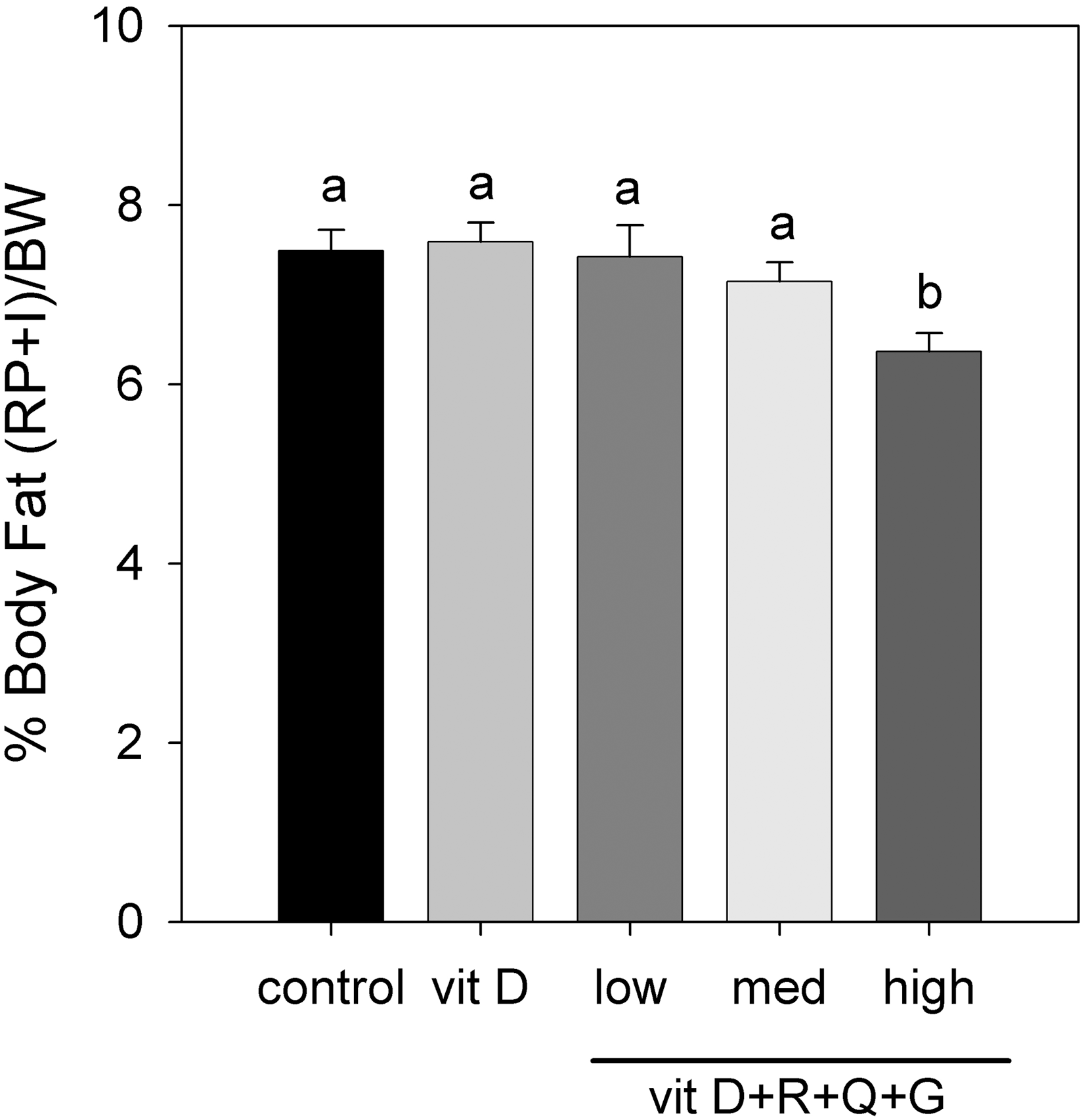
Treatment effect on percentage of fat pad weights. Weights of RP and I fat pads are given as a percentage of final body weight (BW). Data are mean±SEM values (n=10). abColumns without a common letter are different, P<.05. BW, body weight.
Serum concentration of biomarkers
Rats given the High treatment had significantly increased serum IGF-1 levels compared with control rats (P<.05) (Table 3). However, there were no significant treatment effects on the other serum biomarkers such as insulin, leptin, and osteocalcin.
Data are mean±SEM values.
Means without a common letter are significantly different, P<.05.
GLP, glucagon-like peptide-1; IGF-1, insulin-like growth factor-1; OPG, osteoprotegerin; RANKL, receptor activator for nuclear factor κB ligand.
Bone densitometry
Results from the dual-energy X-ray absorptiometry analysis showed that the High treatment was associated with higher femur BMD, although the results were not different from those found with vitamin D alone. There were no significant differences in bone mineral content between treatments (Fig. 3); however, bone mineral content corrected for body weight was 17.1% and 10.4% greater with the High treatment compared with control and vitamin D alone, respectively (P<.05) (Fig. 4).
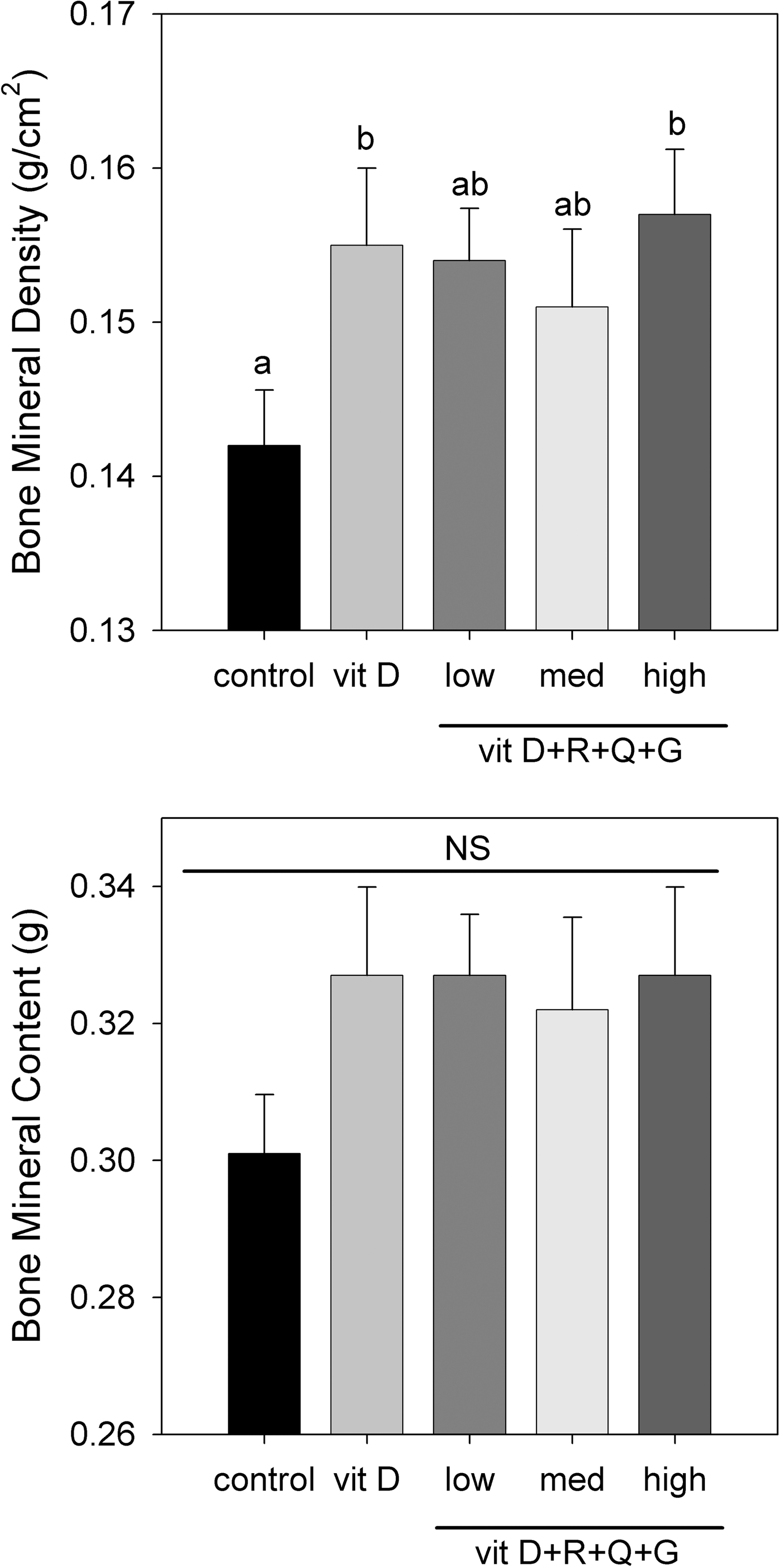
Treatment effect on bone mineral density and bone mineral content. measured in the right femur by dual-energy X-ray absorptiometry (PIXImus system, GE Lunar Corp.). Data are mean±SEM values (n=10). abColumns without a common letter are different, P<.05.

Treatment effect on adjusted bone mineral content (BMC). Right femur BMC is corrected for final BW. Data are mean±SEM values (n=10). abcColumns without a common letter are different, P<.05.
Micro-computed tomography analysis
After 8 weeks of treatment, the trabecular bone in the distal femur was markedly diminished in the control group compared with the High treatment group after ovariectomy (Table 4). Administration of the High treatment to the ovariectomized rats retained most of trabecular bone as revealed by the three-dimensional pictures (Fig. 5). The indices of trabecular bone microarchitecture, including BV/TV, BS/TV, Tb.Th, and Tb.N, in the rats given the High treatment were significantly higher than those of the control and vitamin D groups. Rats receiving the High treatment had significantly higher BV/TV and BS/TV compared with control and vitamin D–treated rats (P<.01 for both) (Table 4). Furthermore, Tb.Th (P<.05) was 25% higher and Tb.Sp was 40.4% lower in the rats that received the High treatment compared with control rats (P<.05). Structure model index and degree of anisotropy were decreased with the High treatment (P<.001 and P<.002, respectively).
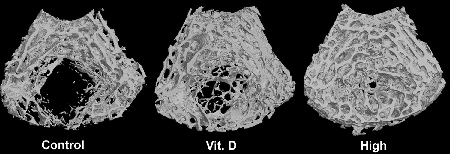
Microcomputed tomography images. Representative three-dimensional images are given of distal femur trabecular bone in rats treated with control, vit D, and the high-dose combination by scanning micro-computed tomography.
Data are mean±SEM values (n=4).
Means without a common letter are significantly different, P<.05.
BS, bone surface; BV, bone volume; DA, degree of anisotropy; NS, not significant; SMI, structure model index; Tb.N, trabecular number; Tb.Sp, trabecular separation; Tb.Th, trabecular thickness; TS, tissue surface; TV, tissue volume.
Bone histomorphometry
Histomorphometry data from the right tibia showed that rats receiving the High treatment had a significant decrease in bone marrow adipocyte number by 40.9% and 27.7% compared with the control and vitamin D alone, respectively (P<.05) (Fig. 6). TRAP-stained sections indicated that numbers of osteoclasts along the endosteal surface were significantly lower in rats receiving the Medium and High treatments by 43% and 47.7% compared with the control and vitamin D alone, respectively (P<.05) (Fig. 7).
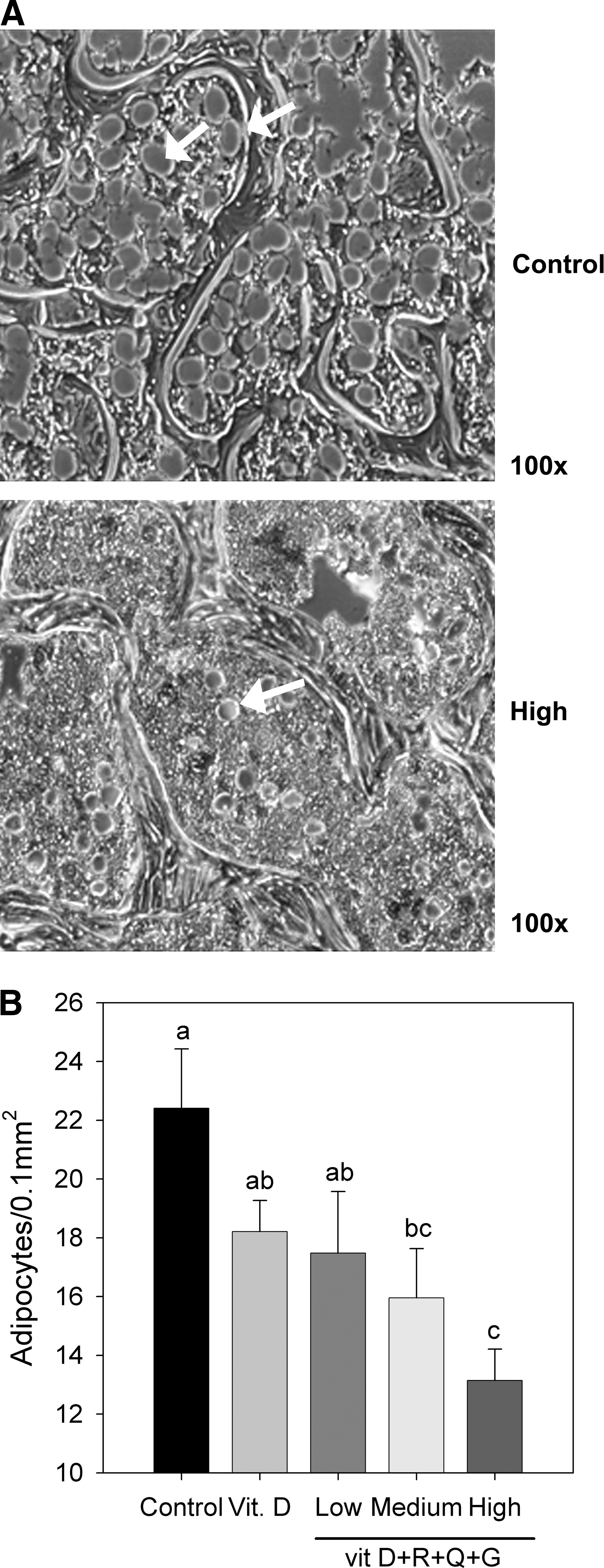
Treatment effect on number of adipocytes in the bone marrow. Bone marrow adipocytes were examined for the effect of 8 weeks of treatment. (
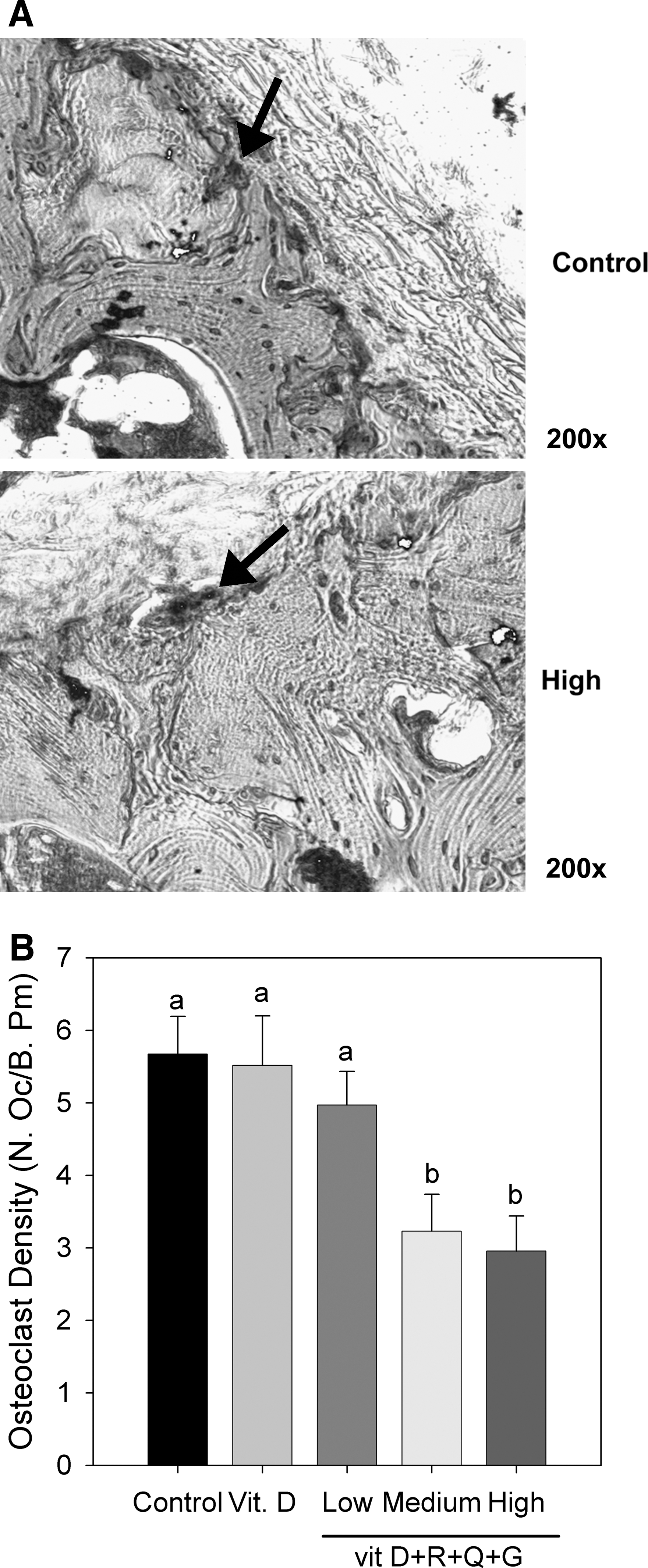
Treatment effect on number of osteoclasts in bone marrow. Bone marrow osteoclasts were examined for the effect of 8 weeks of treatment. (
Discussion
A decrease in fat pad weights with dietary supplementation of phytochemicals is consistent with a recent study demonstrating that genistein has antilipogenic effects in ovariectomized mice, which primarily reflects decreases in adipose tissue mass. 43 Naaz et al. 43 showed that 500–1,500 ppm of dietary genistein produce dose-dependent decreases in adipose tissues of 37–57% after a 12-day treatment and that the antilipogenic effect of genistein was similar in subcutaneous (inguinal) and visceral (retroperitoneal) fat pads. In the current study, we did not find a significant decrease in individual or combined fat pad weights in rats receiving the combination treatments compared with the control. The difference between our study and that of Naaz et al. 43 could be due to the difference in the age of rodents used. We used 12-month-old rats rather than juvenile mice (25–27 days old), which were used in the study by Naaz et al. 43 The effect of the treatment is more prominent in the juvenile model than in the aging ovariectomized model, possibly because of the sensitivity of the estrogen receptor. Nevertheless, the high-dose combination treatment significantly reduced the weights of retroperitoneal plus inguinal adipose tissue as expressed as a percentage of final body weight.
Our data demonstrated that the high-dose combination–treated rats gained less weight compared with the control and vitamin D–alone groups. The beneficial effects of treatment on body weight and adipose tissue were not a result of decreased food intake, suggesting that the treatment increased energy expenditure and modulated metabolic homeostasis. Among the phytochemicals used in the study, resveratrol at 22.4 mg/kg/day has been demonstrated to increase mitochondrial number, which consequently affects energy expenditure, by activating SIRT1, and this effect was reflected significantly in mice fed a high fat diet. 45 Hence, we hypothesize that the decrease in body weight gain and fat mass in rats daily treated with resveratrol at 25 mg/kg might be correlated to changes in energy expenditure. In addition, all three phytochemicals tested in this study have phytoestrogenic properties and, as such, may have had direct effects on lipid metabolism, thus contributing to reduction in body fat content in the rats receiving the high-dose combination treatment. For example, certain phytoestrogens, including soy isoflavones, quercetin, and resveratrol, have been shown to stimulate lipolysis and inhibit lipogenesis in adipocytes. 46 –48
The current data also indicate that BMD was significantly increased by the high-dose combination, although the result was not different from those produced by vitamin D alone. Simultaneously, serum IGF-1 concentration was elevated, which stimulates proliferation and differentiation of osteoblasts, as evidenced by the increase in BMD in our study. In this regard, Arjmandi et al. 49 have reported that administration of a daily supplement with 40 g of soy protein for 3 months may positively affect bone mass by promoting IGF-1 production in postmenopausal women, supporting the beneficial effect of the treatment on bone loss in our study. Of great interest is the finding that the high-dose combination treatment also greatly reduced loss of trabecular bone structure in the rats treated with the high-dose combination compared with both control and vitamin D treatments. Furthermore, histomorphological results show loss of bone marrow adipocytes coupled with a decrease in osteoclast number with the high-dose combination treatment in ovariectomized rats. These findings are consistent with the recent report that a treatment with flavonoids at 10 mg/kg/day in ovariectomized rats for 4 months significantly increased the values of dynamic histomorphometric indices for bone formation, decreased bone resorption as measured by the low osteoclast formation, and inhibited the expression of adipogenic genes via down-regulating the activity of peroxisome proliferator-activated receptor γ2 in bone marrow stromal cells. 50 The existence of an inverse relationship between osteogenic and adipogenic differentiation of the MSC supports the hypothesis that the common progenitors are directed towards the osteoblast lineage, reducing the accumulation of adipocytes. This has important implications for the use of phytochemical combinations as a therapeutic strategy in bone disorders such as osteoporosis.
Synergistic interactions with combinations of phytochemicals for the treatment of cancer have been investigated, indicating that vitamin D can synergize with genistein to inhibit the growth of prostatic or breast cancer cells. 51,52 Similarly, we have reported that the combination of genistein and vitamin D caused a significant increase in VDR protein levels in maturing preadipocytes, 36 and these results indicate the potentiality of both the increase in apoptosis and suppression of adipogenesis. Moreover, other studies also demonstrated that resveratrol stimulates VDR expression in bone marrow osteoblast precursors and synergizes with 1,25(OH)2D3 to induce the expression of osteocalcin and osteopontin. 24 Such findings provide information that certain phytochemicals contribute to synergistic activities with 1,25(OH)2D3 through up-regulation of the VDR signaling pathway.
Of all the natural alternatives currently under investigation, phytoestrogens appear to provide the most potential with few adverse effects. A study regarding breast safety suggested that daily consumption of 54 mg of genistein exhibited a promising safety profile with positive effects on bone formation in postmenopausal women with 3 years of treatment. 53 The finding of increased uterine weight in our study suggests a mild uterotropic activity by the combination treatments, but the change did not differ from that of ovariectomized rats that received a diet with vitamin D alone. It is notable that a chronic safety study in rats indicated that there were no treatment-related histopathologic changes in several organs including uterus after 13 weeks of treatment with genistein at dose up to 500 mg/kg/day, even though an increase in the uterine weight was noted. 54 One of the limitations in the current study is the lack of a sham-operated group, which could serve as a reference to compare the relative uterine weight of the ovariectomized rats treated with control diet and further determine the estrogenic effect on uterine weight. Proliferation of uterine endometrium is commonly assessed by weighing the uterus. Because the potential toxicity of this particular combination of phytochemicals has not been previously investigated, further study is needed to determine the safety profile of concentrations that have positive effects on both adiposity and bone.
There are some limitations present in this study that may need to be further addressed. First, although the static histomorphometric and micro-computed tomography evidence showed the antiresorptive effects based on a decrease in bone marrow adipocytes and osteoclasts and maintenance of trabecular structure, we did not find changes in plasma levels of biomarkers related to bone formation. Second, the effect of treatments on intact female rats was not studied. Generally, the efficacy on bones of intact rats should be less detectable, as ovariectomized rats resemble postmenopausal osteoporosis in humans. Even so, the anabolic effects on bones of intact rats should be included in future studies with larger sample size and a longer treatment time. Third, the activities observed in the rodent study may not be more than predictive of the action/synergies of the components in humans. It is also important to consider the bioavailability and the potential interaction of the compounds. Therefore, application of this research to a clinical trial still requires further investigation to determine safety and effectiveness in humans. Although results from rodent experiments cannot be directly extrapolated to human clinical effects, this study revealed the synergistic effects of vitamin D combined with genistein, quercetin, and resveratrol, which may help in developing a new strategy for the prevention of osteoporosis and obesity.
We conclude that supplementation of vitamin D combined with genistein, quercetin, and resveratrol improved bone density and trabecular bone structure and reduced body weight gain in ovariectomized female rats. Most likely, multiple phytochemicals acting on numerous targets on adipocytes and osteoblasts simultaneously can achieve synergistic beneficial effects at a dose known to be well within safe ranges in humans. Although these treatments remain to be tested for efficacy for preventing weight gain and bone loss in menopausal women, we propose that the synergistic effects of a combination of phytochemicals with vitamin D would be effective in preventing or reducing bone loss after menopause.
Footnotes
Acknowledgments
This work was supported in part by a grant from the Georgia Research Alliance and by the Georgia Research Alliance Eminent Scholar endowment held by C.A.B.
Author Disclosure Statement
C.A.B., M.A.D.-F., and M.W.H. are members of the board of directors of AptoTec, Inc. No competing financial interests exist for C.-Y.L., J.-Y.Y., S.R., D.L.H., S.A., or R.D.L.