
Abstract
The effect of HT042, a blend of three herbal extracts, on longitudinal bone growth was investigated in short- and long-term rat models. In the short-term model, we divided female Sprague-Dawley rats (3 weeks old) into six groups, according to treatment: vehicle, HT042 (100 mg/kg), Phlomis umbrosa (100 mg/kg), Astragalus membranaceus (100 mg/kg), and Eleutherococcus senticosus (100 mg/kg) were administered twice daily, and recombinant human growth hormone (rhGH) (1 IU) was subcutaneously injected once daily. Treatments were maintained for 4 days in each case. On day 3, tetracycline (20 mg/kg) was injected intraperitoneally (20 mg/kg) to form the fluorescent band on the growth plates. On days 2–4, 5-bromo-2′-deoxyuridine (BrdU) (50 mg/kg) was injected intraperitoneally to label proliferating cells. On day 5, the tibias were dissected and fixed in 30% sucrose. Dehydrated bone was sectioned at a thickness of 40 μm and observed. The bone growth in groups administered HT042 and rhGH was significantly increased to 433.50 ± 21.61 and 434.49 ± 15.21 μm/day, respectively, from 410.03 ± 17.4 μm/day (control). The height of the growth plates in the HT042 and rhGH groups was also significantly increased to 556.5 ± 21.1 and 544.2 ± 21.1 μm (P < .05), respectively, from 518.1 ± 4.1 μm (normal). The number of BrdU-positive cells in chondrocytes of the HT042 and rhGH groups was increased to 389 ± 36 and 627 ± 39 cells/mm2 (P < .001), respectively, from 264 ± 17 cells/mm2 (control). Insulin-like growth factor-1 and bone morphogenetic protein-2 in the HT042 group were highly expressed in the growth plate. In the long-term rat model, the body weight, nose–tail length, and nose–anus length were measured by microknemometry for 4 weeks. The body weight of the rhGH group was significantly increased. The nose–anus length of the HT042 and rhGH groups was significantly greater at 18.5 ± 0.3 and 18.7 ± 0.3 cm compared to 18.2 ± 0.2 cm (control).
Introduction
G
Bone growth is a result of endochondral cell proliferation in growth plates and conversion of chondrocytes into new bone. 6 To track these cells, cell labeling techniques using 5-bromo-2′-deoxyuridine (BrdU) were developed. BrdU is a thymidine analog that incorporates into DNA of dividing cells during the S-phase of the cell cycle. 7
For this bone growth study based on medicinal herbs we selected three herbs—the root of A. membranaceus, the stem of E. senticosus, and the root of Phlomis umbrosa—from Dongeuibogam and designated the formula HT042. The bone growth effect of HT042 was investigated in adolescent female Sprague-Dawley rats. We measured the bone growth of tibia and BrdU-positive cells on chondrocytes in the growth plate zone. 4 Additionally, the body weight, the nose–tail (N-T) length, and the nose–anus (N-A) length for 4 weeks were measured by microknemometry. 8
Materials and Methods
Plant material
The dried roots of P. umbrosa, the roots of A. membranaceus Bunge, and the stem barks of E. senticosus Harms were used (Omniherb Co., Daegu, Republic of Korea). They were identified by Prof. Dr. Hoyoung Choi, and voucher specimens (numbers HP125, HP123, and HP060) were deposited with the Department of Herbal Pharmacology, College of Oriental Medicine, Kyung Hee University, Seoul, Republic of Korea.
Preparation of HT042
Each herb of HT042 was mixed in the following ratio: P. umbrosa:A. membranaceus:E. senticosus = 19.7:27.0:53.3. At first, the herbs were extracted separately with 70% ethanol for 6 hours at 82°C in a reflux apparatus. After reflux, the extract was filtered, the filtrate was evaporated in a rotary evaporator, and samples were lyophilized in a freeze-dryer (Operon™, Seoul). The extract yield of each of the above herbs was 14.8%, 29.0%, and 7.0%, respectively. Then, the resulting three kinds of powders were mixed for HT042 in the proportion of the raw materials. The quantitative authentication of HT042 was performed on a Waters (Milford, MA, USA) instrument equipped with a Waters 600 pump, a Waters 717 autosampler, and a Waters 996 PDA detector using a Hypersil™ Gold C18 column (particle size, 5 μm; 250 × 4.6 mm; Thermo, Bellefonte, PA, USA). The column was equilibrated with a 5:95 mixture of distilled water containing 0.5% phosphoric acid (solvent A) and acetonitrile (solvent B) at a flow rate of 1.0 mL/minute. The column was eluted as follows: 0–60 minutes, 5–50% solvent B; 60–61 minutes, 50–70% solvent B; 61–80 minutes, 70–70% solvent B. Each extract was analyzed in triplicate.
The high-performance liquid chromatogram of HT042 is shown in Figure 1. The quality of HT042 was standardized using three representative components: 315.46 ± 33.58 mg% of 6,9-epi-8-O-acetylshanzhiside methyl ester for P. umbrosa, 14.32 ± 0.17 mg% of formononentin for A. membranaceus, and 298.73 ± 31.95% of eleutheroside E for E. senticosus. The mixed extracts were used as sample materials and stored at −20°C until used.

Three-dimensional high-performance liquid chromatogram of HT042, a blend of three herbal extracts. Color images available online at
Animals
To investigate the effects of HT042 treatment on longitudinal bone growth, 3-week-old female Sprague-Dawley rats, weighing 60 ± 10 g each, were used (Samtako Co., Osan, Republic of Korea). The experimental procedures were performed in accordance with the animal care guidelines of the Kyung Hee University's Institutional Animal Care and Use Committee [protocol number KHUASP (SE) 2009-007]. Animals were housed under controlled temperature (23 ± 2°C), relative humidity (55 ± 10%), and lighting (lights on 07:00–19:00 hours) conditions, with food and water made available ad libitum. After 1 week of acclimatization, the rats were administered the herbal mixture.
Short-term administration of HT042 and treatment with tetracycline and BrdU
Animals were randomly allocated into six groups according to supplement regimen. Vehicle (nine rats), HT042 (100 mg/kg; nine rats), P. umbrosa (100 mg/kg; nine rats), A. membranaceus (100 mg/kg; nine rats), or E. senticosus (100 mg/kg; eight rats) was administered twice daily at 8:00 and 20:00 hours in a fixed dose volume of 5.0 mL/kg. Recombinant human GH (rhGH) (1 IU; nine rats) (LG, Seoul) was subcutaneously injected once daily.
Treatment was continued for 4 consecutive days in each case. On day 3, all animals were injected intraperitoneally with tetracycline hydrochloride (20 mg/kg, Sigma, St. Louis, MO, USA) in saline for the measurement of bone growth. On days 2, 3, and 4, BrdU (50 mg/kg, Sigma) was intraperitoneally injected to label proliferating cells. Twenty-four hours after the last injection, animals were sacrificed, and then the tibias were dissected free of soft tissue. 4
Tissue preparation and detection of longitudinal bone growth
The dissected tibias were fixed in 4% paraformaldehyde for 48 hours and dehydrated by immersion in 30% sucrose for 1 day. 9 Dehydrated bone was directly sectioned longitudinally at a thickness of 40 μm with a microtome (Leica, Berlin, Germany). 10 The sections were mounted onto gelatinized glass slides and observed by fluorescence microscopy (Olympus, Tokyo, Japan), to measure the longitudinal bone length between the fluorescent line and the epiphyseal end line of the growth plate at three different locations using Optimas version 6.5 (Optimas Corp., Bothell, WA, USA). 11 Data were averaged to calculate the longitudinal bone growth rates.
Detection of chondrocyte proliferation
For detection of cell proliferation in the growth plate, BrdU-specific immunohistochemistry was performed. 12 Dehydrated tibia sections were pretreated in 2 N HCl at 37°C for 1 hour and rinsed twice in 0.1 M sodium borate (pH 8.5). The sections were incubated in phosphate-buffered saline including 3% bovine serum albumin and 0.3% Triton X-100 for 1 hour and incubated with BrdU-specific mouse monoclonal antibody (Santa Cruz Biotechnology, Santa Cruz, CA, USA) (diluted 1:100) overnight at 4°C. Then the sections were washed with 0.05 M phosphate-buffered saline three times and incubated with fluorescein isothiocyanate-conjugated mouse secondary antibody (Jackson ImmunoResearch Laboratories, Bar Harbor, ME, USA) (diluted 1:200) for 4 hours at room temperature. Finally, the sections were washed with phosphate-buffered saline and mounted onto gelatinized glass slides. To measure the number of BrdU-positive cells, fluorescence images of sections were taken by a confocal microscope (LSM-5, Zeiss, Oberkochen, Germany).
The mean density of BrdU labeling cells in each rat was calculated as the number of BrdU-labeled nuclei per area (in mm2) in the growth plate and measured by two observers. 13
Measurement of height of the growth plate
Differential interference contrast images of dehydrated sections were taken by a confocal microscope (model LSM-5, Zeiss), and the length of the growth plate was measured at three different locations by two observers. The resting, proliferative, and hypertrophic zone heights on growth plate were also measured on Ddifferential interference contrast images of a Zeiss confocal microscope.
Measurement of bone morphogenetic protein-2 and insulin-like growth factor-1 in growth plates
For the detection of bone morphogenetic protein (BMP)-2 and insulin-like growth factor (IGF)-1 in the growth plate, the dehydrated tibia sections were incubated in 1% Triton X-100 and incubated with goat BMP-2 antibody or rabbit IGF-1 antibody (Santa Cruz Biotechnology) (diluted 1:500) at room temperature overnight. Then, sections were incubated with anti-goat antibody and anti-rabbit antibody (Jackson ImmunoResearch Laboratories) (diluted 1:200) for 60 minutes, respectively, and stained with 0.05% 3,3-diaminobenzidine. 10
Long-term administration of HT042 and longitudinal bone growth
To investigate the effect of long-term treatment on longitudinal bone growth, rats were randomly divided into three experimental groups. Vehicle (seven rats) or HT042 (100 mg/kg; 16 rats) was administered twice daily at 8:00 and 20:00 hours in a fixed dose volume of 5.0 mL/kg. rhGH (1 IU; eight rats) was injected subcutaneously once daily. This treatment was continued from days 28 to day 56 of each rat's life for 4 weeks. The body weight, N-T length, and N-A length were assessed by measuring with microknemometry weekly for bone growth in animals sedated with N2O/O2 gas and isoflorane. 8
Statistical analysis
The data were expressed as mean ± SD values of the different experiments under the same conditions. Student's t test was used to make statistical comparisons between groups. Differences were considered significant at P < .05.
Results
Short-term effect of longitudinal bone growth
To evaluate the rate of longitudinal bone growth, tetracycline was used to label newly formed bone (Fig. 2). We found that the longitudinal bone growth of normal adolescent female rats was 410.0 ± 17.4 μm/day. The rhGH (1 IU) group showed a significantly greater rate (434.5 ± 15.2 μm/day; 5.96%) of longitudinal bone growth compared with the control group (P < .01). The HT042 (100 mg/kg) group also showed a significantly greater rate of bone growth (433.5 ± 21.6 μm/day; 5.72%) compared with the control group (P < .05) (Table 1). For each component of the herbal mixture, P. umbrosa alone, A. membranaceus alone, and E. senticosus, alone, the longitudinal bone growth increased by 2.9%, 2.8%, and 4.6%, respectively. Thus, the longitudinal bone growth rate of each herbal component of HT042 was less than that of the herbal combination of HT042 (Fig. 2).
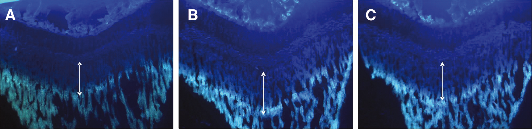
Fluorescent photomicrographs of longitudinal sections at the proximal tibia in the proximal tibia growth plate of
Data are mean ± SD values (n = 8 per group).
P < .05, **P < .01, ***P < .001, significant differences compared with normal control.
rhGH, recombinant human growth hormone injected subcutaneously.
Effects on growth plate thickness and zonal heights
Using histomorphometrical measurements, the overall growth plate zone was divided to the reserve zone (a layer of small, round cells irregularly arranged), the proliferative zone (wherein the cells divide along the long axis of the bone in regular columns), the prehypertrophic zone, and finally the hypertrophic zone (large, glycogen-filled cells). 14,15
The proximal tibia growth plate in normal female rats was approximately 518.9 ± 4.1 μm thick. Following HT042 administration at a dosage of 100 mg/kg, growth plate thickness increased to 544.2 ± 21.1 μm (P < .05). In calculating the each zonal length, the prehypertrophic zone was included in the hypertrophic zone (Fig. 3). The resting, proliferative, and hypertrophic zonal heights increased by 6.9%, 11.2% (P < .01), and 2.9%, respectively. For the rhGH (1 IU) group, growth plate thickness increased to 556.5 ± 21.2 μm (P < 0.01), and the resting, proliferative, and hypertrophic zonal heights increased by 8.0%, 5.9% (P < .001), and 6.2%, respectively. The heights of the proliferative zones of the rhGH and HT042 groups were increased significantly (Table 1 and Fig. 4).

Representative differential interference contrast image of the tibia growth plate in 3-week-old Sprague-Dawley rats. Resting zone, proliferative zone, prehypertrophic zone, hypertrophic zone, and ossification zone are designated.

Representative rat growth plate sections, indicating the three zones in sections of tibial growth plates in the
Effect on chondrocyte proliferation
In the present study, BrdU-labeled cells were observed in the chondrocytes (Fig. 5). The number of BrdU-positive cells in the control group was 264 ± 17 cells/mm2. In the rhGH group, the number of BrdU-positive cells increased to 627 ± 39 cells/mm2. The number of BrdU-positive cells in the HT042 group also increased, to 389 ± 36 cells/mm2. The increased ratios of the rhGH and HT042 groups were 2.38- and 1.47-fold, respectively; differences were significant (P < .001) (Table 2 and Fig. 6).
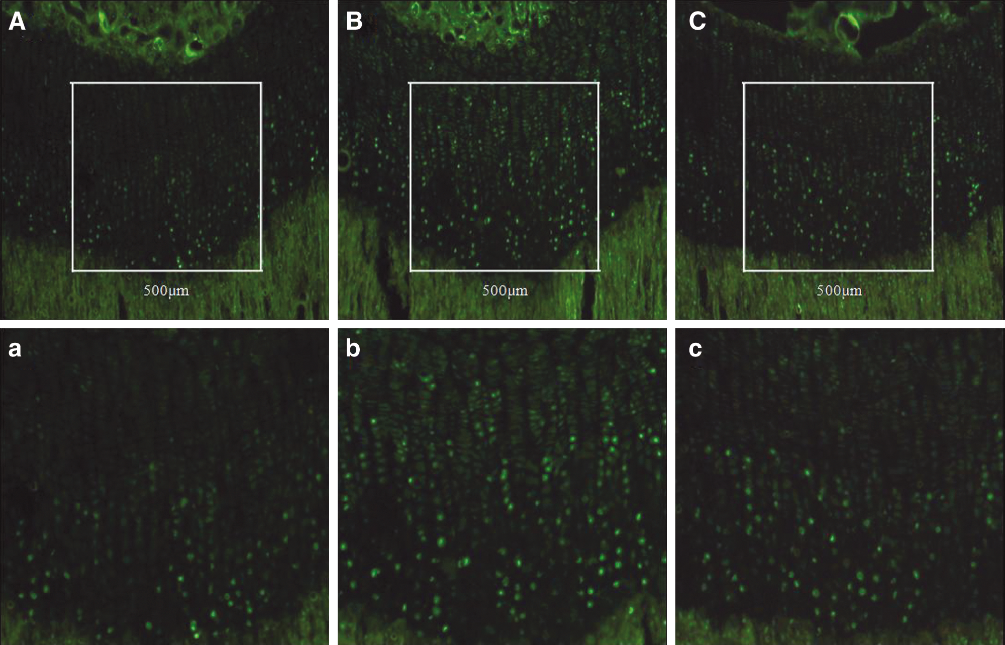
Representative images of 5-bromo-2′-deoxyuridine (BrdU)-labeled chondrocytes in growth plates of the proximal tibiae:
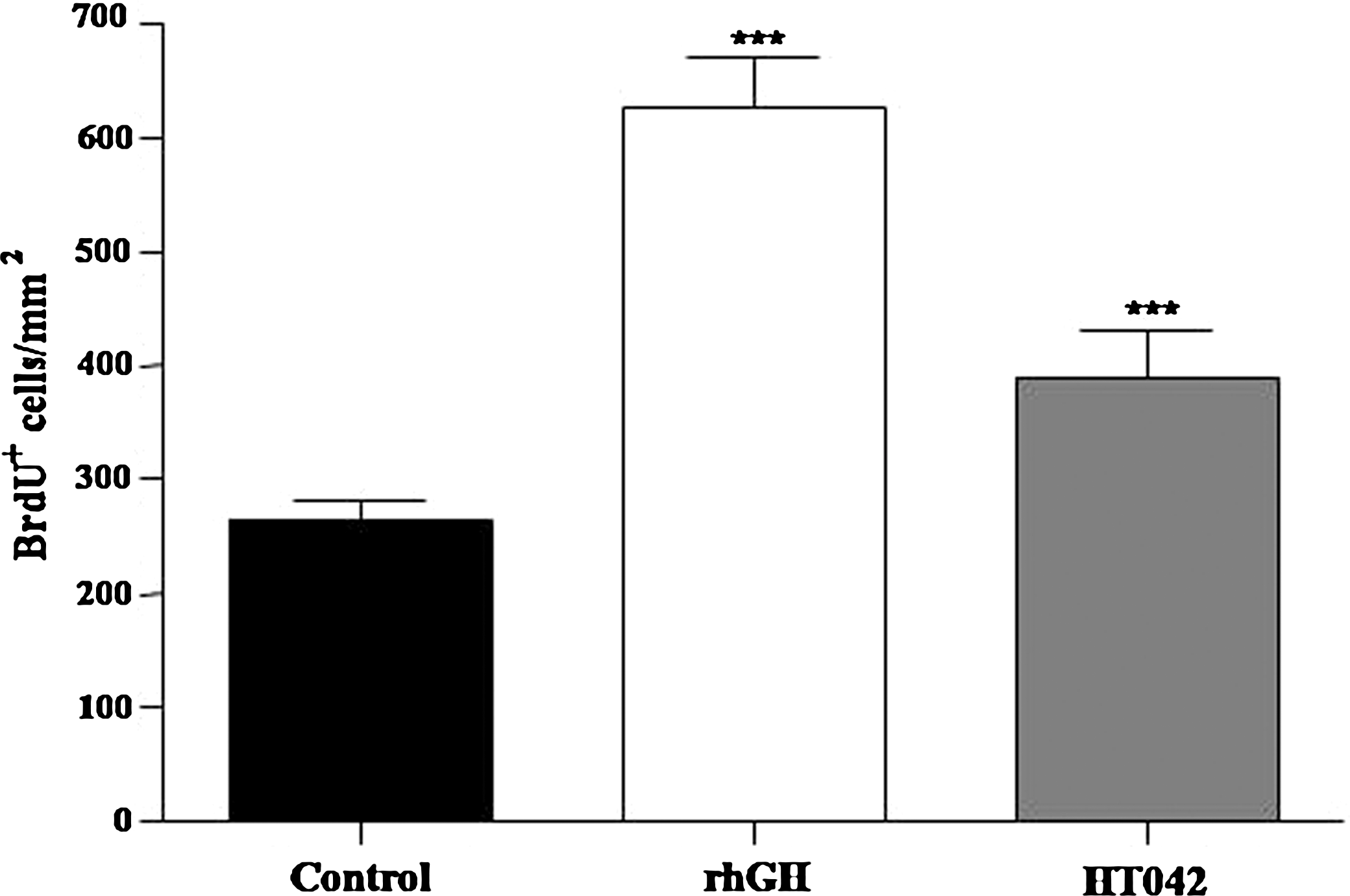
Effects of HT042 on chondrocyte proliferation in growth plates of adolescent female rats. BrdU-positive cells were quantified. Data are mean ± SD values (n = 8). Statistical significance was determined using a t test: ***P < .001, compared with control.
Data are mean ± SD values (n = 8 per group).
P < .001, significant differences compared with control.
BrdU, 5-bromo-2′-deoxyuridine.
Effect on BMP-2 and IGF-1 expression
To investigate the expression of BMP-2 and IGF-1, we performed immunohistochemistry in the growth plate. We found that BMP-2 was highly expressed in the prehypertrophic and hypertrophic zones in the rhGH group but was minimally expressed in the resting and proliferative zones (Fig. 7). IGF-1 expression was relatively higher in the proliferative and prehypertrophic zones than in other zones (Fig. 7). BMP-2 expression of the HT042 group was higher than that of the rhGH group, whereas IGF-1 expression of the HT042 group was lower than the rhGH group but higher than the control group (Fig. 7).

Immunohistochemical localization of
Long-term effect on body weight and axial skeletal growth
To study the long-term effect of HT042 on bone growth, the body weight of the female rats was measured weekly for 4 weeks. The body weight of the rhGH group was significantly increased by day 35 (Fig. 8). At 56 days, body weight increased by 14.5% in the rhGH group and by 7.3% in the HT042 group compared with the control group.

Change of
To evaluate the effect of HT042 on axial skeletal growth, N-T length and N-A length were measured with microknemometry every week for 4 weeks. In the rhGH group, N-T and N-A lengths were significantly increased by day 35. In the HT042 group, the N-A length was significantly increased by day 42 (Fig. 8 and Table 3). The N-A length was increased by 4.2% in the rhGH group and by 2.8% in the HT042 group compared to that of the control group, indicating that HT042 promotes the longitudinal N-A growth through at least 2 weeks of treatment in female rats.
Data are mean ± SD values (n = 8 per group).
P < .05, **P < .01, significant differences compared with normal control.
N-T, nose to tail.
Discussion
We demonstrated that HT042 increases longitudinal bone growth in adolescent female rats. HT042 significantly increased the tibia length by 5.72% compared to normal rats. This rate was nearly the same as the 5.96% of the rhGH group; rhGH is well established to increase longitudinal bone growth. 16,17
The HT042 herbal formula exhibits synergistic effects in promoting longitudinal bone growth rate. None of the individual herbs in HT042 was as effective as the HT042 mixture in eliciting bone growth. Consistent with the previous study, 7 A. membranaceus and E. senticosus increased bone growth rates; however, each of the component herbs of HT042 showed lower efficacy compared with the HT042 mixture.
The linear growth of long bone is achieved at the growth plate, a layer of cartilage situated between the epiphysis and metaphysis. The chondrocytes of the growth plate exist within a cartilaginous matrix and are arranged in specific layers, or zones. In order from the epiphysis to the metaphysis, these are the reserve zone (a layer of small, round cells irregularly arranged), the proliferative zone (wherein the cells divide along the long axis of the bone in regular columns), the prehypertrophic zone, and finally the hypertrophic zone (large, glycogen-filled cells). 14,15,18 The proliferative zone is the driving force behind bone elongation. After several mitoses, the chondrocytes are transformed to the hypertrophic zone, in which the cell division has ceased and become greatly enlarged and transformed to the bone matrix, which becomes calcified. 19 The height of the growth plate is regulated by the proliferative and hypertrophic chondrocyte zones. 20 Therefore, the length of the growth plate or the length of the chondrocyte zone is direct evidence of longitudinal bone growth. In the HT042 group, the heights of the overall growth plate and the proliferative zone were significantly increased compared to controls. This suggests that HT042 can increase the overall growth plate thickness responsible for transformation into bone matrix where longitudinal bone growth occurred. 20
Additionally, the proliferation of chondrocytes is also an important phenomenon in bone growth. 21 In the present study, proliferation was determined experimentally by counting cells positive for incorporation of the thymidine analog BrdU, i.e., cells in S-phase, when exposed to an injection of BrdU. 21 While the number of BrdU-positive cells in the control group was 264 ± 17 cell/mm2, the number of BrdU-positive cells in the rhGH and HT042 groups increased by 627 ± 39 cells/mm2 (2.38-fold) and 389 ± 36 cells/mm2 (1.47-fold), respectively (P < .001). This result suggests that HT042 induces cell proliferation of chondrocytes. Furthermore, BMP-2 expression was high in the prehypertrophic and hypertrophic zones with HT042 treatment. IGF-1 was highly expressed in the proliferative and prehypertrophic zones of the growth plate, indicating that longitudinal bone growth effect of HT042 is stimulated by the local IGF-1 and BMP. 22,23 IGFs have been proposed to be the major determinant of postnatal longitudinal growth and to play an essential role in bone metabolism. 22,23 BMP-2, expressed in the growth plates of long bones, 24,25 accelerates longitudinal bone growth by stimulating growth plate chondrocyte proliferation and chondrocyte hypertrophy. 26
When the long-term effect of HT042 treatment on adolescent female rats was investigated for 28 days and the body weight and N-T and N-A lengths were compared with those of the control group, the rhGH group showed a significant increase in all parameters from day 35. In the HT042 group, the N-A length was significantly increased from day 42 (P < .05). The N-A length is universally used as the index of growth-promoting activities. 27 The results suggest that HT042 promotes the longitudinal N-A growth through at least 14 days of treatment in female rats.
In conclusion, HT042 stimulated chondrocyte proliferation and chondrocyte hypertrophy in the growth plate and directly increased the longitudinal tibia length and the N-A length of female rats. Therefore, HT042 could be helpful for increasing bone growth of children who have growth retardation.
Footnotes
Acknowledgments
This work was supported by the Second Stage of Brain Korea 21 project in 2009 and by grant 2009-0063466 from the Korea Science and Engineering Foundation funded by the Korean Government.
Author Disclosure Statement
No competing financial interests exist.