




Abstract
Postmortem examinations of tissues of humans and rodents with a host of neurodegenerative conditions, including Alzheimer's and Parkinson's diseases, have identified oxidative damage in proteins, lipids, and DNA. The aim of this study was to better understand the cellular mechanisms of neuronal cell degeneration induced via oxidative stress and the protective roles of bioactive substance. In order to achieve this aim, we established a screening program to discover therapeutic agents that exhibit preferential neuroprotective activity in H2O2-treated PC12 cells. During the course of our screening program, we isolated an active compound, SG-168, from Dendrobium nobile Lindley and identified it as a neuroprotective agent. SG-168 was identified as a compound with an acetal skeleton, a prototypical compound, by electrospray ionization-mass spectrometry analysis and various nuclear magnetic resonance spectroscopic methods. The protective effect of SG-168 in PC12 cells with H2O2-induced oxidative damage was investigated by the 3-(4,5-dimethylthiazol-2-yl)-2,5-diphenyltetrazolium bromide reduction assay. As expected, incubation with H2O2 for 2 hours resulted in cell viability of 31.8% compared to the control, while pretreatment of SG-168 increased cell viability by 15–50% compared to the H2O2-stressed control cells. These results showed that SG-168 inhibits H2O2-induced apoptotic cell death. Interestingly, flow cytometric analysis showed that H2O2-treated PC12 cells incubated with SG-168 exhibited greatly suppressed apoptosis. In summation, the results of this study suggest that SG-168 has potential as a new antioxidant agent against neuronal diseases.
Introduction
With the aim of adding to this knowledge base, we established a screening system to discover therapeutic agents that can scavenge free radicals and protect cells from H2O2. Using this system, we isolated an active compound, SG-168, from Dendrobium nobile Lindley, which can protect PC12 cells against H2O2-induced apoptosis. By examining its chemical structure and physicochemical properties through liquid chromatography–mass spectrometry analyses and various nuclear magnetic resonance spectroscopic methods, we were able to identify SG-168 as a 3-[[(6-methoxy-10-methyl-1H,3H-benzo[h]furo[4,3,2-de]-2-benzopyran-1-yl)oxy]methyl]-5-methylnaphtho[2,3-b]furan-4,9-dione skeleton as an acetal, a prototypical compound. However, the neuroprotective activity of SG-168 has not been previously reported.
D. nobile Lindley (known as Seokgok in Korea and Shi Hu in mainland China) is widely used in both traditional Chinese and Korean folk medicine as an analgesic, an antipyretic, a tonic to nourish the stomach, and an anti-skin-aging agent and for treating diabetes and cardiovascular disease. 21 Pharmacological studies have revealed that the phenanthrenes isolated from D. nobile Lindley exhibit antifibrotic activity. 22 Besides containing bibenzyl derivatives and fluorenones, D. nobile Lindley extracts have been shown to possess antioxidant activity. 23 This suggests that crude extracts of D. nobile Lindely are nontoxic or only slightly toxic to humans and animals. Considering its low toxicity and potent neuroprotective effect, the crude extract of D. nobile Lindely, which contains SG-168, might prove to be a valuable therapeutic agent for neurodegenerative diseases such as Parkinson's disease and Alzheimer's disease.
In this study, we present results on the antioxidant and neuroprotective effect of SG-168 against H2O2-induced cell death in PC12 cells. Furthermore, we suggest that SG-168 may possess potential as an antioxidative therapeutic agent for neuronal diseases.
Materials and Methods
Materials
Dried D. nobile Lindley was obtained from Kumkang Pharm. Co. Ltd. (Masan, Republic of Korea). SG-168 isolated from D. nobile Lindley was prepared as a stock solution of 10 mg/mL in dimethyl sulfoxide (DMSO) and stored at −20°C. SG-168 was added to cell culture medium such that the DMSO made up less than 1% of the volume of the culture medium. H2O2 solution was purchased from Sigma-Aldrich (St. Louis, MO, USA) and dissolved in sterilized phosphate-buffered saline (PBS) at a stock concentration of 50 m
Purification of SG-168 from D. nobile Lindley
The neuroprotective compound was isolated and purified from D. nobile Lindley as follows. The dried D. Lindley (250 g) was percolated overnight with methanol (5 L). The extracts were concentrated in vacuo to eliminate the methanol. The resulting aqueous solution was extracted with diethyl ether. The organic layer was concentrated, applied to a silica gel column, and eluted with chloroform–methanol (50:1 to 1:1 [vol/vol]). Finally, the pure compound was obtained by reverse-phase column chromatography and eluted with 60% methanol to give a dark green powder (4 mg).
The active compound, SG-168, prevented H2O2-induced cytotoxicity in PC12 cells. Using liquid chromatography–mass spectrometry spectroscopy, the molecular formula of SG-168 was found to be C30H22O7. The structure of SG-168 was determined from its physicochemical properties and with various nuclear magnetic resonance spectroscopic methods. Using background literature research, SG-168 was determined to have a 3-[[(6-methoxy-10-methyl-1H,3H-benzo[h]furo[4,3,2-de]-2-benzopyran-1-yl)oxy]methyl]-5-methylnaphtho[2,3-b]furan-4,9-dione skeleton as an acetal, which had previously been reported as a prototypical compound (Fig. 1). 24
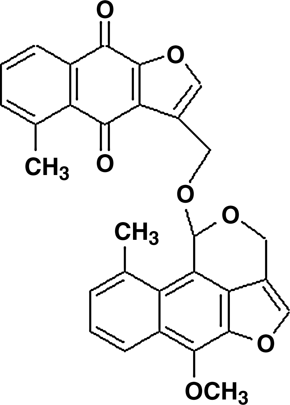
Chemical structure of SG-168.
Measurement of 2,2-diphenyl-1-picrylhydrazyl radical scavenging ability
Eighty microliters of 0.2 mM 2,2-diphenyl-1-picrylhydrazyl (DPPH) ethanol solution was added to 20 μL of sample solution of different concentrations and allowed to react at room temperature. A control, containing 20 μL of DMSO and 80 μL of 0.2 mM DPPH, was prepared. The mixture was held at room temperature for 10 minutes, and then the absorbance was measured at 492 nm. The ability to scavenge the DPPH was calculated as percentage radical scavenging activity (RSA) using the following equation: RSA (%) = (1 −A/B) × 100, where A is the absorbance of the sample and B is the absorbance of the control. Vitamin C was used as the positive control.
Measurement of SOD-like activity
The reaction mixture contained 50 μL of Tris-HCl buffer and 7.2 mM pyrogallol, with or without sample. After incubation at 25°C for 45 minutes, the absorbance at 405 nm was determined against a blank. Vitamin C was used as the positive control in this assay. The SOD-like activity was calculated using the following equation: SOD-like activity (%) = (1 − A/B) × 100, where A is the absorbance of sample and B is the absorbance of the control.
Cell culture
Rat pheochromocytoma PC12 cells were obtained from the Korean Cell Line Bank (Seoul, Republic of Korea) and were routinely grown in Dulbecco's modified Eagle's medium (Invitrogen, Grand Island, NY, USA) supplemented with 10% fetal bovine serum, 5% horse serum, penicillin (100 U/mL), streptomycin (100 μg/mL), and 3.7 g/mL NaHCO3 and were maintained in a 37°C humidified 5% CO2 atmosphere. In all experiments, cells were treated with SG-168 before treatment with H2O2 for the indicated times. All in vitro experiments were performed using exponential growth-phase cells and were repeated at least twice.
Morphological analysis
PC12 cells in Dulbecco's modified Eagle's medium containing 10% fetal bovine serum and 5% horse serum were seeded into six-well plates (2 × 10⁵ cells/mL) and incubated overnight in a humidified atmosphere of 5% CO2 at 37°C. The cells were pretreated with SG-168. After incubation for 30 minutes, the cells were treated with 0.5 mM H2O2 for 2 hours. The cellular morphology was observed by using a phase-contrast microscope (Nikon, Tokyo, Japan) at × 100 magnification.
3-(4,5-Dimethylthiazol-2-yl)-2,5-diphenyltetrazolium bromide reduction assays
Cell viability was measured with blue formazan that was metabolized from colorless tetrazolium dye [3-(4,5-dimethylthiazol-2-yl)-2,5-diphenyltetrazolium bromide (MTT)] (Sigma-Aldrich) by mitochondrial dehydrogenase, which is active only in live cells. PC12 cells were seeded in 96-well plates at a density of 1 × 10⁵ cells/mL and allowed to attach overnight at 37°C. H2O2 was used to induce apoptosis at the doses indicated. Pretreatment with SG-168 was done for 30 minutes, and then the cells were treated with 0.5 m
Lactate dehydrogenase release assay
The cytotoxicity was determined by measuring the release of lactate dehydrogenase (LDH). An LDH release assay kit was purchased from Wako Pure Chemical Industries (Osaka, Japan). PC12 cells were seeded in 96-well plates (1 × 10⁵ cells/mL), and the cells were pretreated with SG-168. After incubation for 30 minutes, the cells were treated with 0.5 m
Cell staining
Apoptosis was investigated by staining the cells with Hoechst 33342 (Sigma-Aldrich). PC12 cells were washed twice with PBS and then fixed in PBS containing 10% formaldehyde for 4 hours at room temperature. Fixed cells were washed with PBS and stained with Hoechst 33342 for 30 minutes at room temperature. PC12 cells were examined under a fluorescence microscope (Nikon) for nuclei showing typical apoptotic features such as chromatin condensation and fragmentation. Photographs were taken at a magnification of × 400.
Flow cytometric analysis
PC12 cells were harvested and fixed with ice-cold 70% ethanol. The fixed cells were stained with 50 μg/mL propidium iodide at room temperature in the dark for 30 minutes. PC12 cells were analyzed by cytometry (Beckman Coulter, Fullerton, CA, USA) with a laser excitation wavelength of 488 nm and an emission wavelength of 620 nm (FL3) to quantify the red propidium iodide fluorescence. The percentages of cells in various phases of the cell cycle—namely, sub-G1, G1, S, and G2/M—were assessed using WINCYCLE 32 software (Beckman Coulter). Every measurement typically counted at least 10,000 events. Apoptotic rates were determined by the percentage of nuclei accumulated at the peak of the sub-G1 phase.
Statistical analysis
All data are expressed as the means of three determinations, and the differences were analyzed for significance using the SPSS Package for Windows (Version 14.0; SPSS Inc., Chicago, IL, USA). The data were evaluated by one-way analysis of variance followed by Scheffé's test. The differences were considered significant at P < .05.
Results
H2O2-induced cytotoxicity in PC12 cells
Neuronal cell death has been shown to be elicited by increases in various kinds of reactive oxygen species.
25
In order to determine the cytotoxicity underlying neuronal loss we established a model of oxidative stress by exposing PC12 cells to H2O2. PC12 cells were incubated with various concentrations of H2O2 for 2 hours. Morphological alteration was assessed by phase-contrast microscopy. As expected, the control group exhibited round cell bodies with clear edges and a fine dendritic network. However, incubation with 0.1, 0.5, and 1 m
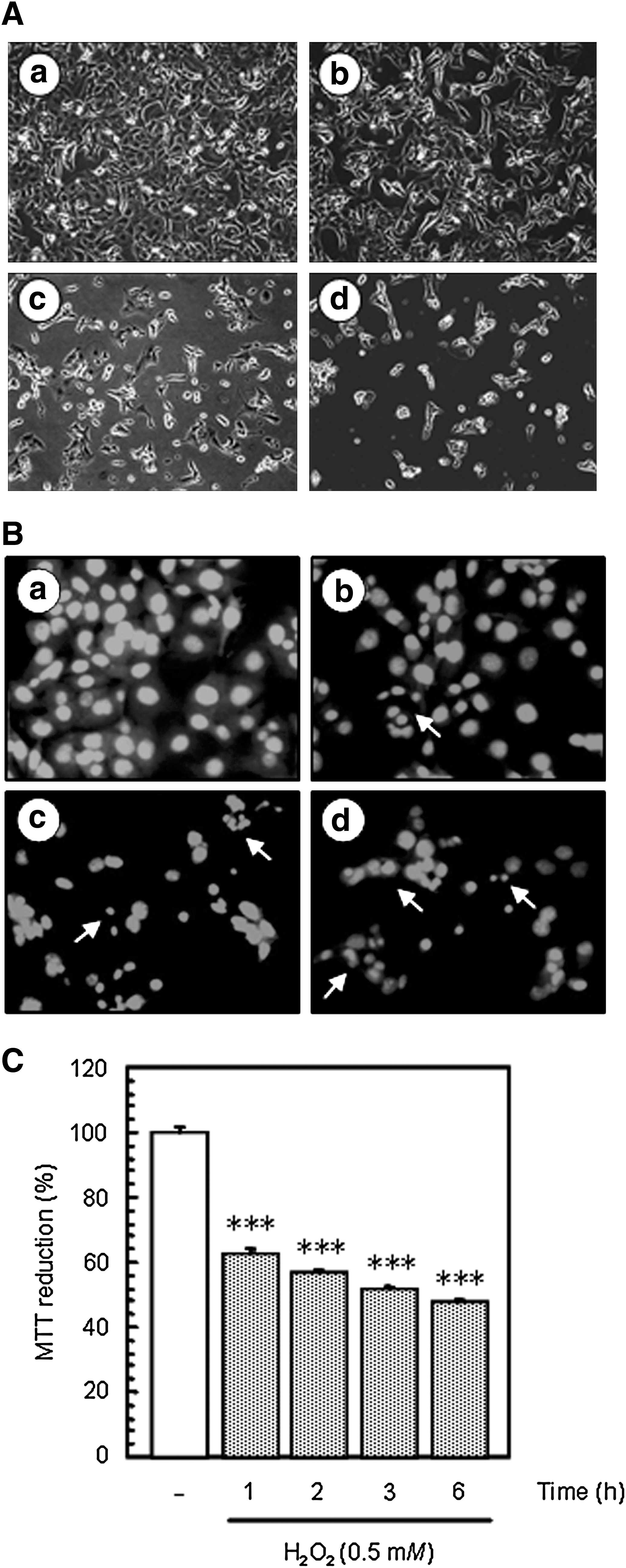
Hydrogen peroxide (H2O2)-induced cell death in PC12 cells.
These results indicated that H2O2 induced cytotoxicity and apoptosis in the PC12 cells.
Antioxidant activity of SG-168
The DPPH RSA and SOD-like activity of SG-168 were determined, and the results are shown in Table 1. SG-168 exhibited good scavenging activity by its capacity to reduce the stable radical DPPH to yellow-colored diphenylpicrylhydrazine in a dose-dependent manner, and it reached up to 30.8% at the concentration of 2,000 μg/mL. The SOD-like activity of SG-168 showed trends similar to the DPPH RSA. The effective concentration at which radicals were scavenged by 50% for DPPH RSA (3.16 mg/mL) and SOD-like activity (2.39 mg/mL) of SG-168 was relatively lower than those of vitamin C (0.66 and 0.61 mg/mL, respectively), a known antioxidant used as the positive control (data not shown).
Data are mean ± SE values of triplicate experiments.
Radical scavenging activity (RSA) (%) = (1 – absorbance of sample/absorbance of the control) × 100.
Superoxide dismutase (SOD)-like activity (%) = (1 – absorbance of sample/absorbance of the control) × 100.
Values not sharing the same letter are significantly different from one another by Duncan multiple range test (P < .05).
Protective effect of SG-168 against H2O2-induced cytotoxicity and apoptosis of PC12 cells
To assess the neuroprotective effect of SG-168, we investigated for morphological alterations such as cell shrinkage and membrane blebbings that are normally associated with apoptotic cell death. PC12 cells treated with 0.5 m
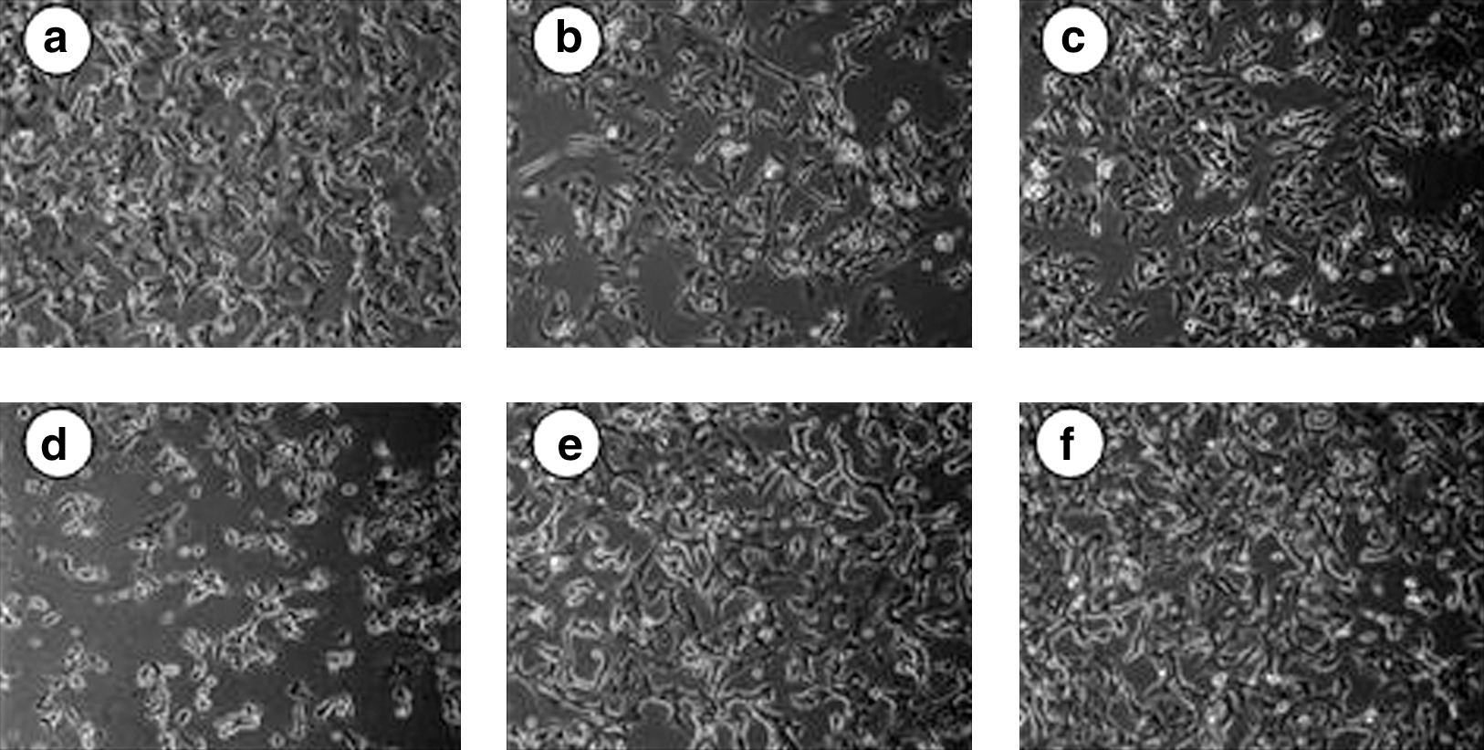
Effect of SG-168 on H2O2-induced morphological alterations of PC12 cells for 2 hours:
As shown in Figure 4A, incubation with 0.5 m

Effects of SG-168 on cell viability of H2O2-challenged PC12 cells:
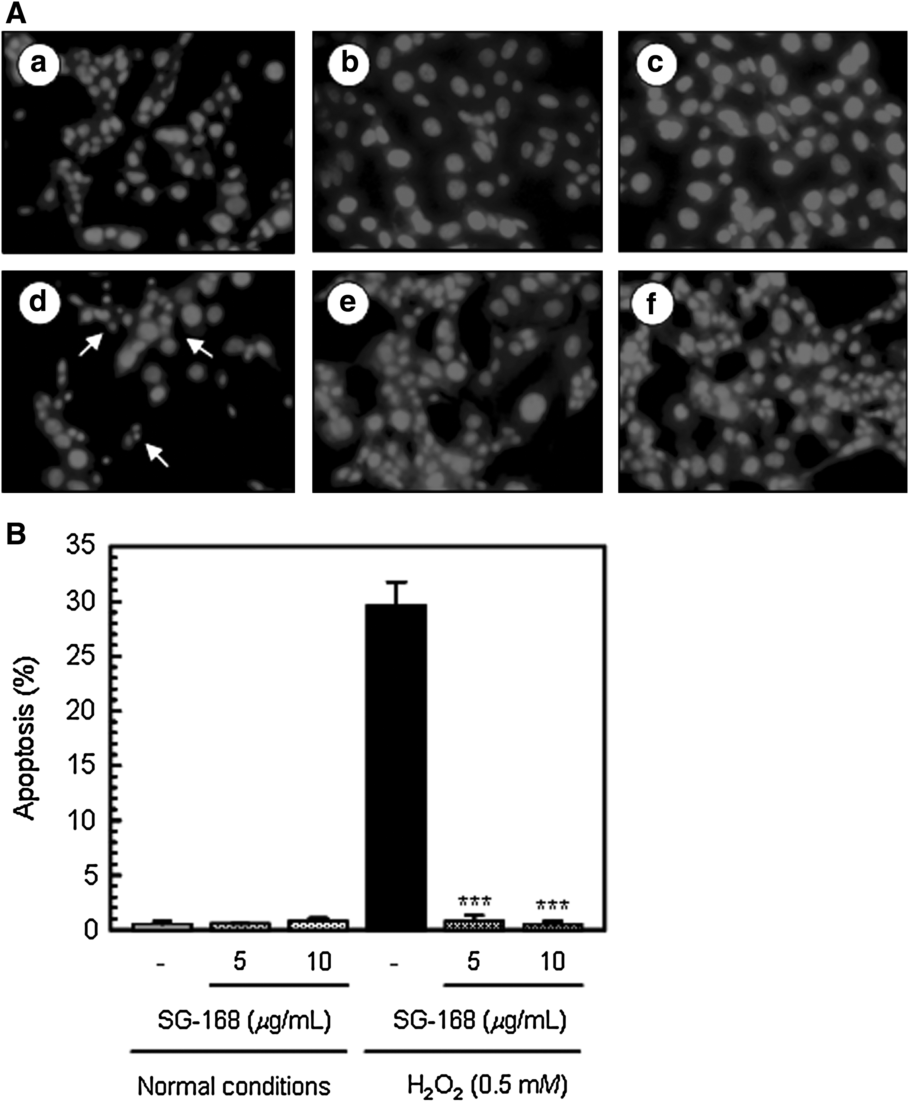
SG-168 inhibition of H2O2-induced apoptosis in PC12 cells.
Taken together, these results lead to the conclusion that SG-168 with its acetal skeleton was effective for the protection from H2O2-induced cytotoxicity in PC12 cells.
Discussion
Oxidative stress-induced cell damage has long been implicated in the physiological process of aging and in a variety of neurodegenerative disorders such as Parkinson's and Alzheimer's diseases. 27 However, the exact role of oxidative species in these disorders remains unclear, and little is known regarding the complex series of events that lead to neuronal death. 28 In this study, we used the rat pheochromocytoma PC12 cell line to examine oxidative stress-induced cell death. Initially, we verified that treatment of PC12 cells with H2O2 induces oxidative stress through morphological assessment. We confirmed that treating cells with H2O2 resulted in a dose-dependent viability loss (Fig. 2). The detailed mechanisms by which H2O2 initiates apoptosis are not fully understood. In this regard, H2O2 effects may be either direct or indirect, or both. We also explored whether H2O2 induces neuronal cell apoptosis. H2O2-treated cells stained with the fluorescent DNA-binding dye Hoechst 33342 displayed typical morphological features of apoptosis with sickle-shaped nuclei. In order to explore the role of H2O2-induced cytotoxicity in PC12 cell apoptosis, we observed changes in the MTT reduction assay. Our data showed that H2O2 at 1, 2, 3, and 6 hours significantly induces a decrease in MTT reduction.
One of the markers of neurodegenerative disorders in neurons is impaired antioxidant potential due to damage to the defense mechanisms responsible for maintaining intracellular redox homeostasis. 29 More recently, the search for natural antioxidants with neuroprotective potential has gained significant momentum. SG-168, a major active ingredient occurring naturally in D. nobile Lindley, has been reported to be a prototypical compound. 24 The free radical scavenging activities of SG-168 were measured in terms of hydrogen donating or radical scavenging ability using the stable radical DPPH. 30 The DPPH assay has been widely used to provide basic information on the antioxidant activity of plant extracts or of single compounds because this method is a simple, rapid assay that has been shown to be accurate and reliable. 31 The SOD-like activity reveals the cumulative capacity of antioxidants to scavenge superoxide anion radicals by nonenzymatic and by SOD action. 32 The latter catalyzes the dismutation of two superoxide radical anions into H2O2 and oxygen, and its essential role is connected with scavenging damaging reactive oxygen species from the cellular environment. 33 Hirayama et al. 34 investigated the singlet oxygen quenching abilities of 18 carotenoids and reported that the conjugated keto group enhanced quenching. Therefore, the radical scavenging effect of SG-168 could be attributable to the chemical structure of its conjugated keto group and its electron transfer/hydrogen donating ability. The results suggested that the SG-168 displays DPPH RSA and SOD-like activity that could help prevent or ameliorate oxidative damage. However, its antioxidant activity was much lower than that of plant-derived compounds with hydroxyl groups. The 50% inhibitory concentrations of single compounds with hydroxyl groups such as quercitrin, caffeic acid, and rosmarinic acid for DPPH RSA were 12.9 μg/mL, 5.3 μg/mL, and 10.0 μg/mL, respectively. 35,36 Wolfe and Liu 37 reported that single non-phenolic compounds with a keto group have lower antioxidant activity than single compounds with a hydroxyl group.
The protective effect of SG-168 on H2O2-reduced cell viability was determined in PC12 cultured cells by morphological observation. Our investigations confirmed that treating cells with H2O2 induced significant cell death. However, pretreatment with 5 and 10 μg/mL SG-168 greatly increased cell viability, which was further confirmed by the MTT reduction assay (Figs. 3 and 4A). In agreement with theses findings, we also found that its cytotoxic effects were attenuated in the presence of SG-168 in the PC12 cells. These results indicate that SG-168 protects PC12 cells from H2O2-induced cytotoxicity (Fig. 4B). Excessive reactive oxygen species ultimately lead to apoptotic or necrotic cell death. Therefore, we further explored whether SG-168 has a protective effect against neuronal cell apoptosis. H2O2-treated cells stained with Hoechst 33342 displayed morphological features of apoptosis such as chromatin condensation, whereas SG-168 pretreatment mitigated these morphological changes (Fig. 5A). Apoptosis was also quantitatively analyzed by flow cytometry, which detects apoptotic cells with fragmented nuclei. As shown in Figure 5B, SG-168 treatment significantly reduced the numbers of apoptotic cells induced by H2O2. Based on this finding, we propose that SG-168 protects cells from H2O2-induced apoptosis. SG-168 is a furonaphthalene derivative, structurally similar to cacalol, cacalone, and maturone. The potent antioxidative sesquiterpene cacalol was isolated from Cacalia delphiniifolia and has been reported to exhibit antioxidative activity in a rat brain homogenate model (50% inhibitory concentration = 40 nM) and also proved to be a potent neuroprotective substance by protecting neuronal hybridoma N18-RE-105 cells from
In summary, the results of the current study suggest that SG-168 is able to protect cultured PC12 cells against damage in a challenge by H2O2. Taken together, these data suggest that SG-168 has potential for protecting against free radical and oxidative damage, thus helping to treat or delay the progression of neurodegenerative diseases.
Footnotes
Acknowledgment
This work was supported by a grant in 2010 from the Kyungnam University Foundation.
Author Disclosure Statement
J.-H.H. is an employee of Advanced Industrial Science & Technology. No competing financial interests exist for M.-Y.Y., J.-H.P., M.-R.L., H.-J.K., E.P., and H.-R.P.