
Abstract
Acute necrotizing pancreatitis is characterized by focal macroscopic or diffuse necrosis, hemorrhage, and vascular thrombosis of the pancreas. Current treatment options are limited to supportive and symptomatic interventions. A large amount of experimental work is ongoing to identify novel therapeutic agents for acute pancreatitis. The present study was carried out to explore the beneficial effects of Emblica officinalis, a medicinal plant of India, on acute pancreatitis. Ascorbic acid is one of the major chemical components of E. officinalis, so a vitamin C group was included for comparison. Acute pancreatitis was induced by
Introduction
As therapeutic agents targeting the underlying pathology of acute pancreatitis are lacking, a large amount of ongoing experimental work is seeking to identify compounds with potential efficacy. After an acute attack of pancreatitis, the pancreatic tissue undergoes spontaneous repair and regeneration. Recently several studies have investigated this repair and regeneration process in experimental models. 4 Usually complete pancreatic regeneration is seen within 2 weeks of induction of pancreatitis. 5 Several studies 6,7 have explored the mechanisms of pancreatic regeneration, and several mediators have been identified that play an important role in the regeneration process. Pancreatic stellate cells have been shown to play an important role in regeneration process. It has been hypothesized that during pancreatic injury, pancreatic stellate cells are activated and participate in the regeneration and remodeling of the injured tissue. 8 These cells, once activated, proliferate and transform into a myofibroblastic phenotype. It has been observed that the regeneration process is a balance between newly synthesized and deposited extracellular matrix and degradation of extracellular matrix. Thus extracellular matrix creates a platform for pancreatic regeneration, and pancreatic stellate cells have been shown to be a major source of extracellular matrix components.
Emblica officinalis is a medicinal plant, described in Ayurveda, the traditional medicinal system of India. 9 It is commonly known as amla or Indian gooseberry. Amla is native to tropical southeastern Asia, particularly in central and southern India. 9 Fresh or dried whole fruit is usually used for its medicinal properties. E. officinalis has been traditionally used to treat several diseases. In Unani medicine the dried amla fruits are used to treat hemorrhage, diarrhea, and dysentery. 10 Amla is a very rich source of ascorbic acid. 11 Apart from ascorbic acid, it contains fat, phyllemblin, and tannins. Amla fruits also contain minerals such as phosphorus, iron, and calcium.
Amla has been shown to possess antioxidant 12 and anti-inflammatory 13 properties. Its antioxidant property has been evaluated in many pathological conditions. 14 –16 One preliminary study has also evaluated the effects of E. officinalis in acute pancreatitis. E. officinalis decreased serum amylase levels and improved histopathological scores; 17 the most probable mechanism proposed was its antioxidative activity.
Thus the present study was planned to evaluate effects of E. officinalis on the pancreatic repair process in a rat model of acute pancreatitis. As vitamin C is one of the major active component of E. officinalis, an additional vitamin C group was included in the study to explore if the beneficial effect observed are solely due to ascorbic acid or if other components are also important.
Materials and Methods
Wistar rats of either sex weighing 150–200 g were housed under controlled conditions of temperature (22 ± 2°C) and 12-hour light/dark cycle. All the animals were kept in quarantine for 1 week before any experiment. They were fed standard laboratory chow, given water ad libitum, and fasted overnight before each experiment. The study was approved by the Institute Animal Ethics Committee. E. officinalis (voucher number BPN 11) used in the present study was obtained (batch number F06006G) from Himalaya Drug Company (Bangalore, Karnataka, India). The total tannin content was ≥30% (wt/wt) (measured with a spectrophotometer), and gallic acid content was ≥10% (wt/wt) (measured by high-performance liquid chromatography) in the E. officinalis extract used.
Experimental protocol
Rats were divided into the following treatment groups: control group, to which only saline was given;
Acute pancreatitis was induced by the noninvasive
On the day of sacrifice rats received their respective drugs and 2 hours later were injected with thiopentone (50 mg/ kg i.p.), and blood was withdrawn by cardiac puncture and allowed to clot. Pancreatic tissue was identified and removed from its attachment with the spleen, stomach, and duodenum, and excess fat was trimmed. The tissue was divided into four parts: one for pancreatic amylase and total protein determination, the second portion for pancreatic nucleic acid content determination (these two were immediately stored at −20°C), the third portion was immediately put in formalin for histopathological and immunohistochemical evaluations, and the remaining tissue was rinsed with saline and immediately put in Hanks' balanced salt solution for determination of rate of DNA synthesis.
Serum amylase, lipase, and interleukin-10
The blood was allowed to clot, and serum was separated. Serum was then divided into three portions and used for amylase, lipase, and interleukin-10 (IL-10) estimations. Serum amylase activity was determined by the amylocastic method. 22 Serum amylase levels were expressed as SU/100 mL. Serum lipase levels were determined by a method using olive oil as substrate. 23 Units of serum lipase were expressed as mL of sodium hydroxide consumed. Serum IL-10 levels were analyzed using a rat IL-10 enzyme-linked immunosorbent assay kit. 24 IL-10 estimations were done according to the protocol provided by the manufacturer (Ray Biotech, Inc., New Delhi, India).
Pancreatic amylase and total proteins
Pancreatic tissue was weighed, put into Tris-HCl buffer (pH 8), and homogenized. The homogenate was then centrifuged at 12,000 g for 15 minutes at 4°C. The supernatant was collected and divided into two parts. Pancreatic amylase content was determined using the same method as for serum amylase. 22 Pancreatic amylase was expressed as SU/g of pancreatic tissue. Pancreatic total proteins were determined using the method of Lowry et al. 25 A standard curve was plotted using bovine serum albumin. The amount of protein in samples was determined at 750 nm. The amounts of proteins were expressed as mg/g of pancreas.
Pancreatic nucleic acid content
Pancreatic tissue was weighed and homogenized in phosphate-buffered saline (pH 7.4). The homogenate was centrifuged (402 g for 10 minutes), and the pellet was washed twice with cold 0.2 N perchloric acid. The pellet was again suspended in 0.3 M potassium hydroxide and allowed to incubate for 2 hours at 37°C. Then, cold 1 N perchloric acid and 70% perchloric acid were added, and the sample was centrifuged for 20 minutes. The supernatant, which contained RNA, was removed. The amount of RNA was determined using orcinol reagent at 665 nm. 26 Proteins in the pellet were precipitated using 0.5 N perchloric acid. Samples were again centrifuged for 20 minutes. Supernatants were separated, and the amount of DNA was determined using the diphenylamine method at 600 nm. 27 A standard curve was plotted using calf thymus.
Rate of DNA synthesis
Pancreatic tissue was rinsed in Hanks' balanced salt solution, blotted on paper, and weighed. The tissue was cut into 1-mm-thick blocks and incubated with [ 3 H] thymidine at a concentration of 8 μCi/mL of Eagle's medium for 2.5 hours on a rotary shaker at 37°C. 28 The reaction was stopped by the addition of carrier thymidine in 0.4 N perchloric acid, and tissue was homogenized. The homogenate was centrifuged (402 g for 10 minutes), and the pellet was washed twice with cold 0.2 N perchloric acid. The pellet was again suspended in 0.3 M potassium hydroxide and allowed to incubate for 2 hours at 37°C. Then, cold 1 N perchloric acid and 70% perchloric acid were added, and the sample was centrifuged for 20 minutes. The supernatant was discarded, and proteins in the pellet were precipitated using 0.5 N perchloric acid. Samples were again centrifuged for 20 minutes. Supernatant was separated, and the radioactivity of the supernatant was determined by counting 0.1 mL of supernatant using a liquid scintillation counter. DNA synthesis was expressed as dpm/g of pancreas.
Histopathological examination
Samples of pancreatic tissues were fixed in buffered 10% formalin for at least 24 hours before processing followed by embedding in paraffin. Paraffin sections were cut into 4-μm-thick sections. The sections were taken onto albumin-coated slides. Hematoxylin and eosin staining of the slides was done for examination of morphological changes. Histological grading was done according to Schmidt's scoring system with modification. 29
Edema was graded on the following scale: 0, absent or rare; I, edema in interlobular space; I+, edema in intralobular space; and I++, marked and diffuse edema, greater than I+.
Inflammation was graded as follows: 0, absent; I, mild inflammatory cell infiltration; II, moderate inflammatory cell infiltration; and III, heavy inflammatory cell infiltration.
Acinar cell degeneration was graded on the following scale: 0, absent; I, focal (<5% of pancreatic acinar cells); II, same as I and/or sublobular (<20% of pancreatic acinar cells); and III, same as II and/or lobular.
Hemorrhage was graded as follows: 0, absent; I, mild; II, moderate; and III, severe.
Fibroblastic proliferation was graded on the following scale: 0, absent or rare; I, small foci in few areas; II, larger foci in few areas; III, larger foci in multiple areas; and IV, extensive and diffuse.
Fat necrosis was graded as follows: 0, absent; I, small foci; II, small multiple foci; III, larger confluent foci; and IV, extensive and diffuse.
Status of islet cells was graded on the following scale: 0, no change; I, (a) mild edema or (b) mild hemorrhage; II, degenerative changes of (a) shrinkage or (b) vacuolization with edema; and III, diffuse degenerative changes.
Immunohistochemistry
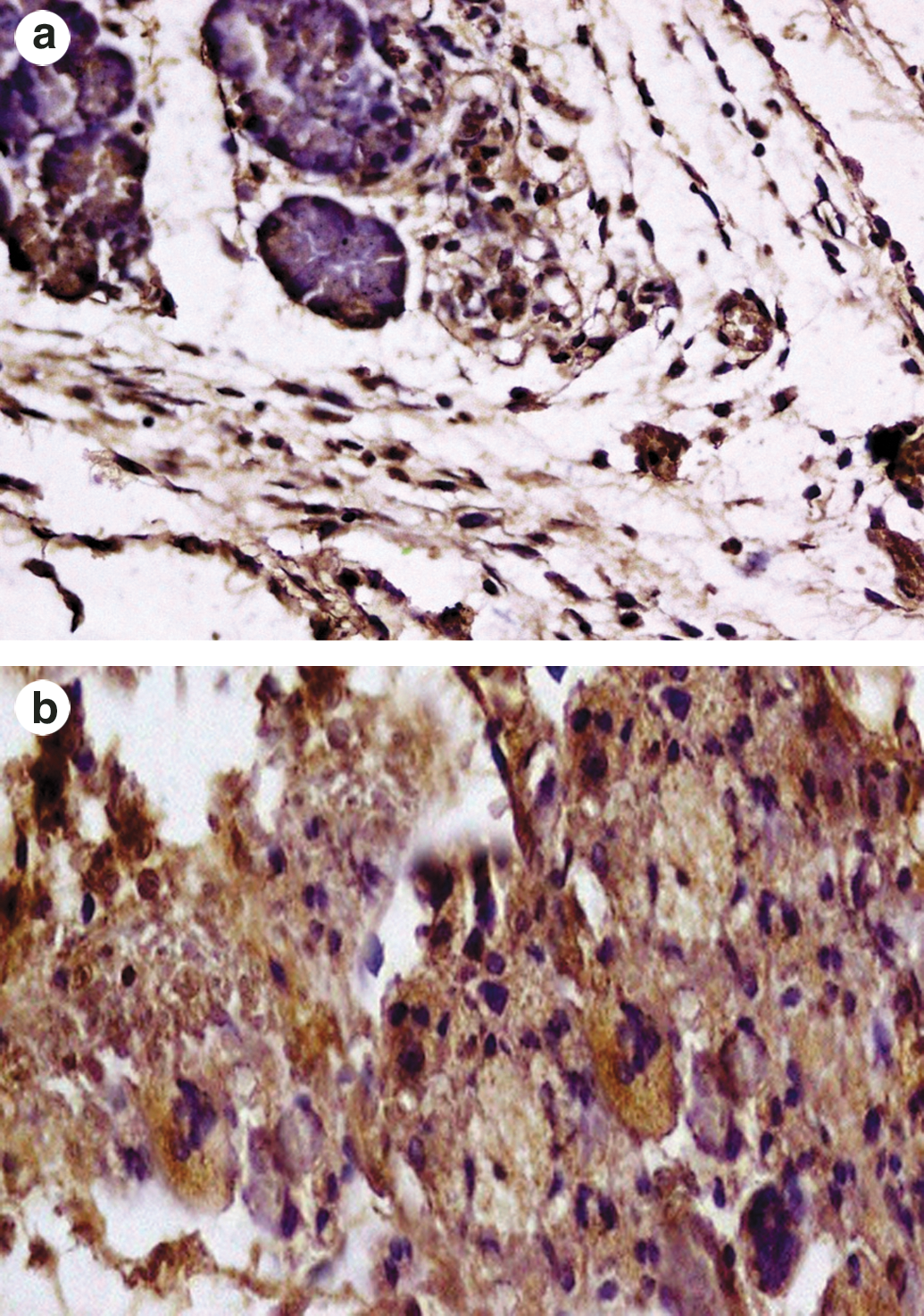
Immunohistochemistry was carried out with 2-μm-thick paraffin sections mounted on polylysine-coated slides. Pancreatic stellate cell activation was evaluated using mouse anti-actin, a smooth muscle monoclonal antibody. Primary antibody was used in a dilution of 1:50 (anti-actin, a smooth muscle monoclonal antibody [Millipore, Bedford, MA, USA]). The sections were incubated with primary antibody for 2 hours in a humidified chamber followed by addition of secondary antibody, which was added onto the sections and allowed to incubate for 40 minutes in the humidified chamber. Then slides were treated with chromogenic substrate of peroxidase in a solution of 0.05% 3,3′-diaminobenzidine tetrachloride, 0.03% H2O2, and 10 mM imidazole in 0.05 M Tris buffer (pH 7.6) and counterstained with hematoxylin. For evaluation, sections were scanned at low magnification for the most densely and evenly labeled areas. Unequivocal staining was regarded as positive reactions, regardless of the staining intensity.
Statistical analysis
The data are presented as mean ± SD values. Comparative analysis was done by analysis of variance followed by post hoc Scheffé's test to detect differences between individual groups. Histopathological data are represented as mean total score and were also analyzed by analysis of variance followed by post hoc Scheffé's test. P < .05 was considered statistically significant.
Results
Serum amylase, lipase, and IL-10
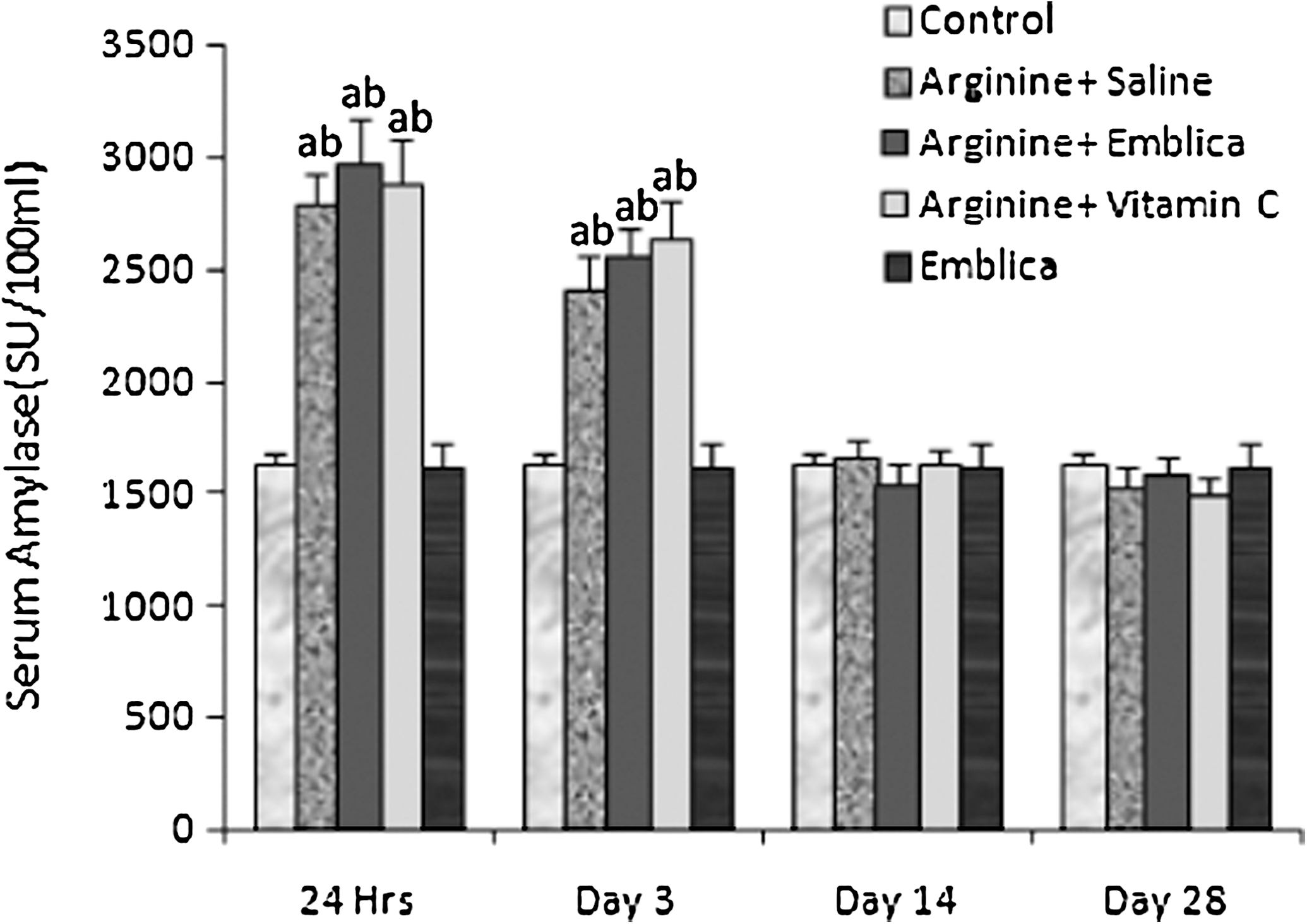
Effect of E. officinalis and vitamin C on serum amylase in
A significant increase in serum lipase levels was observed in the arginine group from Day 3 onwards compared to the control group (Fig. 2), although with time, lipase levels were decreasing but were still significantly higher compared to the control until Day 28. Serum lipase levels were significantly lower in the Emblica group compared to the arginine group. Peak serum lipase levels were seen at Day 3 in the Emblica group, which was significantly higher compared to the control group (control vs. Emblica, 1.08 ± 0.38 vs. 2.17 ± 0.41 mL of NaOH consumed). After the third day, lipase levels declined and by Day 14 were not significantly different from the control group (P = .258 at Day 14 and .667 at Day 28). When the Emblica group was compared to the arginine group, the levels were lower at all time points. This difference was statistically significant at Day 3 and Day 14. As with the Emblica group, when the vitamin C group was compared to the control, a significant difference was observed at 24 hours and Day 3, but by Day 14 no significant difference was seen (P = 1.000 at Day 14 and Day 28). The Emblica group tended to have lower serum lipase than the vitamin C group, but the difference was not significant.
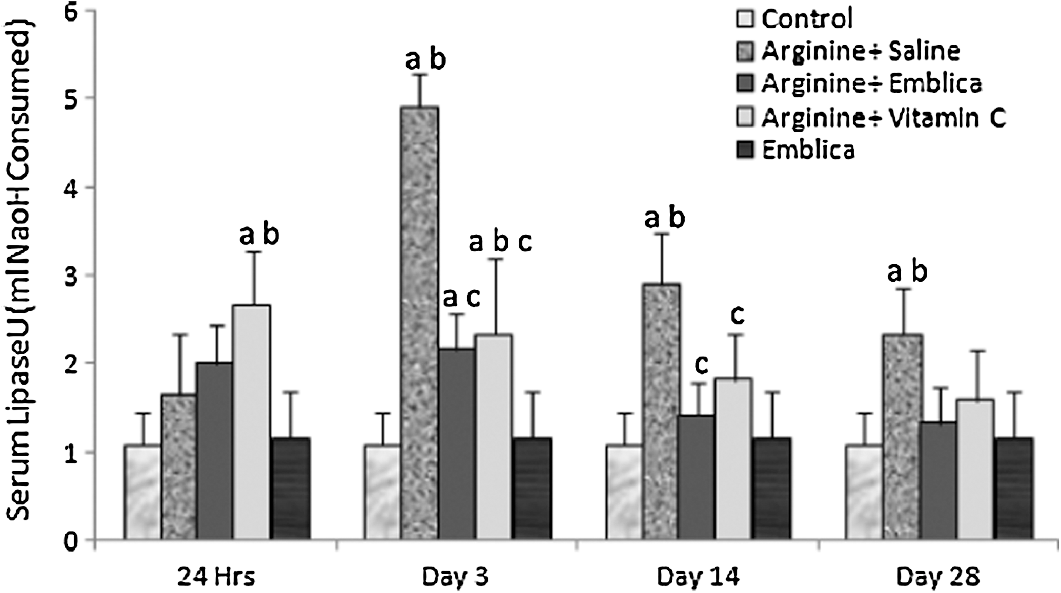
Effect of E. officinalis and vitamin C on serum lipase in
Induction of experimental pancreatitis caused a significant rise in serum IL-10 levels. The arginine group had significantly higher serum IL-10 levels at all time points compared to the control. With time, as the severity of pancreatitis subsided, a gradual decrease in serum IL-10 levels was observed in the arginine group. The Emblica group had significantly lower serum IL-10 levels compared to the arginine group throughout the study period. Thus E. officinalis treatment limited the severity of acute pancreatitis. The serum levels were significantly lower compared to the arginine group, but when compared to the control group serum levels were higher (Table 1).
Data are mean ± SD values.
Statistically significant differences of P ≤ .05 are indicated: *versus control, #versus arginine + saline.
Pancreatic amylase and total proteins
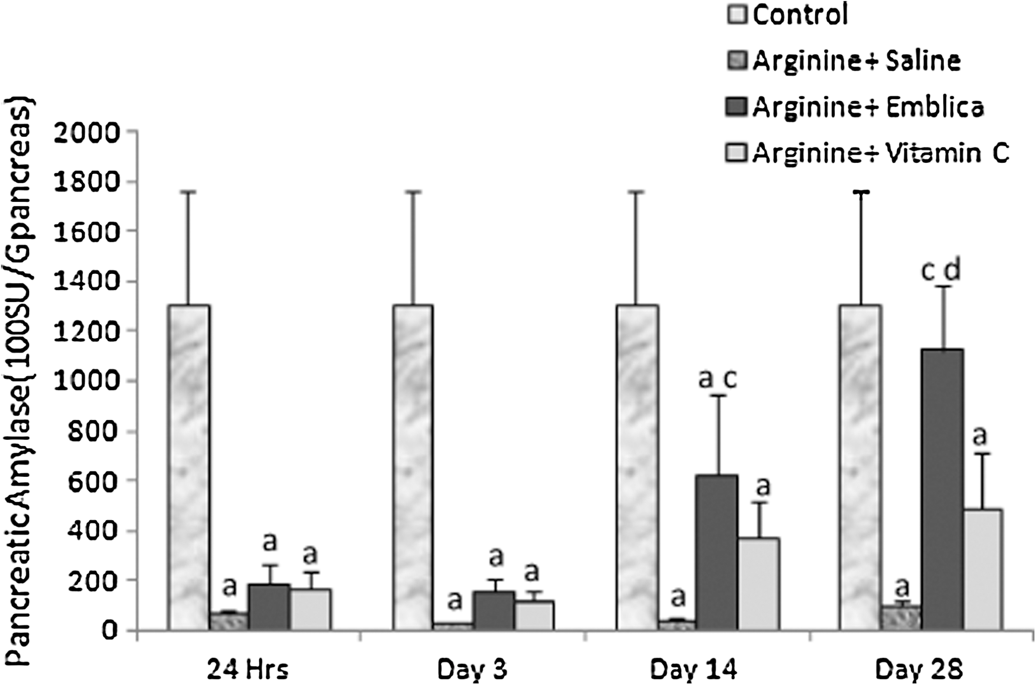
Induction of pancreatitis caused a significant decrease in the amylase content of the pancreas (Fig. 3). Amylase activity was significantly lower in the arginine group compared to the control group until Day 28. Improvement in amylase content of the pancreas was seen in the Emblica group. By Day 14, pancreatic amylase was significantly higher in the Emblica group compared to the arginine group, and it was maintained until Day 28 (P = .018 and .000, respectively). Although in the Emblica group the levels of pancreatic amylase were increasing with time, they remained significantly lower compared to the control group until Day 14; at Day 28 the difference was not statistically significant (P = .870). Treatment with vitamin C did not have much effect on amylase content. Although levels were increasing with time and were higher than in the arginine group, the difference did not attain statistical significance at any time point. Compared to the control the levels were significantly lower until Day 28. Compared to the Emblica group levels were low at all time points, but statistical significance was seen at Day 28 only (P = .018).
Effect of E. officinalis and vitamin C on pancreatic amylase content in
Like pancreatic amylase, total pancreatic proteins were also reduced after induction of pancreatitis. A significant fall in protein content was observed in the arginine group compared to the control group, and the difference was significant at all time points (Fig. 4). E. officinalis treatment improved the pancreatic content of total proteins compared to the arginine group. Although E. officinalis treatment improved protein content, the extent of improvement was lower compared to the control group. The Emblica group compared to the arginine group had significantly higher total protein levels from 24 hours onwards. The vitamin C group compared to the arginine group had a higher protein content, but compared to the Emblica group the levels were significantly lower at all time points.

Effect of E. officinalis and vitamin C on pancreatic total proteins in
Pancreatic nucleic acid content and [ 3 H]thymidine uptake
Nucleic acid content of the pancreas decreased after
Data are mean ± SD values.
Statistically significant differences of P ≤ .05 are indicated: *versus control, #versus arginine + saline, †versus arginine + vitamin C.
Data are mean ± SD values.
Statistically significant differences of P ≤ .05 are indicated: *versus control, #versus arginine + saline, †versus arginine + vitamin C.
Induction of pancreatitis in the arginine group reduced the [ 3 H]thymidine uptake compared to the control group (Table 4) at both Day 3 and Day 7, and the difference was significant. E. officinalis treatment increased the [ 3 H]thymidine uptake compared to the arginine group, and this difference was significant at both time points. Compared to the control group the Emblica group had lower uptake, but the difference was statistically significant only at Day 3 (P = .029 and .154). Vitamin C treatment also increased [ 3 H]thymidine uptakes compared to the arginine-alone group, but statistical significance was attained at Day 3 only (P = .028 at Day 3 and .095 at Day 7). The vitamin C group compared to the Emblica group had lower uptake at both days, and the difference was significant at Day 7 (P = .014).
Data are mean ± SD values.
Statistically significant differences of P ≤ .05 are indicated: *versus control, #versus arginine + saline, †versus arginine + vitamin C.
Histopathological evaluation
The results are presented as total histopathological score. The histopathological score for the control group was defined at 0. The histopathological score in the Emblica group was lower compared to the arginine group throughout the study period (Fig. 5).
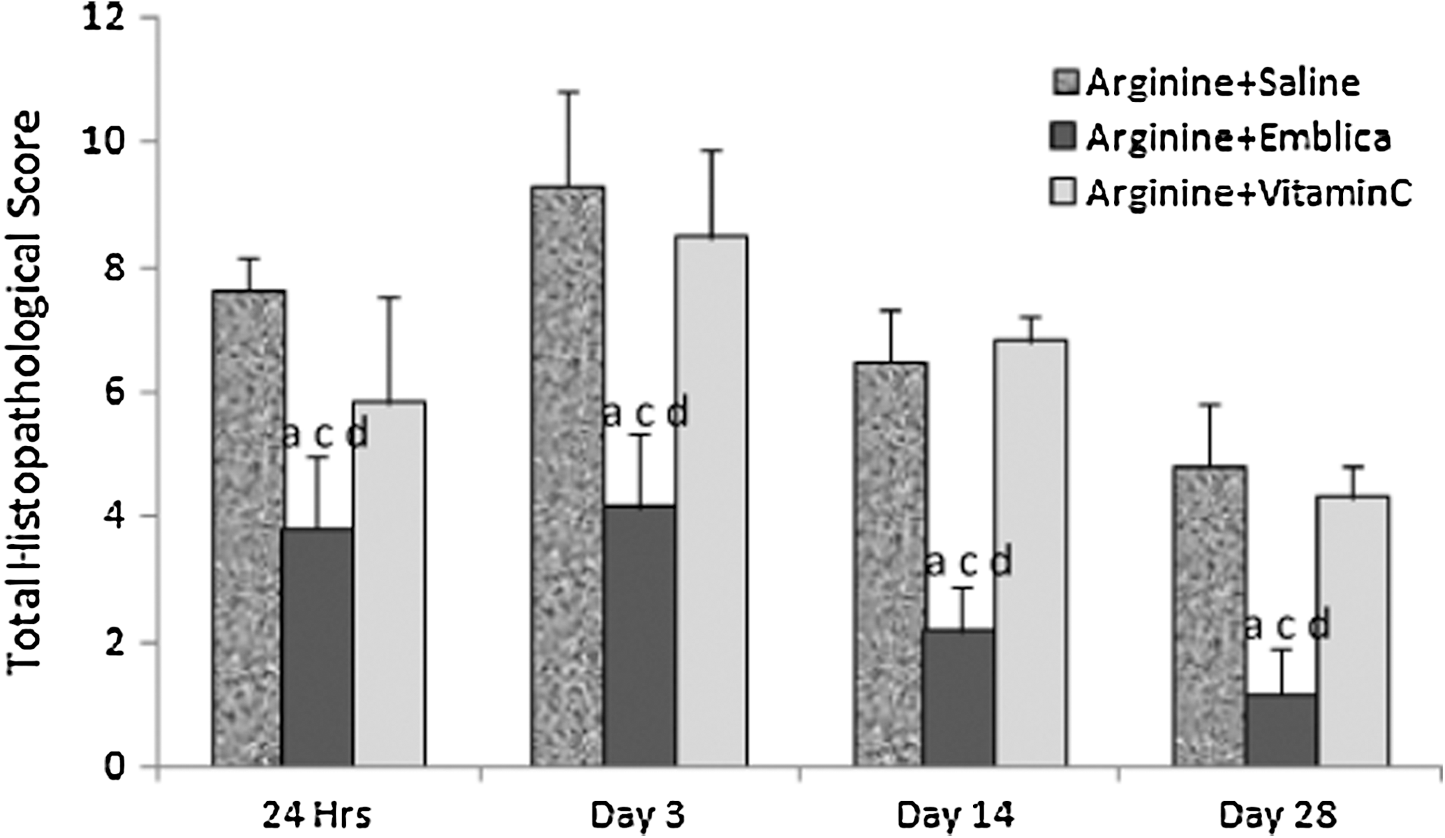
Effect of E. officinalis and vitamin C on total histopathological score in
In the control group, normal pancreatic parenchyma was observed (Fig. 6a). Induction of acute pancreatitis in the arginine group resulted in marked histopathological changes compared to the control group. At 24 hours after
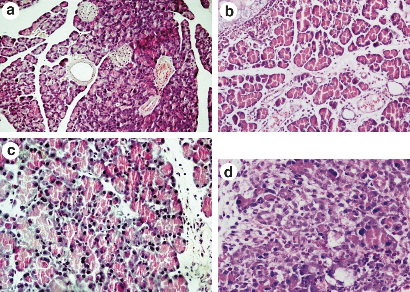
(
Immunohistochemistry
Pancreatic stellate cells have been shown to play an important role in pancreatic regeneration after an acute attack of pancreatitis. Activated stellate cells participate in the inflammatory organization and regeneration process. In the present study the pancreatic stellate cells having acquired myofibroblastic quality stained positive for α smooth muscle actin. Arginine group animals stained positive for α smooth muscle actin, indicating spontaneous regeneration (Fig. 7a). E. officinalis-treated animals also stained positive for α smooth muscle actin (Fig. 7b).
(
Discussion
The results of the present study show that treatment with E. officinalis reduced severity of acute pancreatitis induced by
Treatment with E. officinalis did not significantly modulate the elevations in serum amylase levels at 24 hours and Day 3, which were similar to those of the arginine group, but histological examination at Day 3 showed marked pancreatitis-like changes in the arginine group, whereas in the Emblica group few histopathological changes were observed. By Day 14 the serum amylase levels of the arginine group were comparable to that of the control group, but histological findings still showed marked injury. The results of the current study are in agreement with previous findings that no correlation exists between serum amylase and severity of pancreatitis. 30 Moreover, normalization of serum amylase can occur despite underlying pancreatitis. This may be due to increased urinary clearance or extensive destruction of pancreas with cessation of pancreatic amylase production. 31 Also, conditions other than pancreatitis can cause increased amylase, so this is not a very specific test. 30 In contrast to serum amylase, no elevation in serum lipase was seen at 24 hours. Peak elevations were present on Day 3. This is the usual course that amylase follows. During acute pancreatitis the lipase levels are elevated after 72 hours and unlike amylase remain elevated for a longer duration. Increased permeability in the basal pole of the acinar cells accounts for the pronounced rise of the enzyme in serum. 29 Treatment with E. officinalis prevented the rise in serum lipase levels.
As acinar cells are the protein factory of the pancreas, pancreatitis leads to a decrease in the total protein content of the pancreas.
E. officinalis promoted the spontaneous repair process as the rate of DNA synthesis was significantly higher compared to the arginine group. The pattern of increase in DNA synthesis correlated with the quantification of DNA content of the pancreas. The rate of DNA synthesis was measured on Day 3, after 2 days of treatment, and a significant uptake of [ 3 H]thymidine was seen in the Emblica group compared to the arginine group. Similarly, a significant difference in the accumulation of DNA in the pancreatic tissue was observed; the DNA content in the Emblica group was significantly higher than in the arginine group. Furthermore, accumulation of DNA was in parallel with the total protein content of the pancreas, which was also significantly greater in the Emblica group. Not much difference in [ 3 H]thymidine uptakes was observed between Day 3 and Day 7 in the present study. These results are in agreement with other studies showing an approximately 11–20-fold increase at 3 days versus a two- to threefold increase after 7 days of acute pancreatitis. 32,33
As total pancreatic proteins, nucleic acid content, and rate of DNA synthesis are markers for the regeneration process, these results clearly indicate that E. officinalis promoted the spontaneous regeneration process. These findings were further supported by the results of immunohistochemistry. Numerous α smooth muscle actin–positive myofibroblasts were observed in the Emblica group. As stellate cells and their differentiated myofibroblast forms actively participate in regeneration process, their presence clearly indicated regeneration. The regeneration appeared to be more rapid in the Emblica group as the quantitative markers were significantly higher in Emblica group.
The sequence of events during the regeneration process is activation of pancreatic stellate cells, proliferation of myofibroblasts, and deposition of extracellular matrix and proliferation of acinar cells. 6 So the beneficial effects of E. officinalis observed in the present study could be due to promotion and activation of pancreatic stellate cells. The probable mechanism by which E. officinalis showed beneficial effects in the present study could be due to modulation of mediators that cause activation of stellate cells or by effecting regeneration sequence by other means. So further studies are required to explore the exact mechanism of action by which E. officinalis promotes the spontaneous regeneration process.
Conclusions
Our results show that E. officinalis treatment potentiates the spontaneous recovery of the pancreas after an attack of acute pancreatitis. Further studies are required to explore the exact mechanism by which E. officinalis treatment was shown to be beneficial, and studies are also required to explore the chemical component of E. officinalis responsible for this effect.
Footnotes
Author Disclosure Statement
No competing financial interests exist.