
Abstract
The present study aimed at isolating and elucidating the structure of the main components of Pistacia khinjuk L. and exploring its potential anti-inflammatory effect in different experimental models. The extract was evaluated for anti-inflammatory activity by measuring paw volume in three experimental models. Then, prostaglandin E2 (PGE2) level, ear edema, tissue myeloperoxidase (MPO) activity, histopathology, nitric oxide (NO) level, and tumor necrosis factor-α (TNF-α) level were assessed. Seven phenolic compounds, mainly flavonoids and galloylated compounds, were isolated from the aqueous methanol extract: gallic acid (
Introduction
P
Inflammation is an essential protective process that preserves the integrity of the organisms against physical, chemical, and infective insults. 13 However, the inflammatory response to several insults could erroneously lead to normal tissue damage. 14 Prostaglandins (PGs), thromboxanes, and leukotrienes belong to the class of prostanoid fatty acid derivatives of arachidonic acid. 15
One of the early cellular events in inflammation is the margination of leukocytes, primarily neutrophils. This response can be measured by using the neutrophil-specific enzyme myeloperoxidase (MPO), an indicator of neutrophil accumulation. 16 In addition, nitric oxide (NO) plays an important role in inflammation, and NO synthase inhibitors can reverse several classic inflammatory symptoms. 17 Tumor necrosis factor-α (TNF-α), a cytokine, plays an important role in inflammation because of its ability to stimulate cytokine release from neutrophils and biosynthesis of chemokines. 18
Currently, both steroidal and nonsteroidal anti-inflammatory drugs (NSAIDs) are used in the treatment of inflammatory disorders. Steroids have an obvious role in the treatment of inflammatory diseases but, because of rate-limiting toxicities, are only prescribed over short periods except in very severe cases where the risks are acceptable. Prolonged use of NSAIDs is also associated with severe side effects, notably gastrointestinal haemorrhage. 19,20 The recently developed cyclooxygenase-2-selective drugs have been introduced into therapy but, however, do not seem to be free of risk. 21 Consequently, there is a need to develop new anti-inflammatory agents with minimal side effects. In this regard, natural products have long gained wide acceptance among the public and scientific community. 22 Several natural compounds have been proven to show anti-inflammatory effects.
In particular, polyphenolic compounds have been given the most attention. 23 There is a paucity of information regarding the phytochemistry of the polyphenolic content and biological effects of P. khinjuk L. Therefore, the present study was designed to isolate and elucidate the structure of the main components of the aqueous methanol extract of P. khinjuk L. and to explore its potential anti-inflammatory effects in different experimental models.
Materials and Methods
Chemicals
Croton oil, hexadecyltrimethylammonium bromide, o-dianisidine, indomethacin, lipopolysaccharide (LPS) from Escherichia coli serotype O111:B4, modified Griess reagent, sodium nitrite, and gallic acid were purchased from Sigma-Aldrich (St. Louis, MO, USA). Inflammatory-grade carrageenan was purchased from FMC (Rockland, ME, USA). The PGE2 kit was purchased from R&D Systems Inc. (Minneapolis, MN, USA). The TNF-α kit was purchased from Assaypro LLC (St. Charles, MO, USA). Acetone, ethanol, ether, and pyridine were purchased from SDS (Peypin, France). The solvents methanol, water, and acetic acid of high-performance liquid chromatography (HPLC) grade were purchased from Merck Ltd. (Mumbai, India). Other chemicals and reagents used were of analytical grade.
Plant materials
P. khinjuk L. leaves were collected in April 2009 from the El-Zohreya Botanical Garden (Cairo, Egypt) and authenticated by Terease Labib, an agricultural engineer at El-Orman Botanical Garden (Giza, Egypt). A voucher specimen was deposited in the Herbarium of the Pharmacognosy Department, Faculty of Pharmacy, Ain Shams University, Cairo.
Instruments and materials for phytochemical investigation
Nuclear magnetic resonance
1H and 13C nuclear magnetic resonance (NMR) spectra were obtained on a JOEL (Tokyo, Japan) ECA 500 spectrometer. 1H chemical shifts were measured relative to the trimethylsilane scale by adding 39.5, and 13C chemical shifts were measured at 100 MHz, relative to dimethyl sulfoxide-d6 (DMSO-d6), and converted to trimethylsilane width equal to 6,000 Hz for 1H and 22,000 Hz for 13C.
Ultraviolet
Ultraviolet (UV) spectroscopy was performed with a Shimadzu (Kyoto, Japan) UV/Vis spectrometer. Paper chromatography (PC) analysis was carried out on Whatman (Maidstone, UK) No. 1 paper, using the following solvent systems: (1) water; (2) 6% acetic acid; (3) n-butanol–acetic acid–water (BAW) (4:1:5 by volume, upper layer); and (4) n-butanol saturated with water. Solvents 2 and 3 were used for preparative PC (PPC) on Whatman No. 3MM paper. The Sephadex LH-20 (25–100 μm) column was from Sigma-Aldrich, and the polyamide 6 S for column chromatography was from Riedel-De Haen AG (Seelze-Hannover, Germany).
Plant extraction, isolation, and purification
Fresh leaves (2 kg) were exhaustively extracted with 10 L of aqueous methanol (75%). Leaves were boiled in double distilled water for exactly 2 hours. The aqueous extract was dried by lyophilization. The dry lyophilized extract was then further extracted with methanol (HPLC grade) for 30 minutes at 40°C to ensure complete extraction of phenolic components. This method helps to avoid extraction of other plant metabolites as carbohydrates, water-soluble protein, inorganics, lipids, or alkaloids, if any. Finally, the extract was completely evaporated in vacuo at low temperature until dryness to give 66 g of solid residue.
Two-dimensional PC of the extract proved the presence of a high percentage of phenolic constituents (green and blue color reaction with ferric chloride TS and ammonia).
Repeated fractionation of the extract (120 g) on polyamide 6S columns, using water followed by water–methanol mixtures of decreasing polarities, yielded eight fractions (I–VIII), which were individually subjected to further purification using subcolumns and PPC. Compounds
Gallic acid (
3-O-Methyl gallate (4,5-dihydroxy-3-O-methylbenzoic acid) (
Quercetin-3-O-β-
Myricetin-3-O-α-
1,6-Digalloyl-β-
1,4-Digalloyl-β-
Chromatographic conditions
A Shimadzu LC-8A HPLC system with a binary solvent delivery system (LC-8A), a column temperature controller (CTO-10AV), a Rheodyne (Rohnert Park, CA, USA) injector with a 20-μL sample loop, and a UV detector coupled with Class VP analytical software were used for analysis. A reverse-phase column (Phenomenex [Torrance, CA, USA] C-18, ODS-2; particle size, 5 μm; 250×4.6 mm) with an extended guard column was used as the stationary phase, and column temperature was maintained at 35°C. The mobile phase was a gradient elution of water containing 2% acetic acid (solvent A) and methanol (solvent B) at a flow rate of 1 mL/minute. The gradient program of solvent A in B (vol/vol) was as follows: 0–5 minutes, 100% A; 5–10 minutes, 90% A; 10–15 minutes, 90%; 15–20 minutes, 80% A; 20–25 minutes, 80%; 25–40 minutes, 40% A; and 40–60 minutes, 0% A. The chromatographic run was scanned at 280 nm.
Preparation of standard solution of gallic acid
Authentic standard gallic acid was dissolved in HPLC-grade methanol to prepare the calibration curve. Several dilutions in mobile phase were made. All standard solutions were filtered (pore size, 0.45 μm) and injected directly. The standard response curve was a linear regression fitted to triplicate values obtained at each of three concentrations (ranging from 0.002 to 0.004 mg/mL). The results, determined by the regression equation of the calibration curve (y=62.8x – 0.67, R 2=0.99), were expressed as milligrams of gallic acid equivalents per gram dry weight of raw material.
Preparation of plant extract for HPLC analysis
The dried methanolic extract of P. khinjuk L. leaves was dissolved in HPLC-grade methanol. The concentrations were determined by calculating the HPLC peak areas, which are proportional to the amount of analytes in a peak.
Animals
Throughout the experiments, adult male Sprague–Dawley rats weighing 150–175 g were obtained from the animal facility of King Fahd Medical Research Center, King Abdulaziz University, Jeddah, Saudi Arabia. Animals were housed at a temperature of 23±2°C with free access to water and standard food pellets. Rats were acclimatized in our animal facility for at least 1 week prior to any experiment. Procedures involving animals and their care were conducted in conformity with the institutional guidelines of King Abdulaziz University.
Experimental protocol
The potential anti-inflammatory activity of P. khinjuk L. extract was examined in three experimental models: the carageenen-induced rat edema model, the croton oil–induced ear edema model, and the rat air pouch model. Collectively, the parameters assessed included paw volume, PGE2 level, ear edema, tissue MPO, histopathology, NO, and TNF-α. The control group animals received the same experimental handling as those of the test groups except that the drug treatment was replaced with appropriate volumes of the dosing vehicle. Indomethacin, a potent NSAID, was suspended in 0.5% carboxymethylcellulose and used as a reference drug. It was administered in doses of 10 mg/kg in rat paw edema and ear edema experiments, respectively. The choice of the doses used and times of measurement and sampling was based on pilot studies in our laboratory.
Measurement of paw volume and PGE2 in carrageenan-induced rat edema model
Twenty-four rats were equally divided into four groups (I–IV). Animals were fasted, with free access to water, 16 hours before the experiment. Using an intragastric tube, Groups I and II were given the vehicle (0.5% carboxymethylcellulose), whereas Group III was treated with P. khinjuk L. extract. Animals in Group IV received indomethacin as a standard anti-inflammatory drug. The dosing volume was kept constant (10 mL/kg) for all the orally treated groups. Thirty minutes after oral treatment, Group I received 0.05 mL of saline, whereas Groups II–IV received 0.05 mL of carrageenan (1% solution in saline) subcutaneously on the plantar surface of the right hind paw. The right hind paw volume was measured immediately after carrageenan injection by water displacement using a plethysmometer (model 7140, Ugo Basile, Comerio, Italy). 28 The paw volume was remeasured 3 hours after carrageenan injection and immediately before decapitation.
The level of PGE2 was then assessed. Right hind paws were removed. A volume of 0.1 mL of saline containing 10 μM indomethacin was injected to aid removal of the eicosanoid-containing fluid and to stop further production of PGE2. Paws were incised with a scalpel and suspended off the bottom of polypropylene tubes with Eppendorf pipette tips to facilitate drainage of the inflammatory exudates. For the purpose of the removal of the inflammatory exudates, paws were centrifuged at 1800 g for 15 minutes. 29 PGE2 was quantified in the collected exudates using a PGE2 enzyme-linked immunosorbent assay kit. This assay is based on the forward sequential competitive binding technique in which PGE2 in a sample competes with horseradish peroxidase–labeled PGE2 for a limited number of binding sites on monoclonal antibodies. PGE2 in the sample is allowed to bind to the antibody in the first incubation. During the second incubation, horseradish peroxidase–labeled PGE2 binds to the remaining antibody sites. Following a wash to remove unbound materials, a substrate solution is added to the wells to determine the bound enzyme activity. The color development is stopped, and the absorbance is read at 450 nm. The intensity of the color is inversely proportional to the concentration of PGE2 in the sample. 30
Assessment of ear edema, tissue MPO activity, and histopathology in croton oil–induced ear edema model in rats
The experiment was performed using a slight modification of the procedure described by Tonelli et al. 31 An irritant solution was prepared by dissolving 4 parts croton oil (the irritant) in a solvent mixture of 10 parts ethanol, 20 parts pyridine, and 66 parts ethyl ether. The P. khinjuk L. extract and indomethacin were dissolved in the same vehicle of the irritant. Twenty-four rats were equally divided into four groups (I–IV). The irritant solution was applied in a volume of 20 μL topically on both sides of the right ear; the left ear was kept untreated to serve as a control. Group I served as the negative control and hence received only the irritant-free solvent mixture, whereas Group II received the croton oil solution. The third and fourth groups received the P. khinjuk L. extract (100 mg/kg orally) and indomethacin (10 mg/kg orally), respectively. One hour later, the solvent mixtures were re-administered to the groups. After 4 hours, animals were sacrificed. An 8-mm cork borer was used to punch out discs from both the treated and the control ears. The two punches were weighed immediately after decapitation, and the difference in weight was used to assess the inflammatory response.
The entire tissue of the right ear was homogenized for 10 minutes in an ice bath (10%, wt/vol) in 50 mM phosphate buffer (pH 6.0) containing 0.5% hexadecyltrimethylammonium bromide with a IKA® (Staufen, Germany) homogenizer. Tissue suspensions were centrifuged at 40,000 g for 15 minutes. An aliquot of 0.1 mL of the supernatant was added to 2.9 mL of 50 mM phosphate buffer (pH 6.0) containing 0.167 mg/mL o-dianisidine dihydrochloride, serving as the MPO substrate, and 0.0005% hydrogen peroxide. The change in absorbance was measured at 460 nm with a Shimadzu UV-1601 spectrophotometer at 25°C. MPO activity was quantified kinetically; change in absorbance was measured over a period of 2 minutes, sampled at intervals of 15 seconds. 32 The maximal change in absorbency per minute was used to calculate the units of MPO activity based on the molar absorbency index of oxidized o-dianisidine dihydrochloride, which equals 1.13×10⁴ M −1 cm−1. One unit of MPO is defined as that degrading 1 μmol of peroxide/minute at 25°C. Results were expressed as units of activity per milligram of protein. Protein content was determined according to the method of Lowry et al. 33
A representative ear tissue from each group was fixed in 10% formol saline for 24 hours. Washing was done in tap water, and then serial dilutions of alcohol (methyl, ethyl, and absolute ethyl) were used for dehydration. Specimens were cleared in xylene and embedded in paraffin at 56°C in a hot air oven for 24 hours. Paraffin beeswax tissue blocks were prepared for sectioning at 4 μm thickness with a sledge microtome. The tissue sections obtained were collected on glass slides, deparaffinized, and stained by hematoxylin and eosin, and examination was done with the light microscope. 34
Measurement of NO and TNF-α in the rat air pouch model
Twenty-four rats uniformly divided in four groups were used in the study. Air pouches were formed by subcutaneous injection of 20 mL of sterile air in the suprascapular area of the back of the animal. Three days later, the pouches were re-inflated with 10 mL of sterile air. After another 3 days, a 100 μg/mL solution of LPS in physiological saline (1 mL/kg) was administered intrapouch to Groups II–IV; the control group (Group I) received physiological saline (1 mL/kg) intrapouch. Thirty minutes later, drugs were injected, also intrapouch. The volume of administered solutions was kept constant at 3 mL. Groups I and II were administered only saline, Group III received the P. khinjuk L. extract (100 mg/kg), and Group IV received indomethacin (10 mg/kg). Eight hours later animals were sacrificed. 35 Each pouch was lavaged using 1 mL of sterile physiological saline. The lavage fluid was then centrifuged at 3,000 g for 5 minutes. Supernatant was used immediately for analysis of NO and TNF-α.
NO assay is based on measuring total nitrite/nitrate level based on reduction of any nitrate to nitrite by vanadium trichloride followed by detection of total nitrite by modified Griess reaction [sulfanilamide and N-(1-naphthyl)-ethylenediamine]. A chromophore is formed by diazotization of sulfanilamide with acidic nitrite followed by coupling with the bicyclic amine N-(1-naphthyl)-ethylenediamine. 36 In brief, 250 μL of vanadium trichloride solution was added to 250 μL of air pouch exudates followed by rapid addition of 250 μL of modified Griess reagent. The mixture was incubated at 37°C for 30 minutes and then cooled. Absorbance was measured at 540 nm using a Shimadzu UV-1601 spectrophotometer against a blank treated in a similar manner to the test solution but using 250 μL of double distilled water instead of sample. The concentration of NO was expressed as micromolar using a standard curve.
The assay of TNF-α depends on a quantitative sandwich enzyme immunoassay technique that measures TNF-α in 4.5 hours. A murine monoclonal antibody specific for rat TNF-α was precoated onto a microplate. TNF-α in standards and samples was sandwiched by the immobilized antibody and a biotinylated polyclonal antibody specific for rat TNF-α, which is recognized by a streptavidin–peroxidase conjugate. All unbound material was then washed away, and a peroxidase enzyme substrate was added. The color development was stopped, and the intensity of the color was measured at 450 nm using a microplate reader. 30
Statistical analysis
Data are presented as mean±SD values. Statistical analysis was performed using one-way analysis of variance followed by Tukey–Kramer multiple comparisons tests. The .05 level of probability was used as the criterion for significance. All statistical analyses were performed using GraphPad (La Jolla, CA, USA) InStat software version 3. Graphs were sketched using GraphPad Prism software version 4 (ISI® Software, La Jolla, CA, USA).
Results
Phytochemical investigation of P. khinjuk L.
Phytochemical investigation of the aqueous methanol extract indicated the presence of phenolic metabolites. Following column chromatographic fractionation of the P. khinjuk L. conventional and spectral analysis mainly by UV, 1H and 13C NMR spectroscopy was applied. As shown in Figure 1, the isolated compounds were gallic acid (
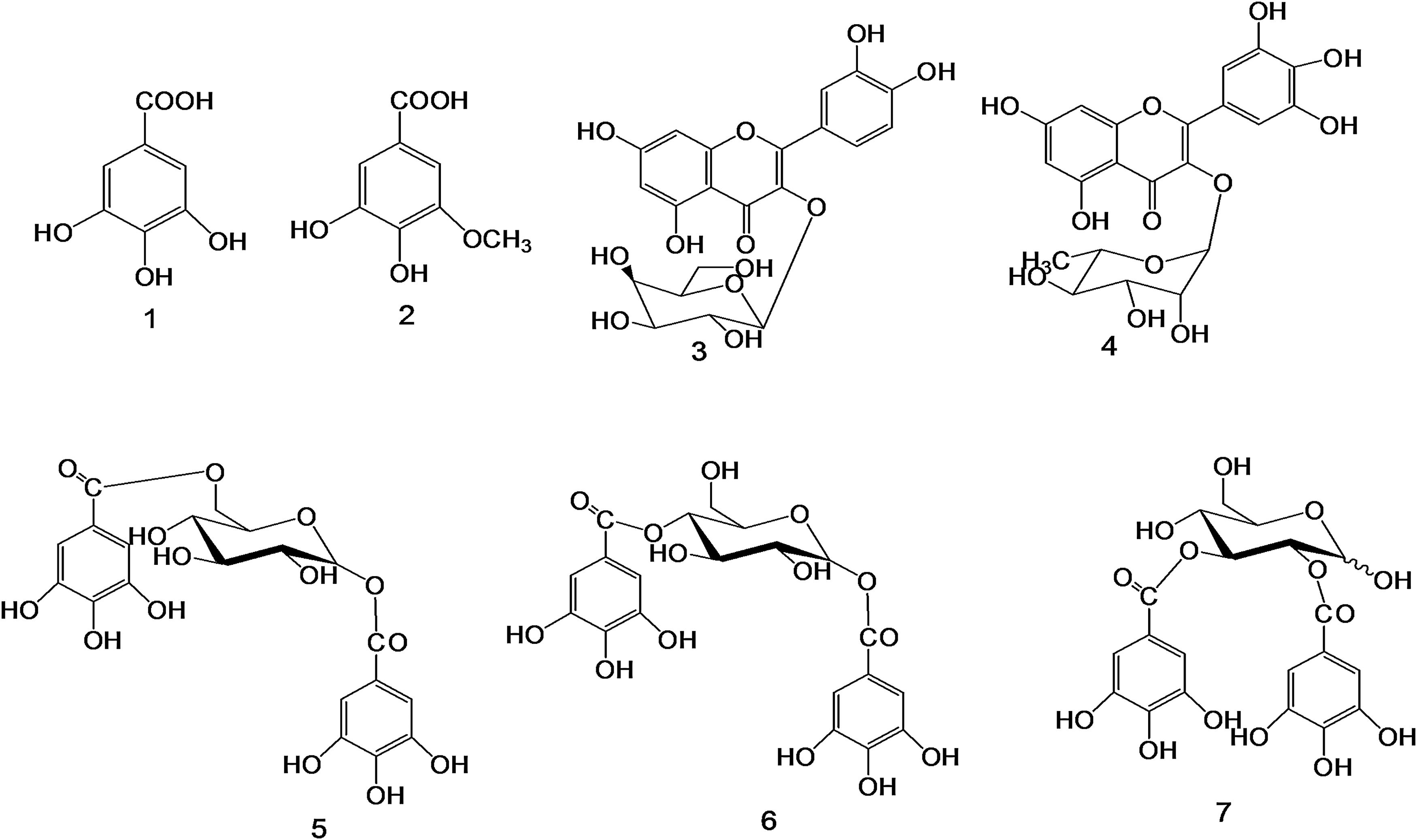
Phenolic compounds isolated from the aqueous methanol extract of P. khinjuk L.: gallic acid (
HPLC investigation of P. khinjuk L.
Reversed-phase HPLC coupled with UV/Vis was used at 280 nm to identify and quantify phenolic compounds in P. khinjuk L. The concentrations were determined by calculating the HPLC peak areas, which are proportional to the amount of analytes in a peak, and are presented as the mean value of two determinations, which were highly reproducible. Gallic acid has been identified in P. khinjuk L. extract according to the retention time and spectral characteristics of its peak against that of the standard gallic acid. The methanol extract of P. khinjuk L. contained approximately 81% gallic acid.
Effect of P. khinjuk L. extract on paw volume and PGE2 in carrageenan-induced rat edema
Intraplantar injection of carrageenan to rats resulted in severe discernible inflammation and significant increase in the mean volume of the challenged paw compared with that of the untreated paws (137.3% of the untreated paws) (Table 1). Pretreatment of rats with P. khinjuk L. extract at a dose of 100 mg/kg significantly inhibited the carrageenan-induced increase in the edema volume of the paws by 46.8%. Indomethacin inhibited the induced edema by 80.8%.
Statistically significant difference from the control group (Group I) at P<.05.
Statistically significant difference from the carrageenan-induced group (Group II) at P<.05.
Carrageenan challenge resulted in a >10-fold increase in PGE2 concentration in inflammatory exudates in Group II compared with unchallenged animals in Group I (Fig. 2). Animals receiving P. khinjuk L. extract showed significant reduction of the PGE2 concentration in exudates by 87.45% of the carrageenan-treated animals. The standard indomethacin lowered PGE2 level in carrageenan-challenged animals, to values approaching normal levels.
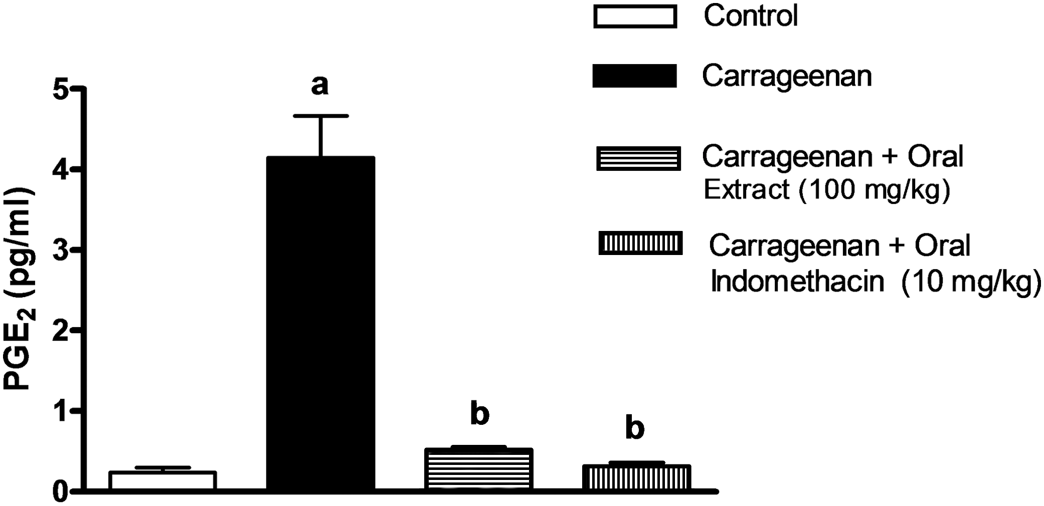
Effect of P. khinjuk L. extract on prostaglandin E2 (PGE2) production in exudates from carrageenan-treated rats. Statistically significant (P<.05) differences are indicated: afrom the control group, bfrom the carrageenan-induced group.
Effect of P. khinjuk L. extract on croton oil–induced ear edema, MPO tissue activity, and histopathological changes
Application of croton oil to rat ears caused a massive increase in the weight of the ear punch from control animals by 83.4%. Pretreatment of rats with P. khinjuk L. extract significantly reduced the increase of punch weight from unchallenged ears to about 52.4%. Indomethacin pretreatment produced significant reduction of punch weight from control values to 37.4%, as shown in Table 2.
Statistically significant difference from the control group at P<.05.
Statistically significant difference from the croton oil–induced group at P<.05.
Assay of MPO activity in rat ears indicated that application of croton oil increased the enzyme activity by more than 25-fold. Pretreatment with P. khinjuk L. extract at a dose of 100 mg/kg, however, reduced MPO activity by 57.53% of the induced ear. Indomethacin pretreatment resulted in significant protection against croton oil–induced enhancement of MPO activity (Fig. 3).
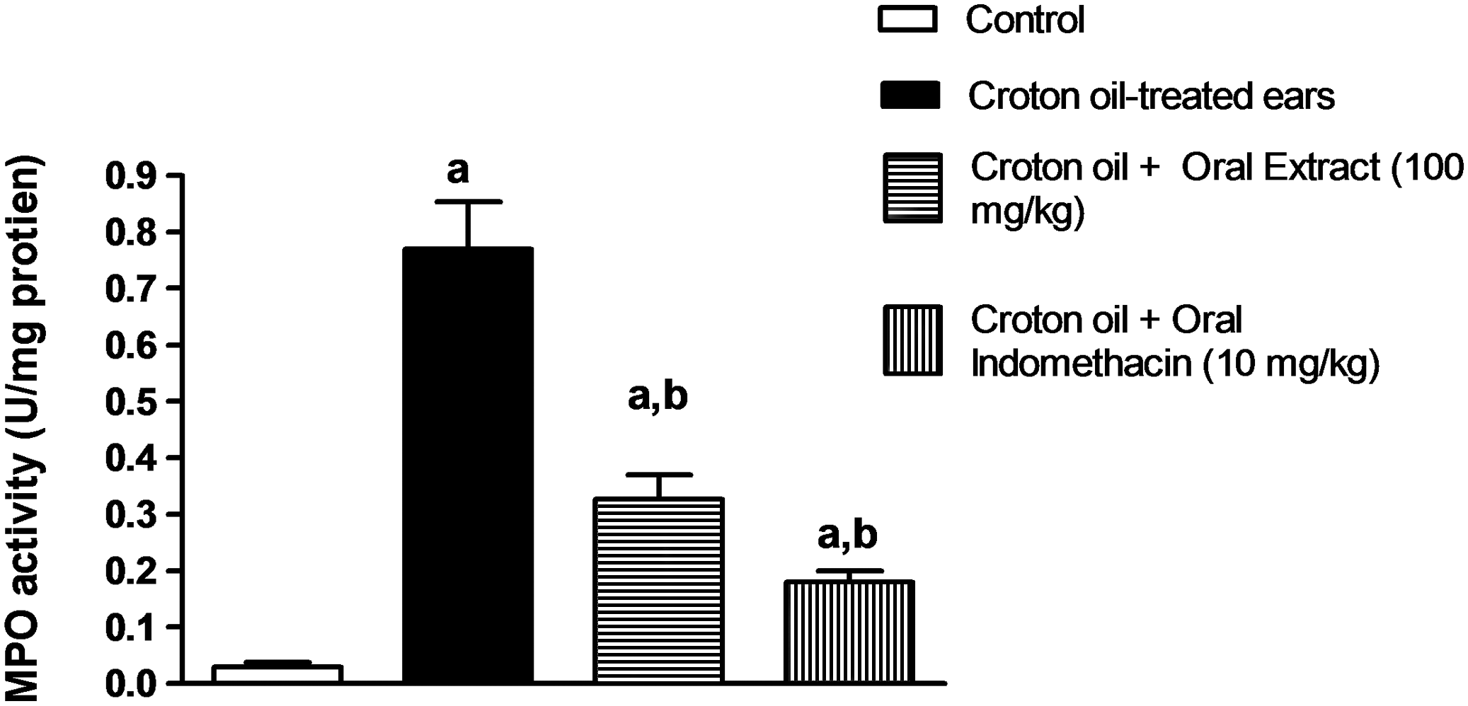
Effect of P. khinjuk L. extract on myeloperoxidase (MPO) activity in croton oil-induced edema. Statistically significant (P<.05) differences are indicated: afrom the control group, bfrom the croton oil–induced group.
Histopathological examination of the ear tissue confirmed those results obtained by assessing MPO activity. Figure 4A shows normal histological structure of the epidermal and dermal layers as well as underlying cartilage with no obvious neutrophil infiltration. The croton oil–treated rats showed massive neutrophil infiltration in the dermal layer (Fig. 4B). Figure 4C, which represents ear tissue treated with 100 mg/kg P. khinjuk L. extract, shows less cellular infiltration in the dermal layer compared with the croton oil–treated group. Figure 4D, corresponding to indomethacin treatment, shows almost intact dermal and cartilaginous structures with little neutrophil infiltration.
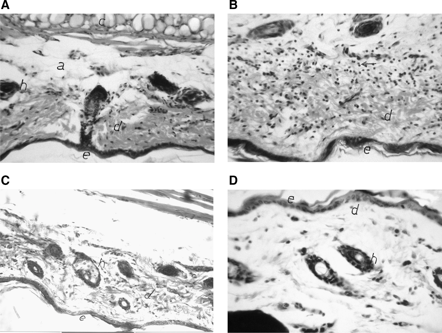
Effect of P. khinjuk L. extract on histopathological changes in the croton oil–induced ear edema experiment. Magnification×64. (
Effect of P. khinjuk L. extract on NO and TNF-α in the rat air pouch model
Injection of LPS into the air pouch caused about a 10-fold increase of NO production compared with that untreated animals (Group I). Injection of P. khinjuk L. extract at 100 mg/kg significantly reduced the NO level by 81.14%, compared with Group II (Fig. 5). Similarly, indomethacin treatment significantly lowered the NO level by 85.3%.
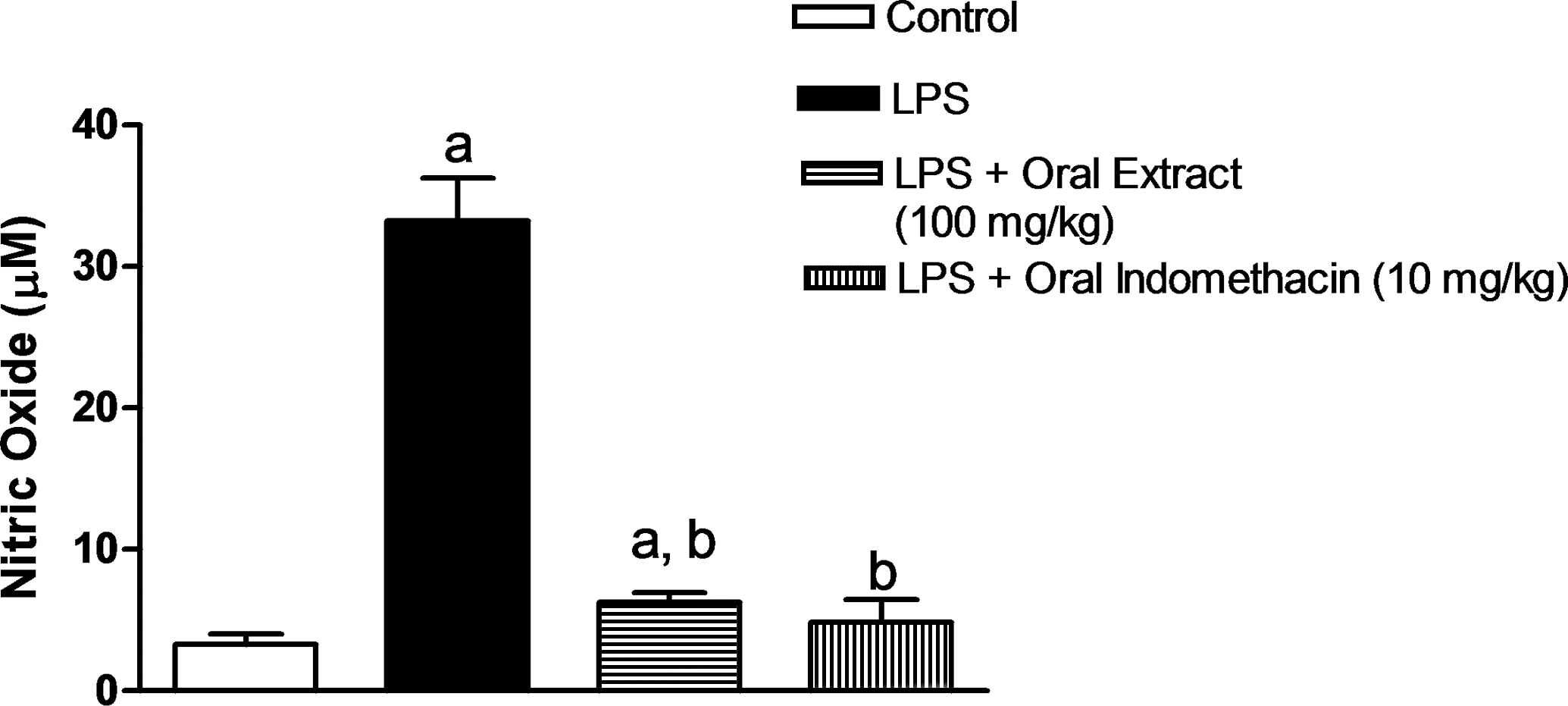
Effect of P. khinjuk L. extract on nitric oxide production in the rat air pouch model. Statistically significant (P<.05) differences are indicated: afrom the control group, bfrom the lipopolysaccharide (LPS)-induced group.
In addition, LPS injection caused about a fourfold increase in TNF-α level in the pouch exudates compared with the untreated group. Treatment with P. khinjuk L. extract at a dose of 100 mg/kg lowered TNF-α release by 32.62%, compared with the untreated group (Fig. 6). Indomethacin could lower TNF-α level in LPS-challenged animals, approaching normal levels.
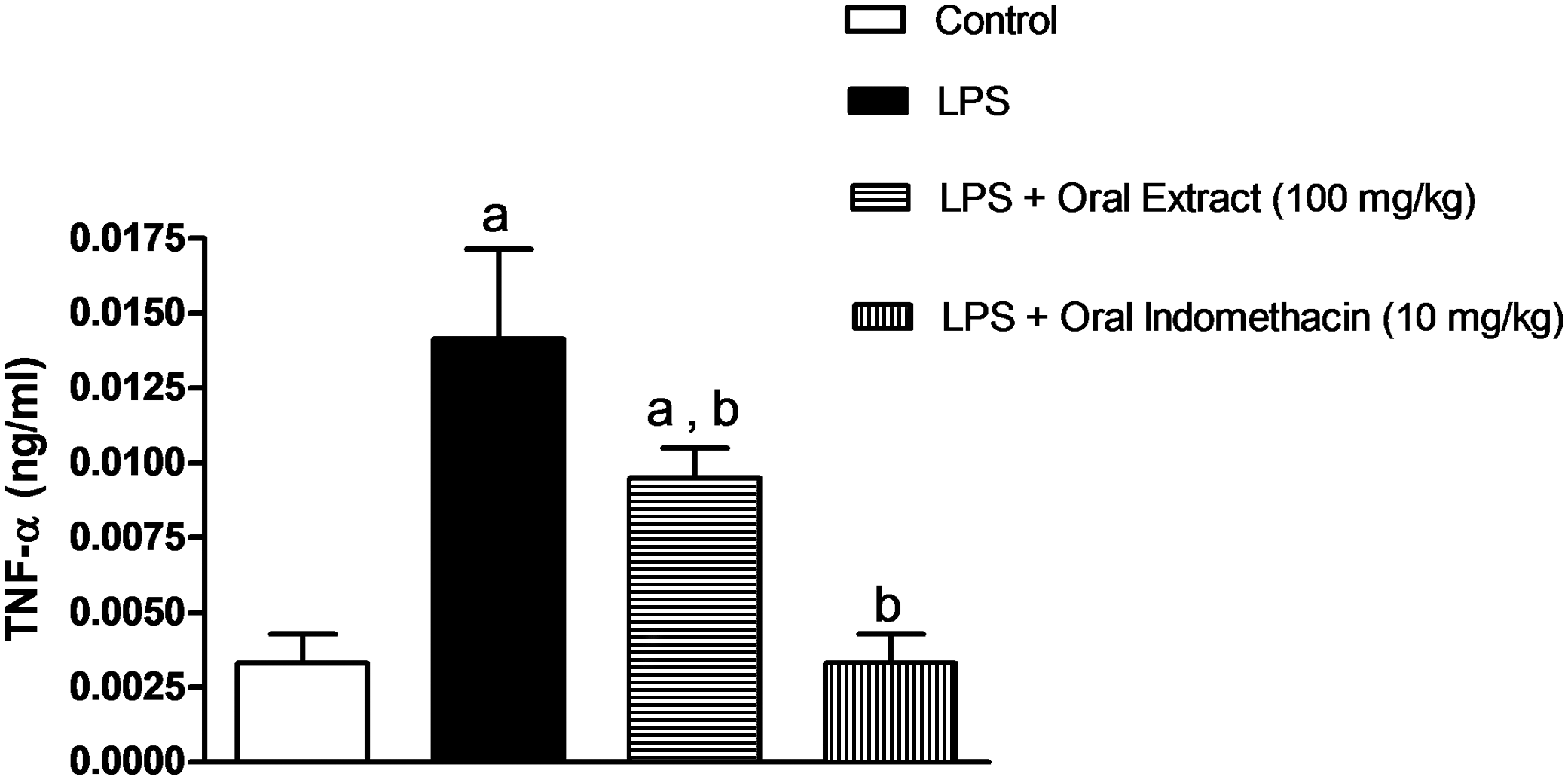
Effect of P. khinjuk L. extract on tumor necrosis factor-α (TNF-α) release in the rat air pouch model. Statistically significant (P<.05) differences are indicated: afrom the control group, bfrom the lipopolysaccharide (LPS)-induced group.
Discussion
Literature review of the genus Pistacia had shown its richness in polyphenolics.
37
Plants containing polyphenolic compounds had shown their efficacy in many pharmacological activities, as anti-inflammatory,
38
antioxidant, hepatoprotective, heart disease protective, and anticancer activities.
39
Therefore, the aim of the present study was to isolate and elucidate the structure of the main components in an aqueous methanol extract of P. khinjuk L. and to evaluate its potential anti-inflammatory effects. In the present study, evaluation of anti-inflammatory effects was undertaken using different animal models to fully investigate the potential of P. khinjuk L. for treatment of inflammatory disorders. Phytochemical investigation of the aqueous methanol extract indicated the presence of phenolic metabolites. These metabolites were gallic acid (
Acute inflammation such as carrageenan-induced paw edema involves the synthesis or release of mediators at the injured site. These mediators include PGs (especially the E series), histamine, bradykinins, leukotrienes, and serotonin and cause fever and pain. 40 Carrageenan-induced paw edema is an experimental animal model for acute inflammation, and it is believed to be biphasic: the early phase (1–2 hours) of this assay is mainly mediated by histamine, serotonin, and an increasing synthesis of PGs in the damaged tissue surroundings; the late phase is sustained by PG release. 41
In the present study, the rat paw edema model showed the ability of P. khinjuk L. extract to significantly reduce paw edema, indicating its potential anti-inflammatory activity. Such a conclusion was further substantiated by the ability of P. khinjuk L. extract to significantly reduce the PGE2 level in the inflammatory exudates in the same model. The results of croton oil–induced ear edema support this finding. A reduction in ear punch weight was shown after pretreatment with P. khinjuk L. extract compared with the croton oil–alone condition. This activity might be justified by the adequate distribution of P. khinjuk L. extract to ear tissues. The findings of the same experiment demonstrated that P. khinjuk L. extract reduced MPO activity in challenged ear tissue. This could be explained based on inhibition of neutrophil infiltration, which was confirmed visually by microscopic examination of the ear tissue. It is noteworthy that the effectiveness of the extract in inhibiting neutophil infiltration was comparable to that of indomethacin. This might imply the potential utility of natural products as substitutes for NSAIDs. These findings are in line with the plant's content of polyphenolics and are supported by the work of Paula et al., 42 who reported that other herbs such as Aegiceras corniculatum rich in polyphenols were able to inhibit the production of eicosanoids in rat neutrophils. Cutaneous inflammation is characterized by the infiltration of polymorphonuclear leukocytes such as neutrophils. 43 MPO is a major component of azurophilic granules of neutrophils. Oxygen-dependent microbicidal activity requires MPO as the critical enzyme for the generation of hypochlorous acid and other toxic oxygen products. 44 The primary function of neutrophils is the release of MPO into the phagosome containing the ingested microorganism, leading to a rapid microbicidal effect. MPO can be released to the outside of the cell, inducing damage to adjacent tissue and thus contributing to the pathogenesis of inflammation. 45
NO is a relatively stable free radical, having numerous promiscuous roles. 46 NO synthesis can be greatly amplified by LPS. 47 Several studies have demonstrated that inflammation correlates with the level of NO. 48 In the current study, the extract of P. khinjuk L. has shown remarkable reduction of nitrite accumulation in the air pouch model. This finding could be attributed to the high content of quercetin flavonoids and phenolic metabolites in the P. khinjuk L. extract observed in the current study. These have been previously shown to possess potent antioxidant and anti-inflammatory activity. 49 Furthermore, the lowering of NO level after P. khinjuk L. extract administration may account for the reduction in PGE2 level because many reports have shown that NO rapidly and strongly stimulates cyclooxygenase enzymes. 50 In addition, NO synthase inhibitors reduce PGE2 biosynthesis. 51
TNF-α is a pleiotropic cytokine that plays a critical role in both acute and chronic inflammation. 52 TNF-α promotes an acute-phase reaction. 53 Several inflammagens have the ability to induce TNF-α synthesis. The formation of several small molecular mediators of inflammation is linked with TNF-α and thus contributes to the range of mediators that critically control inflammation. 54 TNF-α facilitates inflammatory cell infiltration by promoting the adhesion of neutrophils and lymphocytes to endothelial cells. 55 When the TNF-α effect is specifically blocked, the severity of inflammation is reduced. 56 More important is that TNF-α induces the synthesis of PGE2. 53 P. khinjuk L. extract significantly inhibited TNF-α release, which is in agreement with the PGE2 reduction obtained in the current study. The observed anti-inflammatory activity of P. khinjuk L. aqueous methanol extract could be explained by our finding that the extract is rich in flavonoids and phenolic metabolites, represented by the high yield of gallic acid and galloylated sugars. 38
In conclusion, the isolation and NMR spectral data of compound
Footnotes
Acknowledgments
The authors would like to thank Prof. Adel B. Kholoussy, Professor of Pathology, Cairo University, for his help in the histopathological examinations.
Author Disclosure Statement
No competing financial interests exist.