
Abstract
The volatile components from Croton campestris root bark were localized by an anatomical study and analyzed by gas chromatography–mass spectrometry for the first time. The roots of this plant showed secretory cells. These volatile constituents, isolated from the dichloromethane extract by hydrodistillation, were analyzed by gas chromatography–mass spectrometry. We found 69 components. They were characterized, and the major constituents of crude oil root barks were spathulenol (23.3%) and borneol (18.7%). Growth inhibitory activity of the active compounds in solution was evaluated by measuring minimal inhibitory concentrations using a broth micromethod. The minimal inhibitory concentration of root bark volatile constituents was 1.56 μg/mL for Staphylococcus aureus, 3.125 μg/mL for Candida albicans, and 6.25 μg/mL for Aspergillusniger.
Introduction
C
Materials and Methods
Plant material
Roots of C. campestris were collected in Brazil during the flowering stage (in March 1994) and dried at room temperature in Rio de Janeiro before shipment to our laboratory. Voucher specimens of roots were identified by Prof. Isabelle Fouraste (UMR 152, University Paul Sabatier Toulouse III, Toulouse, France).
Preparing slides
Observations are based on microscopic studies of sectioned and stained material of the tissues of root bark. Transverse sections are prepared by hand-cutting, staining in a carmine alum–iodine green reagent or Mirande's reagent 7 for 2–3 min, and then washing with water. Following staining, the transverse sections are mounted on glass slides using glycerin gel. Some observations were made using chloral hydrate solution R. 8 Observations were made with a Leica Microsystems model DMLB microscope, and pictures were taken with a Canon Power Shot S40 digital camera photomicrographic system.
Isolation procedure
We stripped the air-dried roots of the tree C. campestris, and root barks were ground using a blender. First we extracted this biomass (500 g of harvested powdered root barks) with dichloromethane. Then, we subjected the corresponding dried crude extract (20 g) to hydrodistillation at 3–4 mL/min for 4 h on a Clevenger-type apparatus and using the European Pharmacopoeia method. 9 The volatile extract (1.43 mL), which was yellow in color, was stored in vials at low temperature before analysis. An initial extraction with CH2Cl2 allowed us to extract the pigment with wax and volatile organic compounds. This resinoid was then hydrodistilled to recover a pure crude oil denser than water, which was used for this study.
Gas chromatography and gas chromatography–mass spectrometry analysis
The gas chromatography (GC) system used for analyses was from Agilent Technologies (Palo Alto, CA, USA). Analyses were carried out using an HP 6890 gas chromatograph coupled with a HP 5973 mass spectrometer, controlled by HP Chemstation software. GC runs were performed on an HP-5 fused-silica column (30 m, 0.25 mm i.d., 0.25 μm film thickness) and on a HP-Wax column (30 m, 0.25 mm i.d., 0.25 μm film thickness). The injection port was operated in split mode (20:1) with a constant helium carrier gas flow adjusted to 1.0 mL/min. The oven temperature was programmed from 60°C to 260°C at 2°C/min. The injected volume was 1 μL. The scan range was 30–600 atomic mass units. The quadrupole mass spectrometer was operated in electron ionization mode to obtain m/z values at 70 eV and with a connection parts temperature of 250°C.
Identification of components
Retention indices of signals in gas chromatograms were calculated for all volatile compounds using a homologous series of n-alkanes (C8–C32). Constituents were identified by comparing their retention index with those of obtained from the literature 10 –26 and by matching their fragmentation patterns in mass spectra with those of the commercial libraries NIST 02 and Wiley 275. The quantification of spathulenol was performed using geraniol (Extrasynthese, Genay, France) as an internal standard.
Bacterial strains
Staphylococcus aureus C1P483 used as a reference strain. Clinical isolates of the species were obtained from the collection of the Bacteriology Department of Rangueil Hospital (Toulouse). All strains were stored in −80°C and were plated on blood agar before use.
Fungal strains
Candida albicans IP48.72 and Aspergillus niger IP1431.83 were supplied by one of the authors (C.R.)
Assay for antimicrobial activity
The growth inhibitory activity was determined by measuring the minimal inhibitory concentrations (MICs) using a broth micromethod. In brief, 100 μL of specific broths was placed in each well of two 96-well microliter plates, and 100 μL of the test solutions was added to the first column of the first plate. In order to obtain an extended range of tested concentrations, twofold dilutions were carried out from one column to the next, to fill up all the columns of the two microplates except columns 11 and 12, which were used as the control for, respectively, the medium sterility (without test solution and without spores) and growth (without test solution, with inocula). The microplates were inoculated using multipoint inoculators (Denley) to obtain a final concentration of 105 cells/mL and incubated for 24–48 h for bacteria and 5 days for fungi at 30°C. After control of the growth in each well, the MIC was determined as the first concentration corresponding to no visible growth (in micrograms per milliliter). Assays were performed in duplicate.
Results and Discussion
Secretory tissues are present in the root cortex (Fig. 1a). In fact, the cortical parenchyma contains clusters of lignified fibers (Fig. 1b) with a narrow lumen, clusters of pearly fibers (Fig. 1c), cells containing calcium oxalate prisms (Fig. 1d) and starch (Fig. 1e), and an abundant secretory tissue, filled with orange–brown secretion, consisting of single lengthened cells with a slightly thickened wall (Fig. 1f).
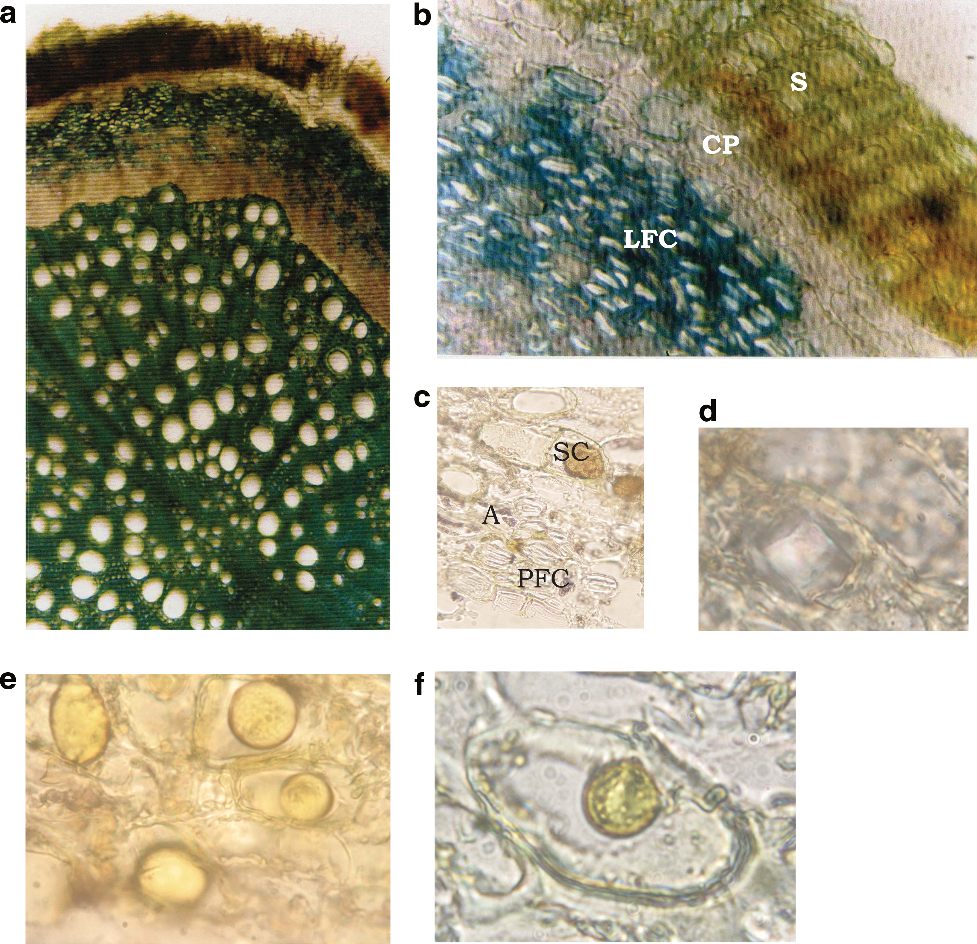
Tranverse sections of root bark of C. campestris St. Hil.
The qualitative and quantitative analytical results of the GC–mass spectrometry analyses of the pure crude oil obtained from root bark resinoid are given in Table 1. Recovery rates for the two steps of the preparation procedure are 4% for dried root extract or resinoid and 7.15% for pure crude oil. More than 90% of the volatile constituents were identified.
Percentages were calculated from flame ionization data.
Sources: Retention index (RI) values are those published by Adams 10 unless indicated otherwise: aYu et al. 11 ; bBorges et al. 12 ; cPino et al. 13 ; dHachicha et al. 14 ; eSylvestre et al. 15 ; fJeong et al. 16 ; gDemirci et al. 17 ; hBaser et al. 18 ; iHymete et al. 19 ; jPaolini et al. 20 ; kLoizzo et al. 21 ; lPaolini et al. 22 ; mMevy et al. 23 ; nSalehi et al. 24 ; oDehghan et al. 25 ; pMondello et al. 26
lit, from literature; MS, mass spectrometry; ND, not detected; Tr, trace (<0.01%).
C. campestris volatile organic compounds comprised 1% monoterpene hydrocarbons, 3.4% aliphatic ketones, 20.7% sesquiterpene hydrocarbons, 27.7% oxygenated monoterpenes, and 47.2% oxygenated sesquiterpenes (Table 2).
Among the 69 compounds isolated from root barks and characterized (Table 1), spathulenol is the major component, present at 23.3%.
By GC–flame ionization detection, with internal standardization, spathulenol was quantified to 3.34 mg equivalents of geraniol/mL of crude oil. Two other constituents, borneol (18.7%) and caryophyllene oxide (5.6%), were also present in significant amounts.
The main compounds identified, using two different types of column, in crude oils were spathulenol, borneol, caryophyllene oxide, and isopathulenol. This is the first report of spathulenol and borneol as the major constituents in C. campestris crude oil. The other components are present at a percentage of <6%.
Spathulenol, an oxygenated sesquiterpene, is present in Croton zambesicus oil in the amount of 14%. 27 The essential oil from Croton ovalifolius contains 6.0% spathulenol and 5.2% caryophyllene oxide. 28 We find spathulenol as the major compound in Croton rosmarinoides 29 at 13.8% and in Croton argyrophylloides 30 at 20.3%. Among the major compounds isolated from Croton flavens, 31 spathulenol reached 3–5%. It may therefore be concluded that C. campestris, which grows widely in Brazil, may be used as a source for the isolation of spathulenol and borneol.
The C. campestris volatile part from bark resinoid was tested for its antimalarial and cytotoxicity activities and proved to be ineffective. However, its crude oil exhibited an MIC of 1.56 μg/mL for S. aureus, 3.125 μg/mL for C. albicans, and 6.25 μg/mL for A. niger.
Footnotes
Acknowledgments
We are grateful to Prof. I. Fourasté, UMR 152, Université Paul Sabatier Toulouse III, for the identification of oregano and Prof. C. Moulis for permitting us to use laboratory materials.
Author Disclosure Statement
No competing financial interests exist.