
Abstract
Acute and chronic inflammation and dyslipidemia play a critical role in the development of various diseases, including cardiovascular disease. Green tea polyphenols possess potent antioxidative and anti-inflammatory properties that contribute to the beneficial effects on heart health. The present study was carried out to determine if administration of a green tea extract (Polyphenon® E [PPE]; Mitsui Norin Co., Ltd., Tokyo, Japan) at 0.2% in the diet reduces cardiovascular risk factors, including dyslipidemia, inflammation, adiposity, and oxidative stress, in rats fed an atherogenic (high fat, cholesterol, and sugar) diet with dextran sodium sulfate (DSS) in drinking water. DSS treatment increased serum total cholesterol, low-density lipoprotein (LDL)-cholesterol, C-reactive proteins (CRP), and markers of liver toxicity and decreased high-density lipoprotein (HDL)-cholesterol significantly. Adding PPE to the atherogenic diet (PPE-diet) was associated with lower total cholesterol and LDL-cholesterol (P<.001) and increased HDL-cholesterol (P=.001). In addition, the PPE-diet was associated with decreased serum CRP concentration (P=.023) and increased total antioxidant capacity (P=.016) and catalase (P=.001) and glutathione peroxidase (P=.050) activities. The PPE-diet significantly lowered epididymal fat pad weight (P=.009). Feeding the PPE-diet also ameliorated some of the DSS-induced lipid, inflammatory, and oxidative symptoms. In summary, green tea supplementation decreased several cardiovascular risk factors, including body composition, dyslipidemia, inflammatory status, and antioxidant capacity, in rats fed an atherogenic diet. This study supports green tea as an effective dietary component for sustaining cardiovascular health.
Introduction
S
The positive effects of green tea are attributed to biologically active polyphenols. Green tea polyphenols make up 10–30% of the dry weight of green tea, with nearly 50% of the total polyphenol content in green tea in the form of epigallocatechin gallate (EGCG). 9 EGCG has strong antioxidant activity and works as a free radical scavenger as well as an inducer of phase II enzyme activities. 9 Free radical damage contributes to several health problems associated with aging such as cancer and cardiovascular and neurological diseases. 10 –12 Antioxidant enzymes such as catalase (CAT), superoxide dismutase (SOD), glutathione peroxidase (GPx), and glutathione S-transferase (GST) neutralize free radicals to help restore cell function and prevent further damage to cell membranes. 13 The strong antioxidant capacity of green tea may help to slow the initial development of atherosclerosis. 14
Progression of atherosclerosis is marked by the release of pro-inflammatory cytokines such as interleukins, C-reactive proteins (CRP), and tumor necrosis factor-α. 15 CRP is a powerful marker for inflammation, yet the exact role of CRP in the atherosclerotic process is still up for debate. CRP binds to blood lipids, activating an inflammatory response that results in endothelial stiffening. 16,17 Thus, CRP not only may be a biological marker for increased cardiovascular risk, but also may be an independent risk factor for atherosclerosis and hypertension. 17 CRP concentration has been shown to be inversely related to consumption of foods high in monounsaturated fats, antioxidants, and tea. 15,18,19
One of the most important risk factors for cardiovascular disease is hypercholesterolemia. 7 High levels of low-density lipoprotein (LDL)-cholesterol, triglycerides (TGs), and total cholesterol (TC) are all associated with increased prevalence and severity of cardiovascular incidence; however, a high level of high-density lipoprotein (HDL)-cholesterol has a favorable effect on heart health. Debate exists whether LDL- or HDL-cholesterol levels are better predictors of cardiovascular risk; 20,21 however, green tea consumption has recently been linked to improving dyslipidemia and vascular function in adults. 7,22
The purpose of the present study was to examine the antioxidant, anti-inflammatory, and cholesterol-lowering effects of a green tea extract enriched in EGCG in rats fed an atherogenic diet. Dextran sodium sulfate (DSS) was administered to induce inflammation to examine possible anti-inflammatory effects of green tea. Body weight, body composition, lipid profile, antioxidant capacity, inflammation, and enzyme activity are hypothesized to improve when compared with rats not treated with green tea polyphenols.
Materials and Methods
Experimental design, animals, and diets
Forty male Sprague–Dawley rats were housed individually in wire-bottomed cages in the research vivarium on the campus of San Diego State University (San Diego, CA, USA). The rats were kept on a 12-h light–dark cycle, and water and food were available to the animals at all times. The temperature and humidity were controlled at approximately 20–24°C and 40–45%, respectively. The animal protocol was approved by the San Diego State University Animal Subjects Committee.
After acclimation, all rats were fed the basal atherogenic diet consisting of 33% sugar, 21% fat, and 3% cholesterol by weight (Table 1). Twenty rats received the diet supplemented with 0.2% Polyphenon® E (PPE) (kindly provided by Mitsui Norin Co., Ltd., Tokyo, Japan) (PPE-diet), and 20 rats were given the diet alone (control). PPE is a standardized green tea preparation consisting of 66.5% EGCG and other catechins (9.7% epicatechin, 7.0% epicatechin gallate, 3.7% epigallocatechin, 3.2% gallocatechin gallate, and 0.9% catechin). Studies in the literature have used green tea extract in the range of 0.12–2% in water or diet. Hininger-Favier et al. 2 has shown that 0.2% green tea extract in diet decreases oxidative stress. Therefore, 0.2% PPE was chosen for this study because the objective of the current study was to determine the antioxidant effect of PPE. Four days prior to the end of the 4-week study, 10 rats in each diet group were administered an inflammation-inducing agent, 3% (wt/vol) DSS (40 kDa) (ICN Biomedicals, Aurora, OH, USA) in drinking water for 72 h followed by normal drinking water for 24 h. Food intake and water consumption were recorded over a 48-h period before and during DSS treatment. Body weight was measured weekly. Liver, kidney, spleen, and epididymal fat pad were weighed upon the conclusion of the study.
PPE, Polyphenon E.
Serum lipids
Rats were euthanized by an overdose of carbon dioxide gas, and blood was collected. Measurements included total cholesterol, HDL-cholesterol, and TGs using kits from StanBio (Boerne, TX, USA). LDL-cholesterol content was calculated by subtracting HDL-cholesterol content from TC; then the value for total TGs divided by 5 was subtracted from the previous number obtained: LDL=(TC–HDL)–(TG/5). 23
CRP
Serum CRP was assessed using a solid-phase sandwich enzyme-linked immunosorbent assay kit from BD Biosciences (San Jose, CA, USA). The enzyme-linked immunosorbent assay kit utilizes an antibody specific for rat CRP coated on a 96-well plate to which rat CRP readily binds. Horseradish peroxidase–conjugated anti-rat CRP was added to generate an antibody–antigen–antibody sandwich, and a substrate solution was added to create a blue color measured at 450 nm.
Antioxidant capacity
Total antioxidant capacity was measured with the antioxidant assay kit from Sigma (St. Louis, MO, USA). This assay is based on the formation of a ferryl myoglobin radical from myoglobin and hydrogen peroxide, which oxidizes 2,2′-azino-bis(3-ethylbenzthiazoline-6-sulfonic acid) (ABTS) to produce the radical cation ABTS+, a soluble green chromogen that can be quantified at 405 nm.
Antioxidant enzyme activity
Serum antioxidant enzyme activities were assessed by measuring activities of CAT, SOD, GST, and GPx with assay kits from Cayman Chemical Co. (Ann Arbor, MI, USA). CAT activity was determined based on a reaction with methanol in the presence of H2O2. The resulting product, formaldehyde, is measured by the addition of a chromogen (Purpald®; Sigma), resulting in a purple color absorbed at 540 nm. SOD activity was measured by adding superoxide radicals in the form of xanthine oxidase and hypoxanthine to serum samples. GST activity was determined by measuring the conjugation of 1-chloro-2,4-dinitrobenzene with reduced glutathione. GPx activity was measured indirectly by a coupled reaction with glutathione reductase. Oxidized glutathione, produced upon reduction of hydroperoxide by GPx, was recycled to its reduced state by glutathione reductase and NADPH. Oxidation of NADPH to NADP+ was accompanied by a decrease in absorbance at 340 nm.
Liver enzyme activity
Alanine aminotransferase (ALT), aspartate aminotransferase (AST), lactate dehydrogenase (LDH), and alkaline phosphatase (ALP) activities were determined using the ALT/GPT (UV-Rate) Kit, the AST/GOT (UV-Rate) kit, the LDH (UV-Rate kit), and the ALP Liquicolor kit, respectively, from Stanbio. ALT and AST activities were measured by the rate of NADH oxidation per minute. LDH activity was determined by the rate in which NAD is reduced to NADH per minute. ALT, AST, and LDH absorbances were measured at 340 nm; ALP was measured at 405 nm.
Statistical analysis
Data are represented as mean±SE values for each test group. Differences between results for the green tea diet and the DSS treatment were analyzed using a two-way analysis of variance using SPSS version 17.0 (IBM, Somers, NY, USA). Comparisons between the different test groups were analyzed using the Student–Newman–Keuls test. An α level of 0.05 was considered statistically significant.
Results
Body weight, organ weights, and food and water intake
Differences in body weight among the four groups were not significant at baseline (data not shown). The DSS treatment resulted in lower body weights. Liver, spleen, and epididymal fat pad weights were significantly lower in the DSS-treated group at the end of treatment (P<.001) (Table 2). The administration of the PPE-diet did not result in a significant effect on body or organ weights. However, the PPE-diet resulted in lower mean fat pad weight (P=.009) compared with rats fed the control diet (Table 2). Prior to the DSS treatment, the 48-h food and water intake was not significantly different among the four groups (data not shown). During the 72-h administration of DSS, the 48-h food (P<.001) and water (P=.036) intake of all treated rats was significantly lower than that of nontreated rats (Table 2).
Data are mean±SE values (n=40, 10 per group).
DSS, dextran sodium sulfate.
Lipid profile
DSS-treated rats exhibited an increase in serum TC (P<0.001) and LDL-cholesterol (P<.001) and a decrease in serum TG concentrations in comparison with control rats (P=.002) (Table 3). No significant treatment effect was found for HDL-cholesterol. Rats fed the PPE-diet had significantly lower mean serum TC (P<.001) and LDL-cholesterol (P<.001) levels and higher mean serum HDL-cholesterol levels (P=.001) (Table 3). DSS-treated rats fed the PPE-diet showed trends to decreased serum TC and LDL-cholesterol and higher serum HDL-cholesterol concentrations compared with rats receiving control diet fed with DSS treatment.
Data are mean±SE values (n=40, 10 per group).
HDL-C, high-density lipoprotein-cholesterol; LDL-C, low-density lipoprotein-cholesterol; TC, total cholesterol; TG, triglyceride.
CRP concentration
Rats given DSS treatment had a significant increase in plasma CRP (P<.001) compared with control-diet fed rats. Rats fed the PPE-diet had a lower mean CRP (P=.023) concentration than the control group and the DSS-treated groups (P=.004) (Fig. 1).
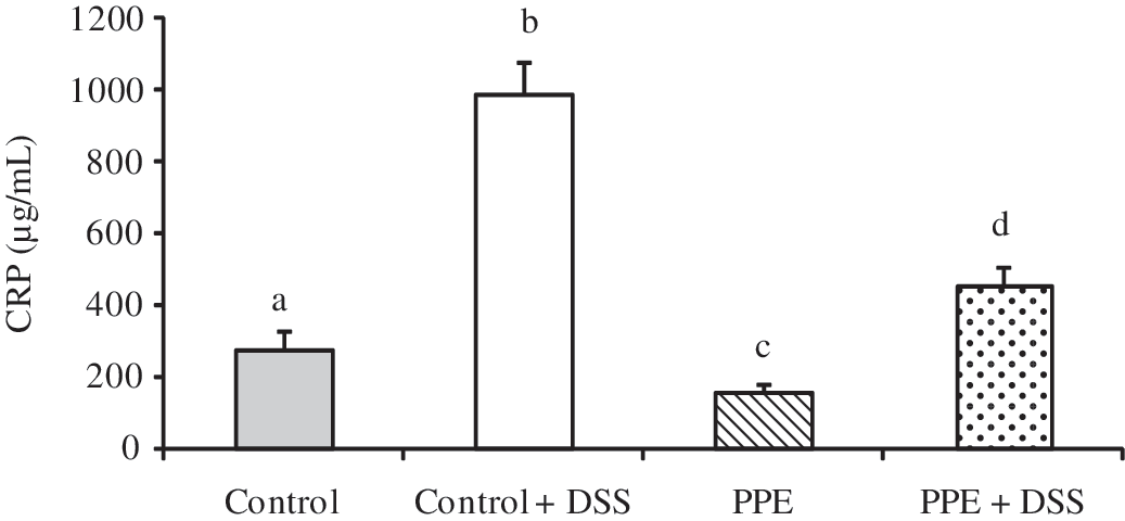
Serum C-reactive protein (CRP) concentration. Green tea-fed rats showed a lower CRP increase than control rats with DSS treatment. Data are mean±SE values. abcdColumns with different superscripts are significantly different at P<.05.
Antioxidant enzyme activity and total antioxidant capacity
DSS treatment increased SOD activity (P=.044) and decreased GST (P=.002) and GPx (P<.001) activities (Table 4). Rats fed the PPE-diet exhibited significantly higher CAT (P=.001) and GPx (P=.050) activities (Table 4). PPE did not have a significant effect on SOD or GST activity.
Data are mean±SE values (n=40, 10 per group).
CAT, catalase (in nmol/min/mL); GPx, glutathione peroxidase (in nmol/min/mL); GST, glutathione S-transferase (in nmol/min/mL); SOD, superoxide dismutase (in U/mL).
Serum total antioxidant capacity was increased in PPE-diet-fed rats compared with control diet groups (P=.016) (Fig. 2).

Antioxidant capacity. Green tea diet increased serum antioxidant capacity. Data are mean±SE values. abColumns with different superscripts are significantly different at P<.05.
Liver enzyme activity
The mean serum activities of the hepatic markers (AST, LDH, and ALT) revealed significant DSS treatment effects but no significant PPE-diet effects. ALT (P<.001), AST (P=.003), and LDH (P=.038) activities were significantly higher in the DSS-treated groups (Table 5).
Data are mean±SE values (n=40, 10 per group).
ALT, alanine aminotransferase; ALP, alkaline phosphatase; AST, aspartate aminotransferase; LDH, lactate dehydrogenase.
Discussion
The aim of the present study was to investigate the protective effect of PPE, a green tea extract, on processes contributing to cardiovascular disease. The inflammation-inducing agent DSS was used to amplify the effects of the atherogenic diet and to increase the expression of inflammatory cytokines near the end of the study period. Lack of appetite and diarrhea caused weight loss and decreased TG levels in the DSS-treated rats. The same DSS concentration, when administered with an AIN 76A-based rodent diet, has not been known for these severe intestinal symptoms. 24 The effects of DSS in combination with an atherogenic diet were dominant and diminished some of the benefits of the green tea extract administration on lipid parameters. Furthermore, the DSS effects on hepatic and antioxidant enzymes were severe; thus the potential benefits of the PPE-diet were likely muted by the severity of the DSS treatment. However, the green tea extract without DSS treatment improved several other cardiovascular risk factors, including hypercholesterolemia, fat pad weight, antioxidant activity, and inflammation status.
As hypothesized, the PPE-diet improved the overall blood cholesterol profile in rats fed an atherogenic diet. Green tea polyphenols were shown to have a significant lowering effect on TC and LDL-cholesterol while improving the HDL-cholesterol in rats with and without DSS treatment. The results in this study are similar to those of previously reported animal studies. 9,14,25,26 Yokozawa et al. 26 also found increased HDL levels associated with green tea supplementation in high cholesterol-fed rats. However, Lee et al. 9 reported lower HDL levels when rats fed a high fat diet were supplemented with either 0.2% or 0.5% EGCG. Although the other plasma lipids (LDL, TGs, and free fatty acids) were reportedly lower with EGCG supplementation in the study by Lee et al., 9 a low level of HDL-cholesterol is a strong predictor of cardiovascular events even when LDL levels are low. 9,20,27 The improved overall cholesterol profile associated with the green tea polyphenol administration in the present study supports the important role of green tea in cardiovascular disease prevention.
Although LDL-cholesterol concentrations are directly associated with risk for cardiac events, the oxidation of the LDL molecules is thought to play a key role in the pathogenesis of atherosclerosis. 28 Many studies have reported that green tea polyphenols suppress oxidation of LDL in vitro and ex vivo and in in vivo models. 29 –32 Antioxidants disrupt the oxidation process and may delay the development of atherosclerotic plaques. 33 In addition to overall antioxidant activity, enzymatic antioxidative enzymes such as SOD, CAT, GPx, and GST play a vital role in scavenging against free radicals that are involved in oxidative injury. Ramesh et al. 14 reported increased CAT, SOD, GPx, and GST activities in cardiac tissue and hemolysate of rats fed an atherogenic diet. Conversely, Khan et al. 34 discovered inconsistencies in enzyme activity dependent on the location of tissue extraction. The present study examined serum antioxidant enzyme activity and revealed inconsistent results as well. The addition of green tea alone significantly increased CAT and GPx activities; however, no changes were seen in GST or SOD. DSS treatment generates oxidative stress, 24 and the current study showed a reduction of antioxidant enzyme activities including CAT in response to the oxidative stress. It has been reported that green tea extracts or green tea polyphenols increase antioxidant enzyme activities. Green tea polyphenol treatment led to a decrease in CAT activity with DSS treatment. Additional research may be needed to further analyze antioxidant enzyme effects. However, total antioxidant capacity was significantly higher in rats fed the PPE-diet. Similar results have been reported in both animal and human studies, 26,35 suggesting that green tea may be an effective treatment for the prevention of oxidative processes involved in atherosclerosis.
Evidence supporting the link between body composition, specifically body fat distribution, and cardiovascular disease continues to grow. Apart from overall obesity, body fat is an important determinant of disease risk and cardiovascular complications. 36,37 Although evidence on body weight is inconsistent, recent studies have shown that tea polyphenols may have an impact on body composition. 38 Possible mechanisms for reducing adiposity include an inhibitory effect of green tea polyphenols on adipogenic genes such as that for fatty acid synthase, along with up-regulation of lipolytic genes and uncoupling protein 2. 9,38 The present study found that although body weights were not significantly different among diet groups, mean epididymal fat pad weight was lower for the green tea group. Additionally, Lee et al. 9 found that 0.5% EGCG supplementation significantly reduced the weight of epididymal, subcutaneous, visceral, and retroperitoneal fat pads in high fat diet-fed mice. The present study revealed significant differences in body fat, yet overall body weight remained consistent among diet groups. The possible mechanisms behind the antilipogenic effects of green tea may call for further investigation as obesity rates continue to increase.
Atherosclerosis is an inflammatory disease that is characterized by the development of plaques and lesions on the inner lining of the arteries. CRP is a biological marker for chronic inflammation, and it is now suggested that CRP may have a role in the pathogenesis of atherosclerosis. 16,17,39 The addition of the DSS treatment in the present study produced a nearly threefold increase in serum CRP concentration. The PPE treatment significantly reduced the CRP levels in both the DSS-treated and nontreated rats. Similar results were reported in a study examining the effects of EGCG on atherogenic and standard diet-fed rats. 39 The reduction of circulating CRP may indicate a slowing of atherosclerosis and reduced risk for cardiac events.
The treatment with a 0.2% PPE-diet did not exhibit any hepatotoxic effect. However, in the DSS-treated rats activities of the hepatic enzymes (ALT, AST, and LDH) were significantly higher. The increase in liver toxicity may have been caused by the hypercholesterolemia associated with the pro-inflammatory treatment. An atherogenic diet has been shown to significantly increase serum AST, ALT, ALP, and LDH activities; however, previous research reported decreased activity associated with EGCG treatment. 40,41 In the current study, the hepatic damage caused by the atherogenic diet coupled with the DSS treatment may have been stronger than the protective PPE effect on liver enzyme activity.
In conclusion, the findings of the present study add to the body of knowledge concerning the protective effects of green tea polyphenols. Green tea plays a positive role in several cardiovascular risk factors, including body composition, dyslipidemia, inflammatory status, and antioxidative capacity. Consumption of green tea in a moderate amount was able to alleviate several symptoms associated with an atherogenic diet. Although feeding 0.2% PPE in the rat diet provides about a twofold higher concentration than can be accomplished by human consumption of brewed tea, 42,43 the results presented provide support for the role of green tea extracts in the prevention of atherosclerosis. Future dose-dependent studies that include a physiologically relevant amount of intake are warranted.
Footnotes
Acknowledgments
The authors thank Mitsui Norin Co., Ltd. (Tokyo, Japan) for the generous donation of Polyphenon E. The authors also thank Ms. Antoinette Averna, Ms. Patcharie Elkins, and the students of the Advanced Nutrition Laboratory classes at San Diego State University for their lab assistance. This study was funded by the San Diego State University Grant Program.
Author Disclosure Statement
No competing financial interests exist.