




Abstract
Acetaminophen (APAP) hepatotoxicity has been related to several cases of hepatitis, cirrhosis, and hepatic transplant. As APAP hepatotoxicity is related to reactive oxygen species (ROS) formation and excessive oxidative stress, natural antioxidant compounds have been tested as an alternative therapy to diminish the hepatic dysfunction induced by APAP. Taraxacum officinale Weber (Family Asteraceae), commonly known as dandelion, is used for medicinal purposes because of its choleretic, diuretic, antioxidant, anti-inflammatory, and hepatoprotective properties. This study evaluated the hepatoprotective activity of T. officinale leaf extract against APAP-induced hepatotoxicity. T. officinale was able to decrease thiobarbituric acid–reactive substance levels induced by 200 mg/kg APAP (p.o.), as well as prevent the decrease in sulfhydryl levels caused by APAP treatment. Furthermore, histopathological alterations, as well as the increased levels of serum aspartate and alanine aminotransferases caused by APAP, were prevented by T. officinale (0.1 and 0.5 mg/mL). In addition, T. officinale extract also demonstrated antioxidant activity in vitro, as well as scavenger activity against 2,2-diphenyl-1-picrylhydrazyl and nitric oxide radicals. Our results clearly demonstrate the hepatoprotective effect of T. officinale against the toxicity induced by APAP. The possible mechanisms involved include its scavenger activities against ROS and reactive nitrogen species, which are attributed to the content of phenolic compounds in the extract.
Introduction
A
APAP hepatotoxicity is related to excessive reactive oxygen species (ROS) production and oxidative stress that can cause cellular damage 5 and hepatocyte death. 6,7
Taraxacum officinale Weber (Family Asteraceae), commonly known as dandelion, has been popularly used for medicinal purposes because of its choleretic, diuretic, antioxidant, anti-inflammatory, and hepatoprotective properties. 8 –12 T. officinale leaf is clinically used in various preparations, including infusions and ethanolic extracts, and is added to salads because of its significant source of the nutrients potassium, vitamins, and minerals. 13 Dried dandelion leaves and roots are also available as herbal teas, and the powdered root is sold in capsule form. 14
Flavonoids and phenolic compounds such as luteolin, caffeic acid, and chlorogenic acid have been detected in extracts of T. officinale. 10,15 These compounds, which are generally found in plants, have been determined to protect cells from oxidative stress by preventing the formation of free radicals or by detoxifying free radicals, resulting in the prevention of a variety of pathophysiological processes. 16
Of particular importance is that recent studies have provided evidence that T. officinale root extract might play beneficial roles against alcoholic liver damage 17 and carbon tetrachloride–induced hepatic fibrosis. 18,19 However, there are no studies on the potential relationship between T. officinale leaf extract and APAP hepatotoxicity.
Considering the previously reported antioxidant and pharmacological properties of the extract and the involvement of oxidative stress in the hepatic disorders caused by APAP, we decided to investigate whether a leaf extract of T. officinale possesses the capacity to prevent the hepatotoxicity induced by APAP. To describe the possible mechanisms through which T. officinale displays hepatoprotective activity, we conducted in vitro experiments to demonstrate the possible antioxidant activity of the extract and its putative scavenger activity against different free radicals such as nitric oxide (NO•), 2,2-diphenyl-1-picrylhydrazyl (DPPH•), hydroxyl (OH•), and H2O2.
Materials and Methods
Chemicals
Thiobarbituric acid, malonaldehyde bis(dimethyl acetal) (MDA), DPPH, and deoxyribose were purchased from Sigma (St. Louis, MO, USA). Sodium nitroprusside was obtained from Merck (Darmstadt, Germany). Iron sulfate (FeSO4) was from Reagen (Rio de Janeiro, RJ, Brazil). All other reagents were obtained from local suppliers.
Plant material
The fresh leaves of T. officinale Weber were used as the plant material and obtained from the campus of Cruz Alta University (Cruz Alta, RS, Brazil). T. officinale leaves were collected from several individual plants at two different periods: during the winter and early spring in 2008. Taxonomic identification was confirmed by the Department of Botany of Cruz Alta University, and a voucher specimen was registered under the number 157 in the Herbarium of Cruz Alta University.
Preparation of the extract
For the preparation of the ethanolic extract, leaves (10 g) were extracted with ethanol (70%) for 2 weeks at room temperature. The extract was evaporated and concentrated to dryness under reduced pressure using a rotary evaporator and redissolved in ethanol (70%) to a concentration of 20 mg/mL.
Animals
Adult male albino mice from our own breeding colony and weighing between 35 and 40 g were kept in cages with continuous access to food in a room with controlled temperature (22±3°C) and a 12-h light/dark cycle, with lights on at 7:00 a.m. All experiments were conducted in accordance with the Guiding Principles of the Animal Care and Wellness Committee of the Federal University of Santa Maria.
In vivo experiments
Animal treatments
APAP was dissolved in warm saline. Thirty animals were divided into the following five groups, with six animals per group: Group I, pretreatment with ethanolic solution (1% ethanol)+vehicle (0.9% NaCl, p.o.); Group II, pretreatment with ethanolic solution (1% ethanol)+APAP (200 mg/kg, p.o.); Group III, pretreatment with extract (0.1 mg/mL, approximately 17.5±0.025 mg/kg/day)+APAP (200 mg/kg, p.o.); Group IV, pretreatment with extract (0.5 mg/mL, approximately 87.5±0.18 mg/kg/day)+APAP (200 mg/kg, p.o.); and Group V, pretreatment with extract (0.5 mg/mL)+vehicle (0.9% NaCl, p.o.).
Mice received extract or ethanolic solution in drinking water for 10 days. These animals were monitored every day for their volume of extract consumed. APAP or vehicle was administered in a single dose on Day 10 of treatment. Twenty-four hours after APAP administration, mice were anesthetized and subjected to a heart puncture. The liver was removed, a portion was reserved for histopathological analysis, and the remaining liver tissue was homogenized (1:10) in 10 mM Tris-buffer (pH 7.4) and centrifuged at 4,000 g at 4°C for 10 min. The low-speed supernatant fraction (S1) obtained was used for biochemical analyses. The APAP dose was chosen after analyses of dose–response curves that were produced from experiments previously conducted in our lab. 20
Biochemical analysis: lipid peroxidation
Lipid peroxidation in mouse liver was assessed by the measurement of thiobarbituric acid–reactive substances (TBARS). 21 The S1 fractions were incubated at 95°C for 1 h in a buffered medium with sodium dodecyl sulfate (8.1%), acetic acid/HCl buffer, and thiobarbituric acid (0.6%). TBARS formation was determined spectrophotometrically at 532 nm, using MDA as a standard. The results were expressed as nanomoles of MDA per milligram of protein.
Biochemical analysis: non-protein thiol content
Liver non-protein thiol (NPSH) content was estimated using Ellman's reagent [10 mM 5,5′-dithiobis(2-nitrobenzoic acid)] after deproteinization with trichloroacetic acid (10%), according with the standard methodology. 22
Biochemical analysis: activities of aspartate and alanine aminotransferases
Serum aspartate aminotransferase (AST) and alanine aminotransferase (ALT) were quantified as liver damage indicators using a commercial kit (LABTEST, Diagnostica S.A., Lagoa Santa, MG, Brazil) and according to the method of Reitman and Frankel. 23
Biochemical analysis: protein measurements
Aliquots from the homogenates (S1) were separated for protein measurements that were assessed according to the procedure of Lowry et al. 24
Biochemical analysis: histopathology
Portions of the livers from treated mice were carefully removed, washed in saline solution, and then immersed in formalin (10%). For light microscopy examination, tissues were embedded in paraffin, sectioned to produce 5-μm-thick specimens, and stained with hematoxylin and eosin (n=3 per group).
In vitro experiments
Determination of total phenolic content
The total phenolic content was determined by adding 0.5 mL of the extract to 2.0 mL of 7.5% sodium carbonate and 2.5 mL of 10% Folin–Ciocalteu reagent. The reaction mixture was incubated at 45°C for 15 min. Afterward, the absorbance was measured at 765 nm using a spectrophotometer. Gallic acid was used as a phenol standard. 25 The total phenolic content was expressed as micrograms of gallic acid equivalent per milligram of extract.
Qualitative and quantitative analysis by high-performance liquid chromatography with diode array detection
High-performance liquid chromatography with diode array detection was performed with a Prominence (Shimadzu, Kyoto, Japan) autosampler (model SIL-20A) equipped with Shimadzu LC-20AT reciprocating pumps, connected to a DGU-20A5 degasser and a CBM-20A integrator. An ultraviolet-visible detector (model DAD SPD-M20A) and LC Solution version 1.22 SP1 software were used. Reverse-phase chromatography analyses were carried out with a Phenomenex (Torrance, CA, USA) C-18 column (4.6 mm×250 mm) packed with 5-μm-diameter particles. Injection volume was 40 μL, and the gradient elution was conducted according to the method of Evaristo and Leitão, 26 slightly modified. Ultraviolet absorption spectra were recorded in the range of 200–400 nm.
The crude hydroalcoholic extracts were individually screened for the presence of the following polyphenolic compounds: gallic acid, chlorogenic acid, caffeic acid, quercetin, rutin, and kaempferol. Identification of the compounds was performed by comparing their high-performance liquid chromatography retention times and ultraviolet absorption spectra with those of the commercial standards. Stock methanolic solutions of standards were prepared in the range of 0.0025–0.045 mg/mL. Quantification was carried out by integrating the peaks using an external standard method at a wavelength of 327 nm for chlorogenic and caffeic acids and 365 nm for quercetin, rutin, and kaempferol. Chromatographic operations were carried out at ambient temperature and in triplicate.
NO• scavenging activity
NO• scavenging activity was determined using the Griess reagent. 27 Sodium nitroprusside (5 mM, in phosphate-buffered saline) was incubated at 25°C with different concentrations of T. officinale (10, 20, 50 and 75 μg/mL). After 120 min, 0.5 mL of incubation solution was sampled and mixed with 0.5 mL of Griess reagent [0.1% N-(naphthyl)ethylenediamine dihydrochloride and 1% sulfanilic acid in 5% phosphoric acid]. The absorbance was measured at 550 nm. The values were compared with the control to determine the percentage of inhibition of the nitrite reaction with Griess reagent, which gave an index for the NO• scavenger activity of the T. officinale extract. 28
NO• scavenging activity
Measurement of the T. officinale scavenger activity against the NO• radical was performed in accordance with the procedure of Cho et al. 29 NO• solution (85 μM) was added to a medium containing different concentrations of the extract (10, 20, 50, and 75 μg/mL) and incubated at room temperature for 30 min. The decrease in the absorbance measured at 518 nm depicted the scavenger activity of T. officinale against NO• radical. The values were expressed as percentage of inhibition of NO• absorbance in relation to the control values without extract.
Determination of oxidative damage to deoxyribose
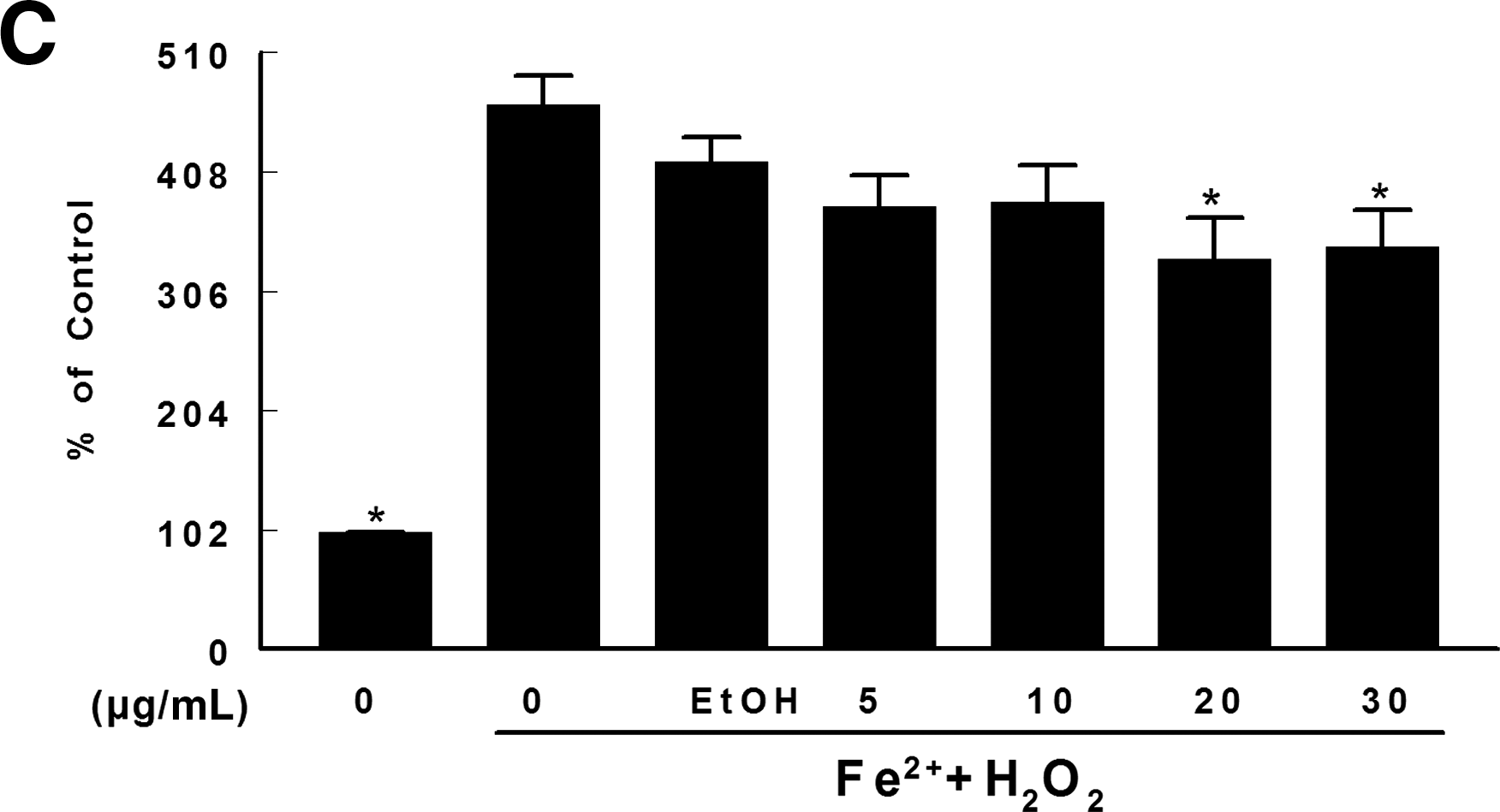
The deoxyribose degradation was induced with both H2O2 and FeSO4 alone and in combination. 30 In brief, the reaction medium was prepared containing the plant extract (5, 10, 20, and 30 μg/mL final concentrations), 3 mM deoxyribose, potassium phosphate buffer (0.05 mM, pH 7.4), 50 μM FeSO4, and 500 μM H2O2. Solutions of FeSO4 and H2O2 were made prior to use. Reaction mixtures were incubated at 37°C for 30 min and stopped by addition of 0.8 mL of trichloroacetic acid (2.8%), followed by addition of 0.4 mL of thiobarbituric acid (0.6%). Next, the medium was incubated at 100°C for 20 min, and the absorbance was recorded at 532 nm. 31 Standard curves of MDA were constructed for each experiment. The values are expressed as percentages of control values.
Statistical analysis
Statistical significance was assessed by one-way analysis of variance, followed by Duncan's test for post hoc comparison. Results were considered statistically significant at P<.05.
Results
Hepatotoxicity induced by APAP (200 mg/kg)
T. officinale or ethanolic solution consumptions and body weights did not differ among groups during the treatment period (data not shown). Administration of APAP at the dose of 200 mg/kg was able to induce lipid peroxidation (P<.05) (Fig. 1A), as well as a lowering of liver NPSH levels in treated mice (P<.05) (Fig. 1B). Also, there was a leakage of ALT and AST activity into plasma, reflecting the liver damage due to APAP treatment (P<.05) (Fig. 2).
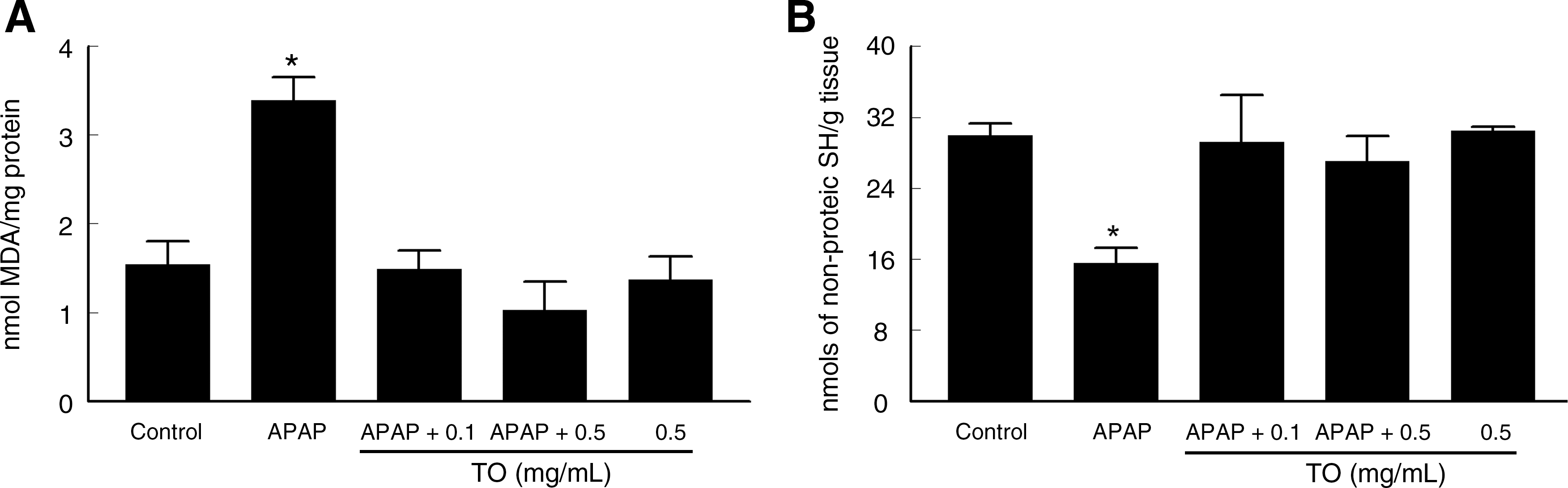
Effects of acetaminophen (APAP) administration (200 mg/kg, p.o.) and/or pretreatment with T. officinale (TO) (0.1 and 0.5 mg/mL) for 10 days on biochemical parameters evaluated in the livers of mice:
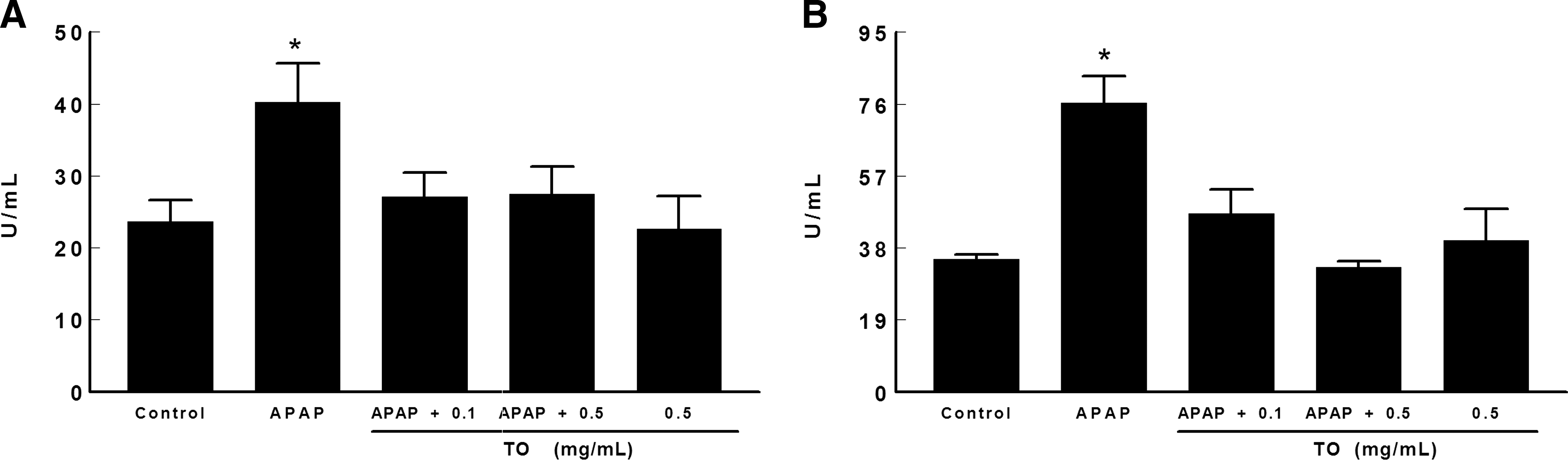
Effects of APAP administration (200 mg/kg, p.o.) and/or pretreatment with TO (0.1 and 0.5 mg/mL) on serum
T. officinale pretreatment was able to protect against APAP-induced hepatotoxicity. Lipid peroxidation was diminished by pretreatment with extract at both doses tested (0.1 and 0.5 mg/mL) (Fig. 1A). NPSH depletion caused by APAP was prevented by extract pretreatment at all tested doses (P<.05) (Fig. 1B). Additionally, the hepatic damage indicated by AST and ALT leakage was prevented by the extract (P<.05) (Fig. 2). The mice treated only with the T. officinale extract did not demonstrate any alterations in the parameters studied.
Figure 3 shows the histopathology analyses from the livers of mice treated with APAP. Light microscopic investigations revealed that control mice and extract-treated mice demonstrated normal histological appearance of the liver with normal polyhedral hepatocytes (Fig. 3A and B). On the other hand, in livers from APAP-treated mice, we observed damage to the hepatocytes that had lost their characteristic appearance, plus manifestations of hydropic degeneration, perinuclear vacuolization, dilatation in blood sinusoids, and perivascular inflammatory leukocyte infiltration (Fig. 3C and D). The pretreatment with 0.5 mg/mL extract resulted in morphological improvement. We observed a normal arrangement of hepatocytes, with decreased dilatation in blood sinusoids, although inflammatory leukocyte infiltrations and vacuoles were present (Fig. 3E and F).
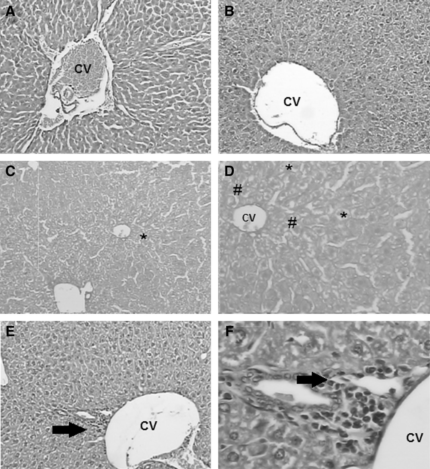
Photomicrography of the hepatic lobe segment of mice.
In vitro experiments
The total phenolic content of T. officinale was 124±0.0157 μg of gallic acid/mg of T. officinale leaf extract. The presence of phenolic compounds including chlorogenic acid, caffeic acid, and quercetin was determined, and the chromatograms are shown in Figure 4. The chlorogenic acid, caffeic acid, and quercetin concentrations were 0.8581±0.02 mg, 0.7526±0.023 mg, and 0.8003±0.036 mg/g of T. officinale, respectively.
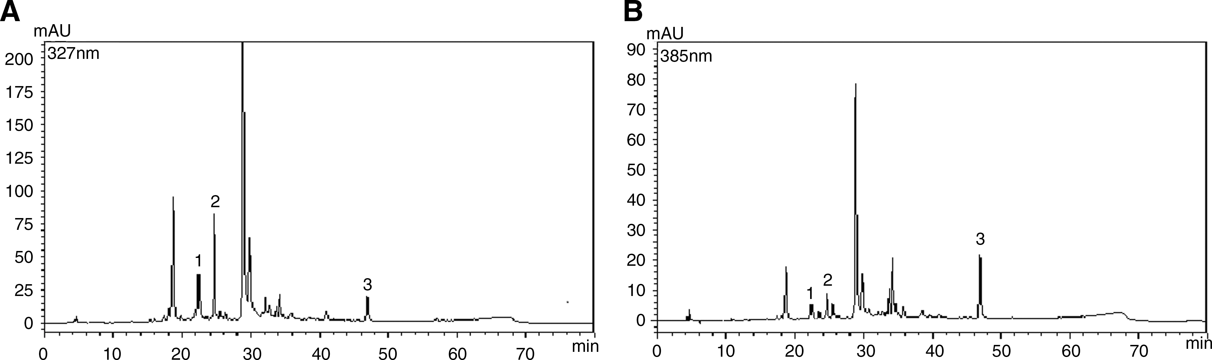
High-performance liquid chromatograms of chlorogenic acid, caffeic acid, and quercetin in TO ethanolic extract.
The capacity of the extract to inhibit deoxyribose oxidation is shown in Figure 5. T. officinale was able to protect against deoxyribose oxidation induced by H2O2 or Fe2+ at the concentrations of 5, 10, 20 and 30 μg/mL (P<.05) (Fig. 5A and B). Furthermore, in the Fenton reaction (Fe2++H2O2), the extract decreased deoxyribose degradation caused by OH• radicals at the concentrations of 20 and 30 μg/mL (P<.05) (Fig. 5C). We also observed a significant NO• scavenging activity of the extract at concentrations between 20 and 75 μg/mL (P<.05) (Fig. 6A) and a significant NO• scavenging activity at concentrations of 10–75 μg/mL (P<.05) (Fig. 6B).
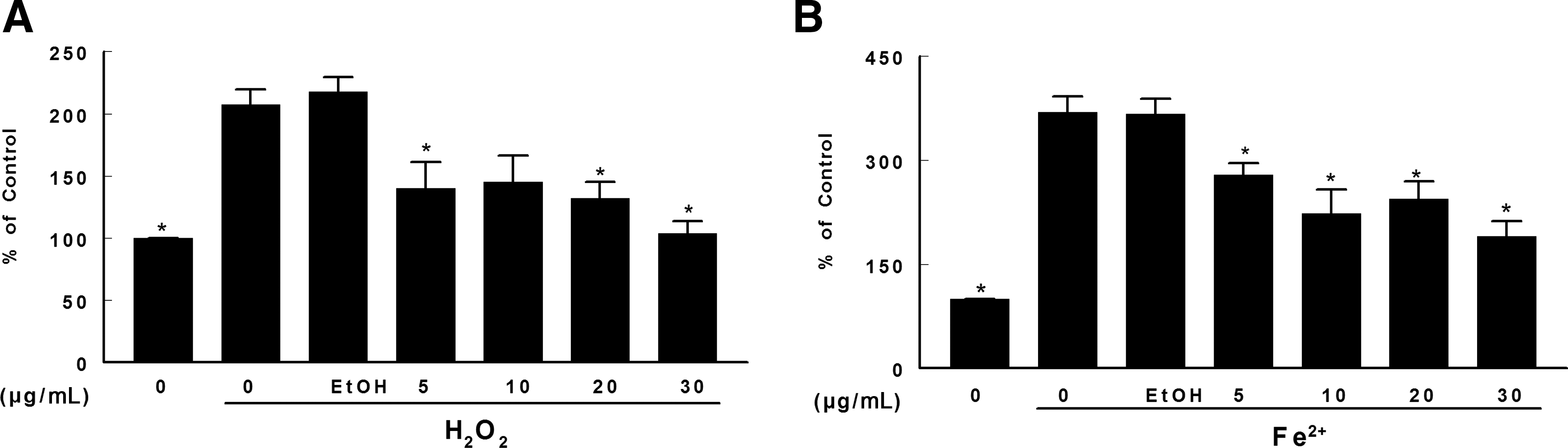
Effect of TO on deoxyribose oxidation induced by
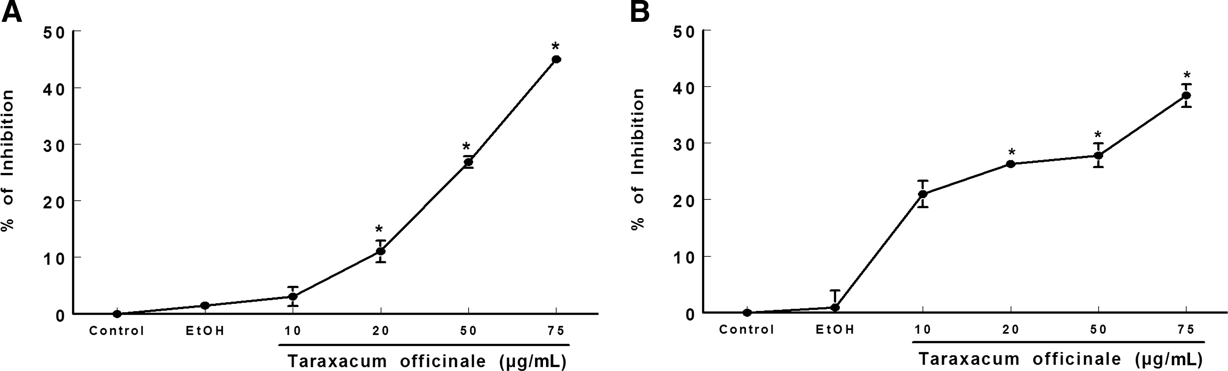
Scavenging activity of TO extract on
Discussion
APAP administered orally to mice at 200 mg/kg caused biochemical and histological damage to hepatic tissue. Pretreatment with T. officinale leaf extract for a 10-day period was able to prevent the damage in liver tissue and alterations in biochemical parameters caused by APAP, demonstrating a significant protective effect against APAP-induced hepatotoxicity.
Oxidative stress is believed to play a major role in the pathogenesis and progression of APAP-induced liver injury. In our exposure protocol, APAP administration caused lipid peroxidation, as well as depletion of NPSH groups. It is known that the APAP metabolite (N-acetyl-p-benzoquinoneimine) rapidly reacts with glutathione, which accounts for 90% of the NPSH groups, 32 which consequently exacerbates the oxidative stress that was evidenced by increased TBARS levels. In addition, APAP toxicity also was observed by the increase in the serum ALT and AST activities, reflecting failure of liver function due to APAP treatment. ALT is a cytoplasmic enzyme that is released into circulation after structural damage to the hepatocytes and is used as a standard biomarker of liver injury. 33 An increase in serum ALT activity is almost always due to hepatocellular damage and is usually accompanied by a similar increase in serum AST activity. 34
Pretreatment with T. officinale was able to prevent the SH depletion, as well as the increase in TBARS levels induced by APAP at both tested doses. This effect could be attributed to its antioxidant activity. Also, ALT and AST activities were maintained at their control levels in animals that received extract and APAP, confirming a protective effect of T. officinale extract.
In line with the biochemical results, histopathological analysis clearly demonstrated that pretreatment with T. officinale leaf extract produced a general protective effect in hepatic tissue compared with the APAP group alone.
In order to investigate the possible antioxidant mechanism by which T. officinale would protect against APAP, we performed some in vitro experiments. APAP metabolization generates a large amount of a variety of reactive species such as H2O2, NO•, and O2–, 35 and the scavenging of such species would be important for the protection against APAP hepatotoxicity. T. officinale extract presented a scavenger activity against NO• and NO• radicals. NO• is a reactive species that combines with superoxide radical (O2–) to form peroxynitrite (ONOO–), a reactive species capable of inducing oxidative stress more pronounced than other reactive species. 36 In our study, the extract effectively reduced the nitrite formation, indicating decreased levels of NO• and suggesting a scavenger activity against reactive nitrogen species. T. officinale extract also presented a capacity to scavenge the NO• radical. The NO• radical is a one electron radical very similar to OH• and can be used to evaluate the OH• scavenging. 37
Moreover, in the present study, low concentrations of the extract were also able to protect deoxyribose from H2O2 and Fe2+-induced oxidation, as well as deoxyribose oxidative degradation induced by the Fenton reaction. The Fenton reaction (Fe2++H2O2→Fe3++OH–+OH•) generates OH• radical, which can induce severe oxidative damage to the cellular components. 38 The mechanism of the antioxidant action of polyphenols has usually been attributed to OH• scavenging activity. 36
In this study, we determined the contents of the phenolic compounds chlorogenic acid, caffeic acid, and quercetin. Chlorogenic acid and caffeic acid have hydroxyl groups on an aromatic residue, and they exhibited antioxidant, antimutagenic, and carcinogenic activities in vitro, which were attributed to their scavenger activities against ROS. 39 Quercetin, a major flavonoid naturally occurring in plants, also deserves attention because of its observed beneficial effects. This flavonoid demonstrated neuroprotective effects, 40 as well as a protective effect against ethanol-induced liver damage. 41
Our results demonstrated for the first time the hepatoprotective activity of T. officinale leaf extract in mice in vivo. We believe that the mechanism by which the extract was able to protect the liver from the oxidative stress generated by APAP is due to its antioxidant activity. The antioxidant mechanism of the extract is probably due to its scavenger activity against several ROS/reactive nitrogen species attributed to the phenolic compounds. These phenolic compounds of the extract act as antioxidants and free radical scavengers and reduce or inhibit the oxidative stress induced by APAP administration.
Footnotes
Acknowledgments
This work was supported by a FINEP research grant, “Rede Instituto Brasileiro de Neurociência (IBN-Net),” number 01.06.0842-00. The National Institute of Science and Technology for Excitotoxicity and Neuroprotection/CNPq also supported this work. D.C. received a fellowship from CNPq/PIBIC/UFSM. N.V.B., J.B.T.R., and F.A.A.S. received a fellowship from CNPq. Additional support was provided by CAPES.
Author Disclosure Statement
No competing financial interests exist.