
Abstract
The essential oil from the leaves of Annona sylvatica (EOAS) was extracted by hydrodistillation, and the analysis was performed by gas chromatography–mass spectrometry. The main compounds identified in the EOAS were sesquiterpenes, such as hinesol, z-caryophyllene, β-maaliene, γ-gurjunene, silphiperfol-5-en-3-ol, ledol, cubecol-1-epi, and muurola-3,5-diene. Oral administration of the EOAS (20 and 200 mg/kg) and subcutaneous injection of dexamethasone (0.5 mg/kg, reference drug) significantly inhibited carrageenan- and complete Freund's adjuvant–induced mouse paw edema. The anticancer activity the EOAS showed growth inhibitory activity on all cell lines when administered in a high concentration. The EOAS inhibited the growth of human cancer cell lines with GI50 values in the range of 36.04–45.37 μg/mL on all of the cell lines tested. This work describes for the first time the anti-inflammatory and anticancer effects of the essential oil of A. sylvatica and its composition. Considering that drugs currently available for the treatment of inflammatory and cancer conditions show undesirable side-effects, the present results may have clinical relevance and open new possibilities for the development of novel anti-inflammatory and anticancer drugs.
Introduction
S
Inflammation is a coordinated response to harmful stimuli aimed primarily at maintaining homeostasis, 6 largely by repairing the tissue injury or the active process of resolution. 7 The inflammatory process plays a role in some diseases, such as atherosclerosis, gout, rheumatoid arthritis, type II diabetes, 8 and cancer. 9 In some types of cancer, the inflammatory process is present before a malignant change occurs; however, in other types of cancer, an oncogenic change induces an inflammatory microenvironment that promotes the development of tumors. 10 In this context, drug discoveries of new agents with anti-inflammatory and anticancer properties have a unique interest for medical care.
Several in vivo and in vitro models of inflammation and cancer have been used for the discovery of new therapeutic agents. One of the most common models is the carrageenan (Cg)-induced inflammation in a rat paw model, which shows the acute efficacy of anti-inflammatory agents against several parameters of inflammation. 11 The model of Cg paw edema has long been used to assess the anti-inflammatory properties of agents, such as nonsteroidal anti-inflammatory drugs (NSAIDs) 12 and other agents. The identification of cytostatic or cytotoxic activity could test the agent in several cancer cell lines with a main objective of separating features associated with antiproliferative activity toward many cell lines from those that affect only a specific cell type. 13
Medicinal plants have been an opportunity in drug discovery for the treatment of inflammation and cancer. Several studies have demonstrated the anti-inflammatory and anticancer activities of products derived from plants, such as essential oils. 14 Essential oils from some Annonaceae species, such as Dennettia tripetala 15 and Annona sengalensis, 16 have shown effectiveness in these processes. Before our work, there have been no reports available on the biological activity of the leaf essential oil of A. sylvatica.
The genus Annona (Annonaceae) consists of ∼250 species distributed across Brazil. Many species, such as Annona squamosa, Annona muricata, Annona reticulata, and Annona cherimola, 17 produce edible fruits. Although Annona are generally consumed as fresh fruits, other parts are also widely used in folk medicine, and several studies have characterized the different biological and pharmacological activities, particularly anti-inflammatory, antinociceptive, 18 –20 and anticancer activities. 21,22 Previous phytochemical studies have attributed these activities to various compounds found in these species, such as acetogenins, alkaloids, 23 and flavonoids. 24
A. sylvatica A. St.-Hil [old name Rollinia sylvatica (A. St.-Hil.) Mart] is a native species from Brazil, found in Minas Gerais and from São Paulo to Rio Grande do Sul states. It is known as araticum, araticum-do-mato, cortiça, and cortiça-amarela. 25 The leaves have been used in folk medicine against fever, cough, ulcers caused by syphilis, muscle spasms, angina, and diarrhea. 26
The popular usage of this species against fever, which is a process regulated by inflammatory mediators, and previous studies that have confirmed anti-inflammatory and anticancer properties in other species of Annonaceae suggest an anti-inflammatory and/or anticancer potential for the essential oil from A. sylvatica. Therefore, the aim of this study was to assess the anti-inflammatory and anticancer activity of the essential oil from leaves of A. sylvatica (EOAS).
Materials and Methods
Plant material
The leaves of A. sylvatica were collected in September 2010 in Dourados, Mato Grosso do Sul, Brazil. An authenticated specimen (DDMS4600) of the plant was deposited in the herbarium of the Faculty of Biological Sciences, Federal University of Grande Dourados (UFGD).
Essential oil extraction and gas chromatography–mass spectrometry analysis
The fresh leaves of A. sylvatica were subjected to hydrodistillation for 3 h using a Clevenger-type apparatus. The oil was dried over anhydrous sodium sulfate and preserved in a sealed vial at 4°C until further analysis.
The analyses were performed on a gas chromatograph (Varian GC 3900) equipped with an ion-trap mass spectrometer detector (Varian Saturn 2100), using a DB-5 (5% diphenyl dimethyl polysiloxane)–fused-silica column (30 m×0.25 mm, 0.25-μm film thickness), under the following conditions: carrier gas helium (99.999%, flow rate 1.0 mL/min); 1-μL injection volume, split ratio (1:20), with initial oven temperature of 40°C, and heating from 40°C to 250°C at 3°C/min. The injector temperature, quadrupole detector, and line-transfer temperature were maintained at 250°C. The mass spectrometry scan parameters included electron impact ionization voltage at 70 eV, a mass range of 40–650 m/z, and a scan interval of 0.5 sec. Temperature-programmed retention indices 27,28 were calculated using a mixture of normal paraffin (C8–C25) as the external references. The identifications were completed by comparing the spectra of the masses obtained with those of NIST 2.0, Saturn Libraries, and literature data. 29 The relative area by gas chromatography–flame ionization detection analyses was determined using a gas chromatograph (Shimadzu GC-17A) equipped with the same DB-5-fused silica capillary column and under the same conditions as the gas chromatography–mass spectrometry.
Solvents and chemicals
Analytical-grade methanol, hexane, anhydrous sodium sulfate, and dimethyl sulfoxide (DMSO) were obtained from Vetec. λ-Cg, Tween 80, dexamethasone, trichloroacetic acid, and doxorubicin were purchased from Sigma Chemical Co.
For the evaluation of anti-inflammatory activity, λ-Cg and dexamethasone were freshly prepared on the day of the experiment in a saline solution (0.9%). The EOAS was dissolved in an aqueous solution with 1% Tween 80 as the vehicle immediately before administration.
Animals
Male Swiss mice (25–35 g) were obtained from UFGD. The animals were kept under standard laboratory conditions, with a constant temperature (22°C±1°C), and a 12-h light/dark cycle with free access to food (Purina®) and water. The Institutional Ethics Committee from UFGD approved the procedures and protocols adopted in the study (authorization number 005/2010).
Cg-induced paw edema in mice
Five groups of mice (n=6), totaling 30 animals, were orally treated (p.o.) with the EOAS (2–200 mg/kg) or vehicle. Another group of mice was treated subcutaneously (s.c.) with the anti-inflammatory drug dexamethasone (0.5 mg/kg). After 1 h, the animals received a 50-μL s.c. injection of Cg (300 μg) dissolved in sterile 0.9% saline into the right hindpaw. The contralateral paw received only saline and was used as the control. The thickness of the paw edema was measured using a digital micrometer 30,31 1 h before any treatment and at several time points (1, 2, and 4 h) after the injection of Cg. The results were expressed in μm, and the difference between basal and postinjection values were quantified as edema.
Complete Freund's adjuvant–induced persistent inflammation in mice
Three groups of mice (n=6), totaling 18 animals, were treated daily for 4 days with the EOAS (200 mg/kg, p.o.), dexamethasone (0.5 mg/kg, s.c.), or vehicle (p.o.). After 1 h, the animals received a 20-μL s.c. injection of complete Freund's adjuvant (CFA; 1 mg/mL of heat-killed Mycobacterium tuberculosis in 85% paraffin oil and 15% mannide monoleate) into the right hindpaw. The contralateral paw received only saline and was used as control. The thickness of the paw edema was measured using a digital micrometer 1 h before any treatment and at several time points (1, 2, and 4 h and 1, 2, 3, and 4 days) after the injection of CFA. The results were expressed in μm, and the difference between basal and postinjection values was quantified as edema.
Anticancer assays
Cell lines
The National Cancer Institute (Frederick MA, USA) kindly provided nine human cancer cell lines: U251 (glioma, CNS), UACC-62 (melanoma), MCF-7 (breast), NCI-ADR/RES (ovarian expressing phenotype multiple drug resistance), 786-0 (renal), NCI-H460 (lung, nonsmall cells), PC-3 (prostate), OVCAR-03 (ovarian), and HT29 (colon). VERO (green monkey kidney cells), a normal cell line, was also used. Stock and experimental cultures were grown in a medium containing 5 mL of RPMI-1640 (Gibco BRL) supplemented with 5% fetal bovine serum (FBS; Gibco BRL).
Cell culture
Stock cultures were grown in 5 mL of RPMI-1640 (Gibco BRL) supplemented with 5% FBS (Gibco BRL). A penicillin:streptomycin mixture (1000 U/mL:1000 μg/mL, 1 mL/L RPMI; Nutricel) was added to the experimental cultures.
Antiproliferative assay
Cells in 96-well plates (100 μL cells/well) were exposed to different concentrations of the EOAS (0.25, 2.5, 25, and 250 μg/mL) in DMSO/RPMI (0.1% v/v) at 37°C and 5% CO2 for 48 h. Final DMSO concentration did not affect cell viability. The cells were then fixed with trichloroacetic acid solution (50%, v/v), and cell proliferation was determined by spectrophotometric quantification (540 nm; Molecular Devices Versa Max Microplate Reader) of cellular protein content using sulforhodamine B assay. 32 Doxorubicin (0.025–25 μg/mL) was used as a positive control. Three measurements were obtained: first at time zero (t 0, at the beginning of incubation) and then 48 h postincubation for both the compound-free (C) and tested (T) cells. Cell proliferation was determined according to the formula ([T−t 0]/[C−t 0])×100. Cytostatic effect was observed when T≥t 0, while cytocidal effect occurred when T<t 0. From the concentration–response curve for each cell line, the TGI (concentration that produces 0% of cell growth or cytostatic effect) value was determined through nonlinear regression analysis using Origin 8.0® software (OriginLab Corp.). The experiments were performed in triplicate.
Statistical analysis
Data are presented as the mean±standard error of the mean. Difference among groups was evaluated by analyses of variance (one-way ANOVA) followed by the Newman–Keuls test. Statistical differences were considered to be significant at P<.05.
Results and Discussion
By hydrodistillation, the fresh leaves of A. sylvatica produced 0.17% (w/w) of essential oil. The components of the studied essential oil are shown in Table 1. The different constituents were identified and quantified by gas chromatography–mass spectrometry procedures, and 36 compounds representing 98.97% of the oil were identified. The analysis of triplicates showed a coefficient of variation of <2% of retention times.
Rt, retention time (min); RI, retention indices on DB-5 capillary column.
The essential oil consisted mainly of oxygenated sesquiterpenes. Hinesol (8.16%), Z-caryophyllene (7.31%), β-maaliene (6.61%), γ-gurjunene (5.46%), silphiperfol-5-en-3-ol (4.75%), ledol (4.43%), cubecol-1-epi (4.36%), trans-muurola-3, and 5-diene (4.33%) were the major components.
Previous studies have shown that β-caryophyllene has anti-inflammatory activity in several animal models, including Cg- and PGE-induced hindpaw edema; this activity does not need the integrity of adrenal glands. 33 Studies evaluating the anti-inflammatory properties of α-humulene and (−)-trans-caryophyllene, isolated from Cordia verbenacea's essential oil, showed that oral treatment with the compounds displayed marked inhibitory effects in different inflammatory experimental models in mice and rats, indicating that substances represent important tools for the management and/or treatment of inflammatory diseases. 34
The injection of Cg in a mouse paw induced an edema that started at 60 min and peaked at 2 h (Fig. 1A). A single dose of the EOAS significantly inhibited Cg-induced paw edema formation in a dose-dependent manner (Fig. 1A, B). The percentage of inhibition was 19%±3%, 27%±7%, and 35%±2% at doses of 2, 20, and 200 mg/kg, respectively (Fig. 1A, B). Moreover, our results showed that the EOAS at doses of 20 and 200 mg/kg presented significant inhibition at time points after its administration, as indicated by the time course analysis (Fig. 1A). In addition, the significant inhibition was observed in the dexamethasone-treated group 2 h after the injection of Cg in the paw (Fig. 1A, B).
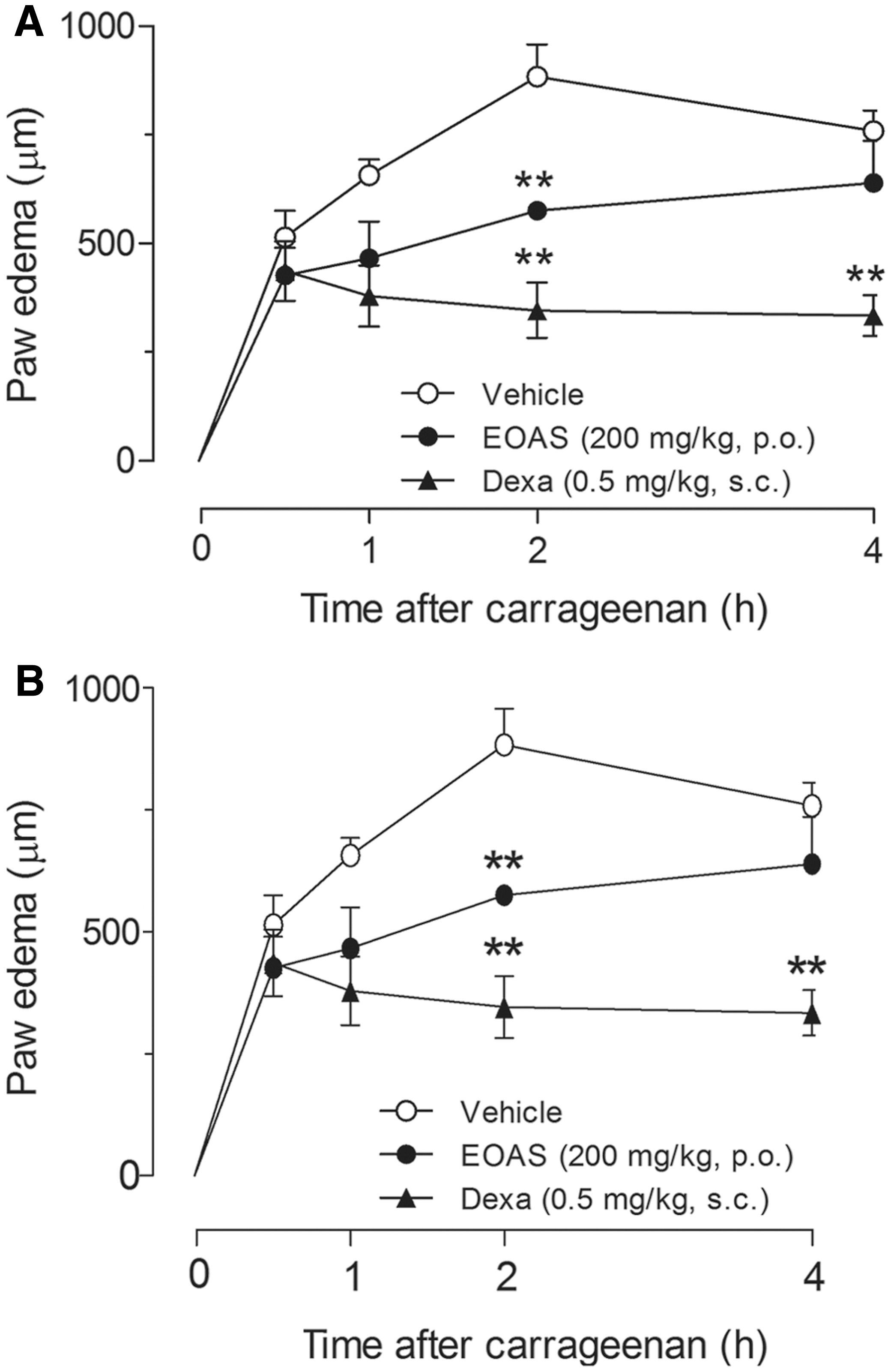
Effect of EOAS on Cg-induced paw edema in mice. Animals received the EOAS (2, 20, or 200 mg/kg, p.o.), DEX (0.5 mg/kg, s.c.), or vehicle, and after 1 h, an intraplantar injection of Cg (300 μg/paw) was performed.
Another acute and persistent inflammation model used was CFA, which is a mixture of oils and water with killed M. tuberculosis that typically elicits a very strong immune reaction and intense inflammation. In the persistent inflammation induced by CFA, daily oral treatment with the EOAS (200 mg/kg) inhibited the edema by ∼39%±1% and 22%±3%, while dexamethasone (0.5 mg/kg, s.c.) reduced the edema 45%±2% and 25%±5% on days 3 and 4, respectively, after injection of CFA (Fig. 2). Thus, the EOAS showed anti-inflammatory activity against CFA-induced persistent inflammation. Taken together, these results demonstrate that oral administration of the EOAS inhibited the persistent inflammation induced by CFA.
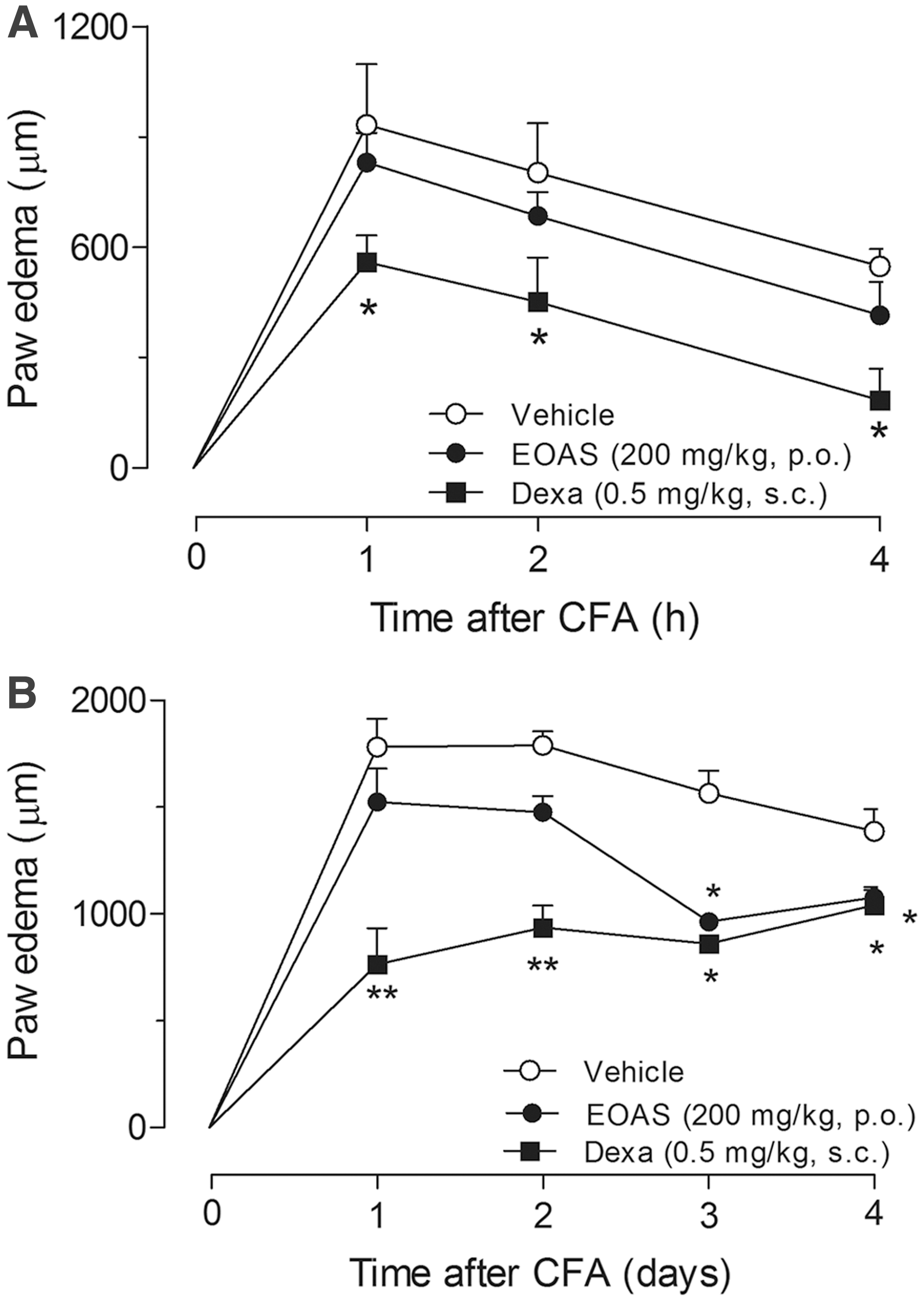
Effect of EOAS on complete Freund's adjuvant (CFA)-induced acute
The antiproliferative activity of the EAOS was evaluated in vitro against VERO and 9 human tumor cell lines: melanoma (UACC-62), breast (MCF-7), lung (NCI- H460), ovarian (OVCAR03), prostate (PC-3), colon (HT-29), renal (786-0), ovarian resistant (NCI/ADR-Res), and glioma (U251). GI50 and TGI values (μg/mL) for EOAS as well as DOX are summarized in Table 2. The results demonstrate that the EOAS possess anticancer activity with GI50 values in the range of 36.04–45.37 μg/mL, but in the highest concentration, cytostatic activity and cytotoxic effects were observed for all cell lines.
Doxorubicin (DOX) was employed as the positive control.
EOAS, essential oil from Annona sylvatica leaves ; GI50, concentrations that elicit inhibition by 50% of the cell growth (in μg/mL); TGI, concentrations that produce 0% of cell growth (in μg/mL).
In conclusion, the present study demonstrates the anti-inflammatory and anticancer effects of the essential oil of the leaves of A. sylvatica, which provides support to the traditional use of this plant from the genus Annona for the treatment of inflammatory processes. Further studies to elucidate the mechanisms of action, and the possible compounds involved in these activities will be undertaken.
Footnotes
Acknowledgments
This work was supported by Fundect (Brazil, MS). We thank CAPES and CNPq for fellowship funding.
Author Disclosure Statement
No competing financial interests exist.