
Abstract
Many researchers have focused on the preventive and curative effects of garlic (Allium sativum), particularly on cardiovascular diseases and cancer. However, its impacts on the male reproductive system have not been clearly defined. In this study, the effect of chronic consumption of two garlic fractions was tested: one soluble in water (aqueous solution obtained by grinding and centrifugation) and the other one precipitated by ethanol (alcoholic precipitate obtained by precipitation of the aqueous solution), on different variables of male rats' reproductive functions. These two fractions were targeted to try to identify the nature of the active garlic compounds responsible for the different modifications observed on testicular parameters. The observation of seminiferous tubules of rats treated with garlic fractions showed an increased number of tubules deprived of spermatozoa. In addition, garlic fractions induced apoptosis of testicular germ cells (TdT-mediated dUTP-X nick-end labeling [TUNEL] approach) and a decrease of serum testosterone levels and seminiferous tubule DNA concentrations. In summary, our histological and molecular results suggest that one or several substances, soluble in water and precipitated by alcohol, impaired spermatogenesis.
Introduction
D
The side effects, particularly on male reproduction, of such a chronic treatment are poorly investigated. To date, it has been reported that garlic juice was effective in recovery of testicular function after experimental testicular hypogonadism 18 and after lindane-induced damages in testes. 19 However, other laboratories found that garlic metabolites such as diallyl trisulfide (DATS) present spermicidal effects. 20,21 Moreover, powder 22 or crude 23,24 garlic preparations may impair testicular and male reproductive-tract functions. In our laboratory, the mechanisms of action of chronic consumption of crude garlic on testicular functions were investigated. The cellular and molecular targets of crude garlic administered in various doses (5%, 10%, and 15%) to adult male rats 23,25 were identified. Garlic had inhibitory effects on Leydig steroidogenic enzyme expression and Sertoli cell markers and induced germ cell death (spermatocytes I and spermatids) by an apoptotic process.
According to our previous results, we asked: What is the nature of molecules in garlic which induce disruption of testicular functions? Are they water-/oil-soluble compounds or proteins, or something else? From these assumptions, crude garlic was fractionated in two phases, an aqueous fraction (containing water- and oil-soluble compounds) and a fraction precipitated by alcohol (containing essentially proteins), and their effects on some variables of male rats' reproductive functions were tested: serum testosterone, sperm density, testicular DNA concentration, and testicular integrity on histological sections. These two fractions were targeted to try to identify the nature of the active garlic compounds responsible for the different modifications observed on testicular parameters. In this preliminary study, we did not isolate the active molecules of garlic, but tried only to determine their nature.
Materials and Methods
Plant preparation
The type of garlic used in the present study was spring garlic. This variety has pink bulbs and is planted between December and March (according to the weather) in Tunisia and collected in July. This type of garlic is composed of 2.1% proteins, 30% carbohydrates, 1.5% fibers, 0.2% fat, 0.015% vitamins, and 0.7% minerals. The garlic plant used in this study was grown in Tunisia and purchased from a local market. In our developed protocol, two garlic fractions were obtained following the different steps described in Figure 1. Briefly, 15 g of garlic were peeled, crushed in distilled water, and the aqueous fraction was obtained after a centrifugation at 3000 g for 20 min at +4°C. This aqueous fraction was divided in two equal volumes. One volume was diluted with distilled water and then mixed with powdered standard rat pellet diet (Industrial Society of Food, Sfax, Tunisia). The second volume was precipitated with cold absolute ethanol (1/3; v/v), and after centrifugation at 3000 g and desiccation, the alcoholic precipitate was mixed with powdered standard rat diet.
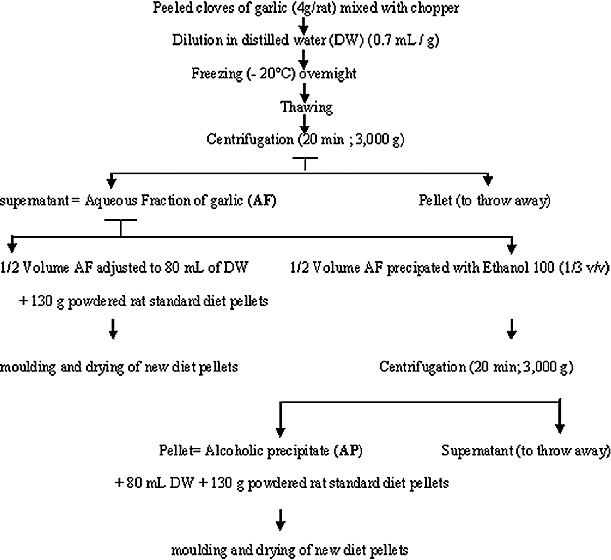
Experimental protocol of garlic fraction preparation.
Animals and treatment
Eighteen adult male Wistar rats (Pasteur Institute of Tunis, Tunisia), with an average weight ranging between 200 and 250 g, were used in this study. The animals were housed with proper aeration at 25°C±2°C, and were given tap water ad libitum. The rats were allowed to acclimatize in the laboratory for a period of 1 week before the beginning of the study. They were divided in three groups of six animals each.
Group 1 (G1): Control rats received a standard pellet diet.
Group 2 (G2): Treated rats received pellets mixed with aqueous fraction (corresponding to 7.5% garlic).
Group 3 (G3): Treated rats received pellets mixed with alcoholic precipitate (corresponding to 7.5% garlic).
Every day, 25 g of food (standard diet or garlic fractions mixed with standard diet) was given to each rat. The animals consumed 25 g of food daily, as no pellet was observed the following day. During the experimentation period, animals consumed the administered diet and were weighed daily. After 48 days of treatment, rats were killed, and a cardiac blood sample was taken from each rat and then put in a sterile tube. Blood was allowed to clot at 20°C. When the clot was retracted, the sample was centrifuged at 3000 g for 15 min at +4°C, and the serum was transferred to a new tube. The serum samples were stored frozen at −20°C until use. All the rats were killed by decapitation the same day. Testes were dissected out and weighed. All studies on animals were conducted in accordance with current regulations and standards approved by the Faculty of Medicine of Tunis animal care committee.
Hormonal analyses
Testosterone contents in serum samples were determined using a radioimmunoassay kit according to the manufacturer's instructions (Biosource). The intra-assay and interassay coefficients of variations were 4.6% and 6.2%, respectively. The detection limit was 0.05 ng/mL.
Histopathological studies
Classic histology
Testes were fixed in a 10% formaldehyde solution, passed through an ascending series of ethanol baths, cleared in toluene, and embedded in paraffin. Tissues were sectioned at 5 μm and stained with hematoxylin and eosin. A slide from each animal was used to evaluate the percentage of seminiferous tubules with a lumen containing at least 2/3 of spermatozoa.
TdT-mediated dUTP-X Nick-End Labeling
Paraffin sections (5 μm) of formaldehyde-fixed testicular tissues were mounted on Superfrost plus slides. The sections were deparaffinized (xylene 5 min, ethanol 100%, 95%, and 70%, 30 sec each), and washed in distilled water before beginning the TdT-mediated dUTP-X nick-end labeling (TUNEL) reaction. The slides were transferred to a plastic jar containing 0.01 M citrate buffer (pH=6), microwaved for 5 min (370 W), and left 20 min at room temperature. After washing with phosphate buffer saline (pH=7.8), the sections were incubated for 60 min at 37°C in a moist chamber with the TUNEL mix, which consists of 0.3 U/μL of calf thymus terminal deoxynucleotidyl transferase, 6.66 μM of biotin dUTP, 1 mM cobalt chloride, 30 mM Tris (pH 7.2), and 140 mM sodium cacodylate. The sections were washed three times in PBS, and then the sections were incubated for 20 min with an Ultraprobe basic reagent at room temperature. The sections were washed three times in PBS and incubated for 5 min in Tris 0.1 M (pH 8.2). Apoptotic nuclei were stained with Fast Red (5 min at room temperature).
Molecular study
Twenty mg of testis was weighed, dissected to eliminate interstitial tissue, and crushed in the digestion solution consisting of 200 μL of nuclei lysis buffer; 50 μL of EDTA, pH=8; 20 μL of proteinase K (20 mg/mL); and 5 μL of RNase A (4 mg/mL). The genomic DNA of all rats used in this study was purified using a commercial Kit (Wizard®SV Genomic DNA Purification System; Promega). Genomic DNA concentrations were evaluated at 260 nm and expressed as mg/g seminiferous tubules.
Statistical analysis
All data are presented as mean±standard deviation. Statistical analyses were performed using SPSS 10.0 for Windows (SPSS). To determine whether there were differences between all groups, a Kruskall–Wallis test was performed and was followed by a Mann–Whitney U-test to determine the significance (P<.05) of the differences between the pair of groups.
Results
Testis weight
There was no significant modification of testis weight of rats treated with aqueous fraction (2.91 g; P>.05) and rats treated with alcoholic precipitate (2.76 g; P>.05) compared to control rats (3.07 g) (Table 1)
All data are presented as mean±standard deviation.
No significant modification of testis weight of rats treated with garlic fractions compared to control rats. In contrast, there is a significant decrease of serum testosterone levels of rats treated with aqueous fraction (G1) and alcoholic precipitate (G2): * P<.05; ** P<.01, compared to the control group.
Hormonal measurement
Table 1 shows the concentrations of serum testosterone of the different groups used in this study. A significant decrease in serum testosterone levels was observed in groups 2 (1.59 ng/mL; P<.05) and 3 (1.37 ng/mL; P<.01) compared to group 1 (1.74 ng/mL).
Testicular morphology
Classic histology
The observation of seminiferous tubule sections of rats treated with aqueous fraction (Fig. 2B) and alcoholic precipitate (Fig. 2C) showed normal stages of spermatogenesis compared to control rats (Fig. 2A). Spermatogonia and spermatocytes I were observed at the periphery of the seminiferous tubules, and none alteration of testicular cells (germ or interstitial) was detected. However, numerous abnormal cells were clustered in the lumen of seminiferous tubule sections of rats treated with the alcoholic precipitate (Fig. 2D), and numerous seminiferous tubules empty of spermatozoa were noted in testis of rats treated with garlic fractions when compared to the control group.
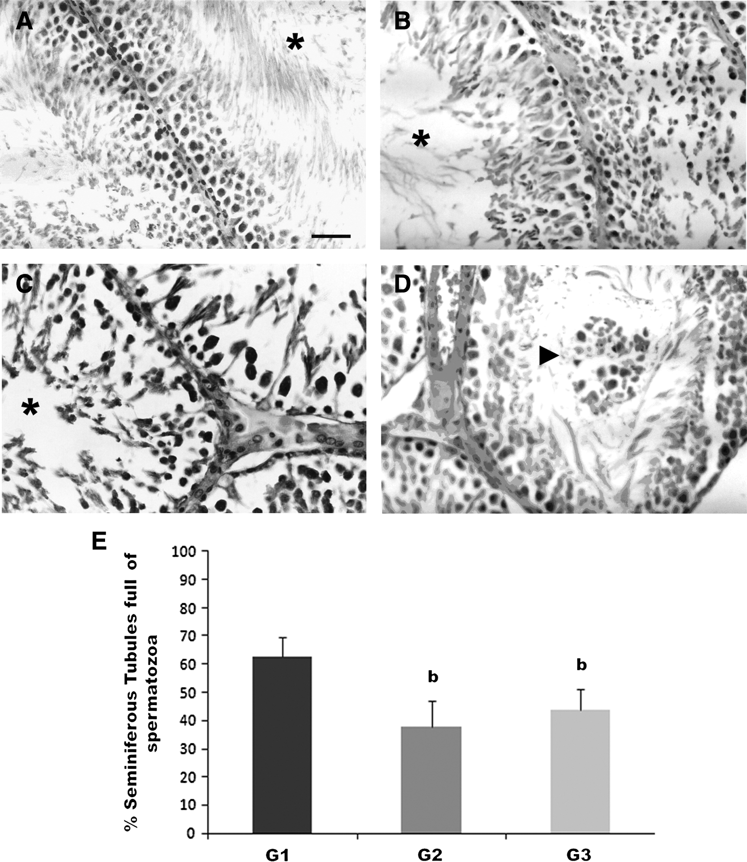
Histology of the rat testis (G×400).
To confirm this result, the percentage of full seminiferous tubules was determined on about 600 tubules for each group of rats. Figure 2E depicts a significant decrease in average percentage of seminiferous tubules full of spermatozoa in both groups 2 (−39.9%, P<.05) and 3 (−30%, P<.05) compared to the control group. The two groups of treated rats did not differ from each other.
Apoptotic effect
The seminiferous tubules of rats treated with garlic fractions showed a cell death process as shown by the TUNEL approach. TUNEL-positive cells were found in rat testis treated with the aqueous fraction (Fig. 3B) and alcoholic precipitate (Fig. 3C). These TUNEL-positive cells were mainly spermatocytes I and spermatids (Fig. 3B, C).
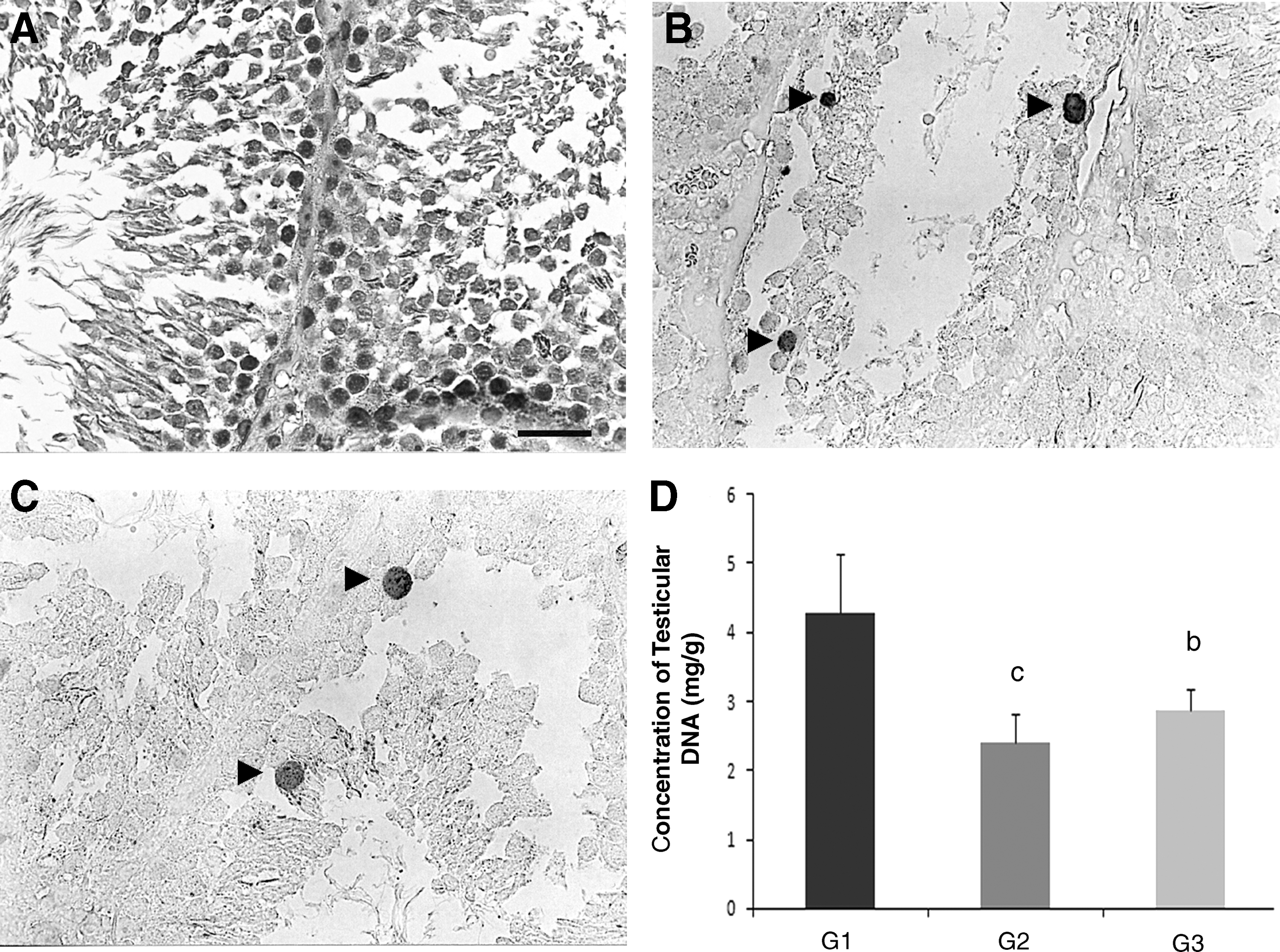
Apoptosis of germ cells induced by garlic fractions (TdT-mediated dUTP-X nick-end labeling [TUNEL]; G×400).
Testicular DNA concentration
As summarized in Figure 3D, the result of testicular DNA concentrations of all rats shows a significant decrease in testicular DNA concentrations of treated rats: −44% (P<.05) for group 2 and −30% (P<.05) for group 3 compared to control rats.
Discussion
In the entire history of human civilization, the medicinal properties of garlic (Allium sativum) have been evaluated and used in the treatment of a wide variety of human diseases, but very few is known about the potential effects of garlic on spermatogenesis. In a previous work, we showed that raw garlic consumption at doses 10%, 15%, and 30% induces disruption of rat spermatogenesis. 23 According to these results, we tried to identify the nature of active molecules in raw garlic responsible for the different modifications observed on testicular parameters. In the present study, we did not isolate the active molecules of garlic, but the garlic homogenate was split in an aqueous fraction (obtained by grinding and centrifugation) and an alcohol-precipitated fraction (obtained by ethanol precipitation from the aqueous solution).
The histological observations showed that chronic consumption of garlic fractions induces a reduction of spermatogenesis suggested by the decrease of seminiferous tubules full of spermatozoa. The seminiferous tubule sections of treated rats showed that the early stages of spermatogenesis are present, including spermatogonia, spermatocytes I, and a few spermatids. The large number of abnormal cells observed in the lumen of seminiferous tubules of rats treated with garlic fractions evoked an apoptotic process. This finding was confirmed by the technique of TUNEL, which demonstrated the DNA fragmentation, one of the best indicators of apoptosis. 26 We showed here that oral administration of garlic fractions induced germ cell death that targeted spermatocytes I and spermatids, whereas spermatogonia and somatic Leydig and Sertoli cells were not affected. The decrease of seminiferous tubule DNA concentrations of rats treated with the garlic fractions confirmed the germ cell death. Moreover, the two percentages of decrease (seminiferous tubules full of spermatozoa and DNA) were similar. Indeed, very few, if any, studies have reported such an apoptotic effect of garlic on nontumor cells.
Therefore, these results confirmed our previous study which demonstrated that feeding raw crude garlic induces the germ cell apoptotic death (spermatocytes I and spermatids only) via activation of caspase 3 and thus a decrease of seminiferous tubules full of spermatozoa. 23,25 Our findings are also in accordance with other studies. Dixit and Joshi reported a spermatogenesis arrest at the primary spermatocyte stage with 50 mg of garlic powder oral administration for 70 days. 22 These authors observed degenerative changes in seminiferous tubules with nuclear condensation of Leydig cells. Abdelmalik also described histological alterations of somatic (Sertoli, Leydig, and myoid cells) and germ cells after feeding crude garlic at 20% dose. 24
In this study, administration of garlic fractions at 7.5% resulted in decreased serum testosterone levels, as we previously showed, 23 suggesting that active molecule of garlic has an inhibitory effect on testosterone synthesis by inhibition of Leydig steroidogenic enzyme expression such as Star, Cyp11a1, Hsd3b5, and Hsd17b3. In this context, the decrease in serum testosterone observed in the present study might explain the decreased percentage of seminiferous tubules full of spermatozoa and the germ cell death via apoptotic way. It has long been known that human spermatogenesis is more sensitive to stress than that of rats, suggesting that concentrations lower than used in the present study (7.5%) might impair human male spermatogenesis, particularly in case of a long-term consumption, for example, to reduce cardiovascular risk or hypertension. In this study, both fractions of garlic were shown to have the same impact on testicular parameters, but the exact composition of these fractions was not determined and is probably complex. It is known that the aqueous extract of garlic contains mainly water-soluble constituents, including SAC, SAMC, and AM 13 ; as well as a variety of oil-soluble sulfur compounds, including allicin and its metabolites. 14 Probably, one or several compounds of this fraction may be responsible for impairment of spermatogenesis in this study. Allicin must be excluded because this molecule is not a biologically active component of garlic, 27 as it is unstable and quickly converted into stable and safe sulfur compounds such as vinyldithiins, ajoene, and sulfides and other constituents of garlic, and no studies reported the effect of allicin on spermatogenesis. One in vitro study has reported the impact of DATS, a metabolite of allicin, 16 on spermatozoa. It was showed that DATS has a spermicidal effect on rat, hamster, and human spermatozoa at dose 7.5 mg/mL. 21
The alcoholic precipitate, obtained by precipitation of the aqueous solution, induced the same modifications as the aqueous fraction. It was rich especially in proteins and adenosine, a nucleoside present in raw garlic and a potent inhibitor of platelet aggregation by inhibition of cyclooxygenase activity, suppression mobilization of intraplatelet Ca2+, and reduction the ability of platelets to bind to fibrinogen.
17
Therefore, two hypotheses may be suggested: (1) One or several molecules present in the aqueous fraction and precipitated with alcohol are responsible for the impairment of testis parameters. Here also, allicin cannot be considered because it is miscible in alcohol. (2) One or several proteins or adenosine, contained in the alcoholic fraction, are the agents responsible for the diminution of testosterone and histological alterations of seminiferous tubules.
In conclusion, this preliminary study does not identify the molecules responsible for impairment of spermatogenesis and metabolism of testosterone. Hence, appropriate methods of extraction, characterization, and identification (such as high-performance liquid chromatography) are needed to specify and characterize the exact molecules responsible for the observed alterations of testicular functions.
Footnotes
Acknowledgment
This work was supported by the Tunisian Ministry of Superior Education and Scientific Research.
Author Disclosure Statement
All authors declare that no competing financial interests exist.