
Abstract
Eucommia ulmoides Oliver is a perennial woody plant distributed widely in China. To characterize some major compounds in E. ulmoides bark extract, six compounds were identified via high-performance liquid chromatography qualitative analysis. E. ulmoides bark extract protects against cadmium-induced oxidative damage in rat kidneys. Two compounds of E. ulmoides bark extract, geniposide and genipin, which were identified both in serum and in kidney tissue, showed inhibitory effects on nitric oxide production. This study provides biological evidence supporting the usefulness of E. ulmoides bark against cadmium-induced toxic oxidative stress in rat kidney tissue.
Introduction
E
Kidney disease is a major health and social problem among all populations. The causes are many and include hypertension, inflammatory disease, diabetes, obesity, and toxic nephropathies from environmental and occupational hazards that include heavy metals, drugs, and chemicals. 16
Several reports have suggested that E. ulmoides is useful for renal protection. The polysaccharide fraction of E. ulmoides was confirmed as a major contributor to the anticomplementary activity. Treatment with polysaccharide fraction protected against glomerular injury from lupus-like syndrome in mice, with reduced immunoglobulin deposition and lowered proteinuria; the increased production of serum autoantibodies and total immunoglobulin G was also inhibited. 17 Another report demonstrated that administration of a mixture of Panax pseudoginseng and E. ulmoides for 8 weeks significantly decreased plasma malonyldialdehyde levels and the overall redox parameters together with a partial mitigation of proteinuria in streptozotocin-induced diabetic rats. Glomerular volume and nephrin and macrophage chemoattractant protein-1 gene expression were also decreased. 18 However, the mechanism and active compounds underlying the kidney-protective effect of E ulmoides remain unclear.
Therefore, we examined the kidney protection mechanisms of E. ulmoides bark using cadmium (Cd)-poisoned rats. Cd is an inorganic toxicant of great environmental and occupational concern, which is classified as a group I human carcinogen. The kidney is the critical target organ for Cd as documented by several studies in humans and animals. Cd can produce a variety of renal effects involving the proximal tubules and the glomerulus, which is believed to be irreversible at advanced stages. 19 Several studies have indicated that oxidative stress and reactive oxygen species formed in the presence of Cd could be responsible for its toxic effects in many organs or cells. 20
Protection against toxic actions of Cd can be accomplished through the antioxidant defense system. For example, naringenin, a naturally occurring plant bioflavonoid found in citrus fruits, showed protective effect against Cd-induced oxidative damage in the kidney of rats. The mechanisms contributing to its effectiveness involve the quenching of free radicals and antioxidant and metal-chelating ability. 21 Several natural herbs, such as onion and garlic extracts, dose-dependently protect against Cd-induced renal oxidative stress in male Wistar rats via decreased lipid peroxidation and enhanced antioxidant defense. 22 The aim of the present study was to investigate the ability of E. ulmoides bark to prevent Cd-induced renal oxidative stress in the rat.
Materials and Methods
Plant materials and extraction
E. ulmoides Oliver bark was purchased from Qixin Traditional Chinese Medicine Co. Ltd. (Anguo, China). Dr. Tianxiang Li, associate professor of the Tianjin University of Traditional Chinese Medicine, Tianjin, China, authenticated the plant material. A voucher specimen (number 20080603-1) was deposited at the Institute of Traditional Chinese Medicine, Tianjin University of Traditional Chinese Medicine. Dried bark of E. ulmoides was macerated using a commercial grinder. The bark mash (1 kg) was then extracted with 70% ethanol (10 L) twice under reflux. The combined 70% ethanol solution was concentrated by evaporation under vacuum at 40°C to obtain a crude extract that was used for downstream experimental work.
Chromatography
The reference standard compounds geniposidic acid, geniposide, syringaresinol diglucoside, genipin, (+)-pinoresinol-di-β-
Ten milligrams of E. ulmoides bark extract was dissolved in 10 mL of methanol/water (6:4, vol/vol) solution, ultrasonicated for 10 minutes, and centrifuged twice at 9168 g for 10 minutes, and the supernatant was transferred into a sample vial for HPLC analysis. HPLC analyses were performed on a Waters 2695 liquid chromatography system (Waters Corp., Milford, MA, USA), which consists of a binary pump, an autosampler, a column compartment, and an ultraviolet (UV) detector. Chromatographic separation was performed on a Diamonsil column (film thickness, 5.0 μm; 250×4.6 mm i.d.; Dikma Technologies, Lake Forest, CA, USA). The mobile phase consisted of acetonitrile (A) and water containing 0.1% acetic acid (B), with a gradient elution of 8–13% (vol/vol) A at 0–12 minutes and 13% A at 12–35 minutes. The flow rate was 1.0 mL/minute, and the column temperature was set at 30°C. UVD wavelengths were as follows: 0–19 minutes, 240 nm; 20–26 minutes, 277 nm; and 26–35 minutes, 240 nm. The injection volume was 10 μL.
Bioassay
Animals
Male Sprague–Dawley rats, 8 weeks of age and weighing 180–220 g, were purchased from Shanchuanhong Laboratory Animal Co., Ltd. (Tianjin). The animals were housed at a temperature of 23±2°C and were fed a standard laboratory chow (provided by The Institute of Laboratory Animal Science, Beijing) and water ad libitum. The animal rooms were well ventilated and had a regular 12:12-hour light/dark cycle throughout the experimental period. The experimental protocol was approved by the Experimental Animal Research Committee at the Tianjin University of Traditional Chinese Medicine.
Nitric oxide production inhibition
Peritoneal exudate cells were collected from the peritoneal cavities of male ddY mice by washing with 6–7 mL of ice-cold phosphate-buffered saline (PBS), and cells (5×10⁵ cells per well) were suspended in 200 μL of RPMI 1640 medium supplemented with 5% fetal calf serum, penicillin (100 units/mL), and streptomycin (100 mg/mL) and precultured in 96-well microplates at 37°C in 5% CO2 in air for 1 hour. Nonadherent cells were removed by washing the cells with PBS, and the adherent cells (more than 95% macrophages as determined by Giemsa staining) were cultured in fresh medium containing 10 μg/mL lipopolysaccharide (LPS) and test compound for 20 hours. Nitric oxide (NO) production in each well was assessed by measuring the accumulation of nitrite in the culture medium using Griess reagent. Cytotoxicity was determined by the 3-(4,5-dimethyl-2-thiazolyl)-2,5-diphenyl-2H-tetrazolium bromide (MTT) colorimetric assay. In brief, after a 20-hour incubation with test compounds, MTT (10 μL, 5 μg/mL in PBS) solution was added to the wells. After a 4-hour culture, the medium was removed, and isopropanol containing 0.04 M HCl was then added to dissolve the formazan produced by the cells. The optical density of the formazan solution was measured with a microplate reader at 570 nm. Each test compound was dissolved in dimethyl sulfoxide, and the solution was added to the medium (final dimethyl sulfoxide concentration, 0.5%). Inhibition (%) was calculated by the following formula, and the 50% inhibitory concentration was determined graphically (n=6):
where A, B, and C represent the
Animals and experimental design
After 1 week of acclimatization, 48 rats were divided into six groups (n=8) as follows: Group 1, normal group, pretreated with distilled water for 5 days, 0.5 mL/100 g, p.o.; Group 2, control group, pretreated with 5% acacia solution for 5 days, 0.5 mL/100g, p.o.; Group 3, positive control group, pretreated with vitamin E in 5% acacia solution for 5 days, 500 mg/kg, p.o.; Group 4, low-dose treatment group, pretreated with E. ulmoides extract in 5% acacia solution for 5 days, 125 mg/kg, p.o.; Group 5, medium-dose treatment group, pretreated with E. ulmoides extract in 5% acacia solution for 5 days, 250 mg/kg, p.o.; and Group 6, high-dose treatment group, pretreated with E. ulmoides extract in 5% acacia solution for 5 days, 500 mg/kg, p.o.
All groups were provided food and water ad libitum during the pretreatment period. One hour after the last pretreatment, rats from Groups 2–6 were intraperitoneally injected with CdCl2 in distilled water solution (2.5 mg/mL) at a dose of 1.0 mL/kg, whereas Group 1 received 1.0 mL/kg distilled water.
Blood samples were collected from the infraorbital venous plexus 2 hours after CdCl2 administration, and the animals were sacrificed under light ether anesthesia. The kidneys were removed, cleaned, and washed with PBS (pH 7.4). The kidney index was calculated by the following formula:
Biochemical analysis
Blood samples were placed in heparinized tubes and centrifuged at 3,000 g (Eppendorf, Hamburg, Germany) for 10 minutes to obtain sera that were used for biochemical variables studies. After removal of the kidneys, the left kidney was homogenized with PBS (10%, pH 7.4). The homogenate was centrifuged (1647 g, 10 minutes) at 4°C. The supernatant obtained was stored at –86°C in aliquots until used for the liquid chromatography–mass spectometry and enzyme biochemical analysis.
ATPase, reduced glutathione (GSH), catalase (CAT), creatinine, and blood urea nitrogen were determined using commercial kits (Nanjing Jiancheng Bioengineering Institute [Janjing, China] or Biosino Bio-technology and Science Inc. [Beijing]).
Liquid chromatography–mass spectrometry analysis of rat serum and renal tissue
Ethyl acetate (600 μL) was added to serum or kidney homogenate (100 μL) to precipitate protein and extract the analyte. After vortex-mixing (2×60 seconds) and centrifugation (17,968 g, 10 minutes at 4°C), an aliquot (0.5 mL) of the supernatant was evaporated to dryness under a nitrogen gas stream in a 40°C water bath. The residue was dissolved in 0.1 mL of acetonitrile-H2O (1:1, vol/vol). After centrifugation (17,968 g, 10 minutes), an aliquot of the supernatant (10 μL) was injected onto the HPLC column.
Mass spectra were acquired using an Agilent 6520 Q-TOF mass spectrometer (Agilent Corp., Santa Clara, CA, USA). Ultra-HPLC analyses were performed on an Agilent 1290 UHPLC instrument (Agilent, Waldbronn, Germany) coupled to a binary pump, a diode-array detector, an autosampler, and a column thermostat. The sample was separated on a Waters Acquity UPLC HSS T3 column (film thickness, 1.8 μm; 100×2.1 mm i.d.). The mobile phase consisted of CH3CN (solvent A) and H2O (containing 0.1% formic acid; solvent B). A gradient program was used according to the following profile: 0–2 minutes, 8% A; 2–2.01 minutes, 8–13% A; 2.01–15 minutes, 13% A; 15–15.01 min, 13–8% A; and 15.01–18 minutes, 8% A. The flow rate was 0.2 mL/minute, and the column temperature set at 35°C.
The Agilent 6520 Q-TOF mass spectrometer was connected to the Agilent 1290 UHPLC instrument via an electrospray ionization interface. The acquisition parameters were as follows: drying gas (N2) flow rate, 7.0 L/minute; temperature, 300°C; nebulizing, 30 psig; capillary, 4,000 V; fragmentor, 200 V; skimmer, 65 V; and octopole radio frequency (OCT RF), 750 V. Each sample was analyzed in both positive and negative ion mode to provide complementary information for molecular formulas and structural identification. Mass range recorded was m/z 100–1700.The acquisition parameters were as follows: collision gas, ultrahigh-purity He; ion spray voltage, 4.5 kV (positive mode), –4.5 kV (negative mode); sheath gas (N2), 50 arbitrary units; auxiliary gas (N2), 0 arbitrary units; capillary temperature, 300°C; capillary voltage, 9 V (positive mode), −10 V (negative mode); and tube lens offset voltage, 5 V (positive mode), −5 V (negative mode).
Histopathological studies in kidney
The kidney tissue was immediately collected after the blood was drained, fixed in 10% neutral formalin solution for 24 hours, and dehydrated with a series of ethanol solutions, from 75% to 100%, before being embedded in paraffin wax. Cross-sections 4 μm in thickness were cut, stained with hematoxylin and eosin dye, and mounted in a neutral deparaffinated xylene medium for microscopic observations.
Statistical analysis
Data are expressed as mean±SD values. All the grouped data were statistically analyzed with SPSS (Chicago, IL, USA) version 11.0. Significant differences between means were evaluated by one-way analysis of variance. P<.05 was considered to indicate statistical significance.
Results And Discussion
A sensitive HPLC method was developed for the simultaneous determination of six major compounds of the E. ulmoides bark extract. The recoveries of the analytes were in the range of 97.6–101.3%, the limits of detection were within 0.4 μg, and all the compounds showed good linearity (r≥0.9990) in a relatively wide concentration range. The intra-day relative SDs (%) ranged from 1.47% to 3.00%, and the inter-day relative SDs (%) were not higher than 3%. The contents of geniposidic acid, geniposide, syringaresinol diglucoside, genipin, (+)-pinoresinol-di-β-
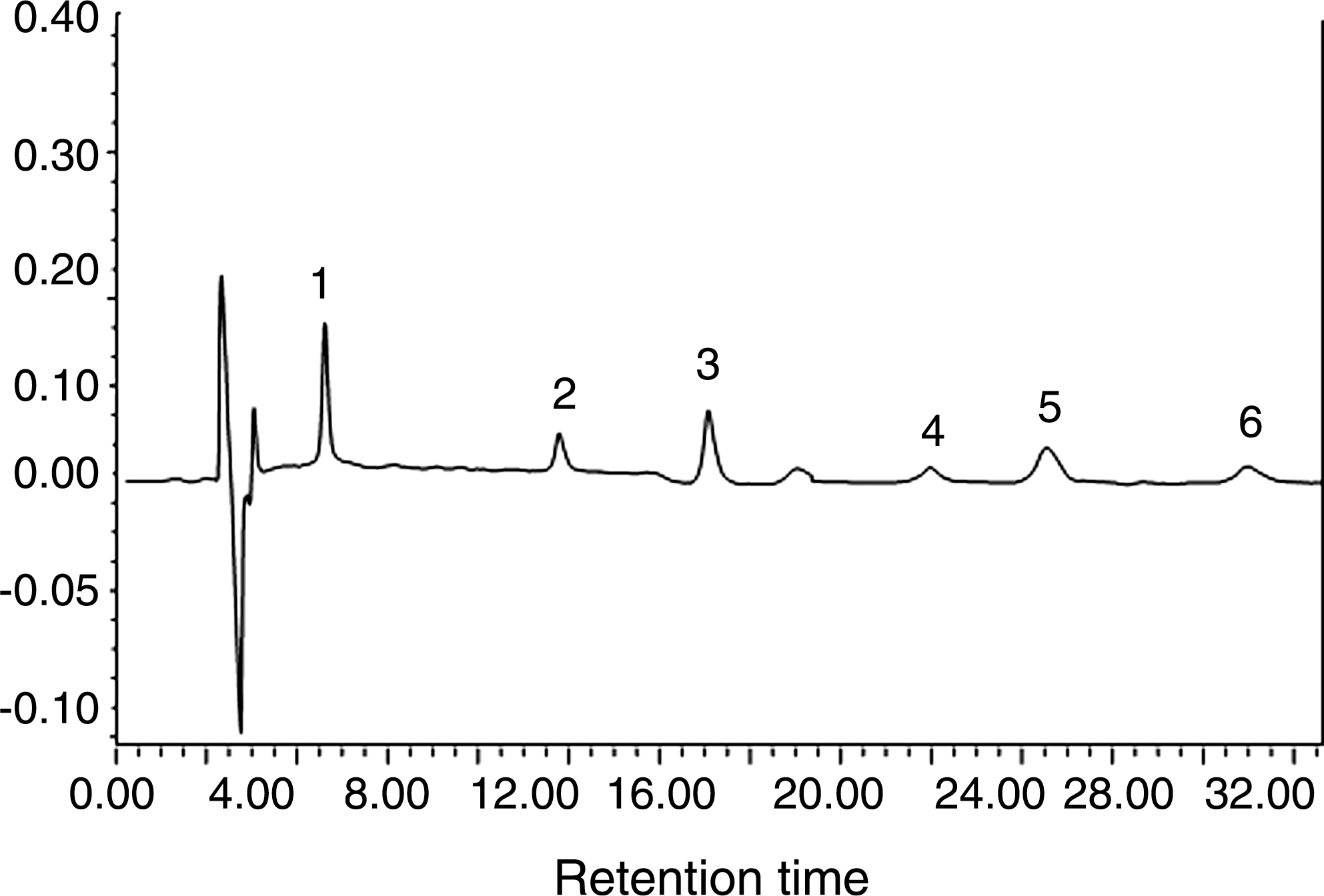
High-performance liquid chromatogram of E. ulmoides extract: peak 1, geniposidic acid; peak 2, chlorogenic acid; peak 3, geniposide; peak 4, (+)-pinoresinol-di-β-
We used a Cd-induced nephrotoxicity model to detect the kidney-protective effect of E. ulmoides extract. Cd is a heavy metal that is present in air, water, soils, and sediments. Soluble Cd salts can cause a number of lesions in many organs, including the liver, kidneys, testes, lungs, heart, and brain. The kidney is highly affected by Cd in both animals and humans, with a slow accumulation causing nephrotoxicity. 23 Acute Cd-induced toxicity may be due to the increase in oxidative stress, and Cd has been shown to induce lipid peroxidation. 24
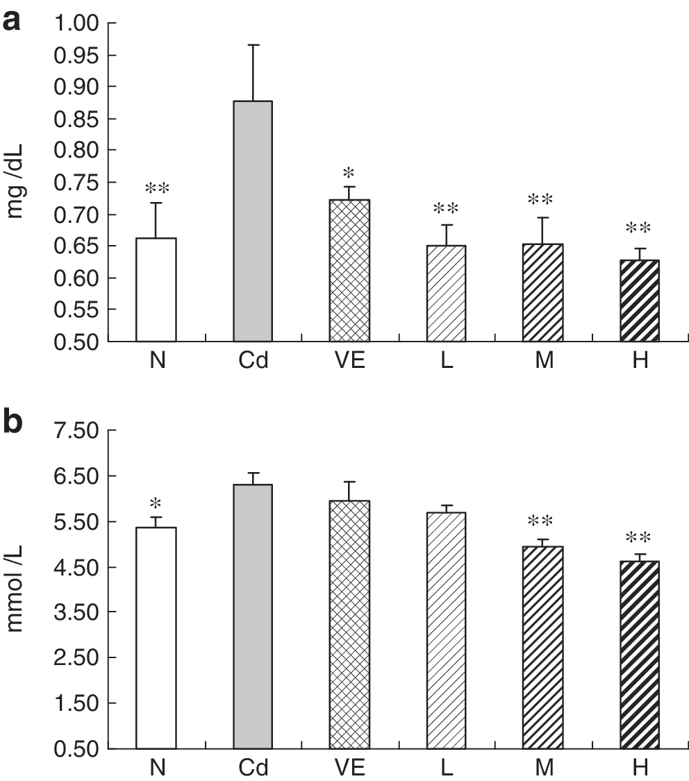
The effect of E. ulmoides bark extract on kidney index change of Cd-exposed rats is presented in Figure 2. During the experiment, the Cd-administered group had a slight decrease in the kidney index. The vitamin E group and the high-dose treatment group of E. ulmoides bark extract showed significant rescuing effects compared with the Cd-treated group.
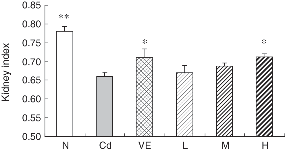
Effects of E. ulmoides extract treatments on kidney index in cadmium-exposed rats: N, normal group; Cd, cadmium-treated group; VE, vitamin E (500 mg/kg)+cadmium-treated group; L, 125 mg/kg E. ulmoides bark extract+cadmium-treated group; M, 250 mg/kg E. ulmoides bark extract+cadmium-treated group; and H, 500 mg/kg E. ulmoides bark extract+cadmium-treated group. The kidney index was calculated by the following formula: kidney index (%)=(weight of kidney/body weight)×100. Data are mean±SD values. *P<.05, **P<.01 for difference from the Cd group.
Renal GSH and CAT activities in kidney tissue are presented in Figure 3. GSH is highly sensitive to oxidative stress, playing a very crucial key role in cellular defense against Cd toxicity. The depletion of the renal level of GSH may be the consequence of enhanced GSH utilization to conjugate Cd and to counteract reactive oxygen species and lipid peroxidative products. 25 CAT is a common enzyme found in nearly all living organisms that are exposed to oxygen, where it functions to catalyze the decomposition of hydrogen peroxide to water and oxygen. 26 Kidney injury induced by Cd has been attributed to an excess of reactive oxygen species. This oxidative damage causes decreased activities of superoxide dismutase, CAT, and other enzymes involved in free radical scavenging. 27 Compared with the normal group, the Cd-treated rats showed significant decreases in renal GSH level and CAT activity. Treatment with the extract at the 125 mg/kg dose prevented the decrease in CAT activity and GSH level (Fig. 3).
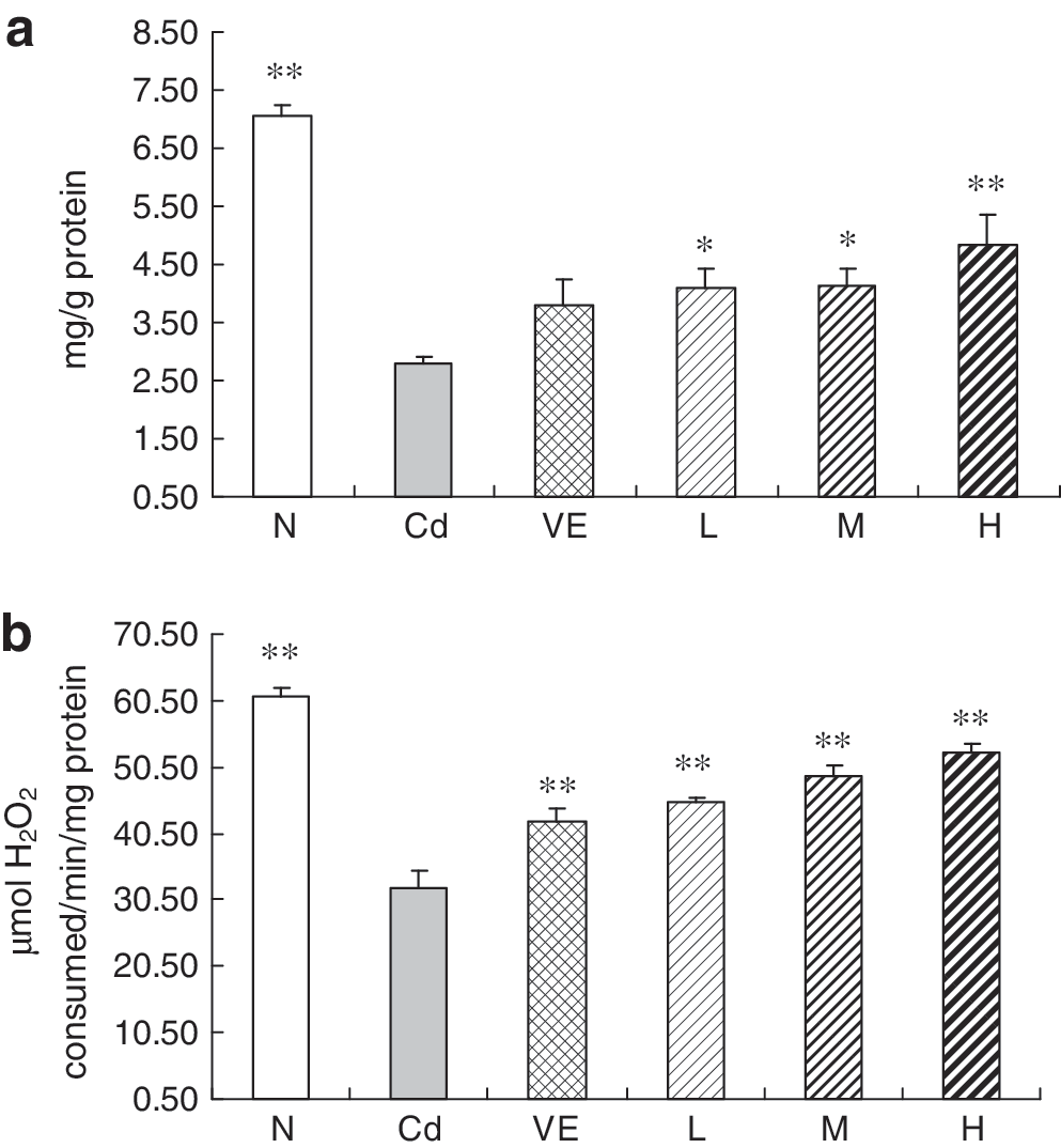
Effects of E. ulmoides extract treatments on level of renal
Na+,K+-ATPase is found in large amounts in the kidney, where it plays a key role in the active translocation of Na+ and K+ across this membrane. Na+,K+-ATPase is an important, complex membrane protein of polarized renal epithelial cells. It exchanges sodium for potassium and functions as the ubiquitous electrogenic sodium pump and is responsible for the active transport of sodium ions out of cells. Na+,K+-ATPase is very highly expressed in renal tubule epithelia, where it is restricted to the basolateral plasma membranes. 28 Inhibition of Na+,K+-ATPase by reactive oxygen species has been described in several tissues, such as heart, kidney, and brain. Na+,K+-ATPase is one cellular oxidative stress target and an important indicator of oxidant damage. The mechanism of free radical inhibition involves preventing increased lipid peroxidation products and lipid bilayer disordering. 29 Rats treated with Cd had significantly decreased renal Na+,K+-ATPase activity relative to the control rats (Fig. 4). The activity of renal Na+,K+-ATPase was significantly enhanced in rats treated with E. ulmoides bark extract in a dose-dependent manner (Fig. 4).
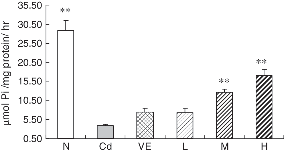
Effects of E. ulmoides extract treatments on renal Na+,K+-ATPase activity in cadmium-exposed rats. Data are mean±SD values. **P<.01 for difference from the Cd group. Pi, inorganic phosphate.
Blood urea nitrogen and creatinine are major measurements of renal function. Blood urea nitrogen is a marker for other nitrogenous wastes. Because urea is cleared from the bloodstream by the kidneys, a test measuring how much urea nitrogen remains in the blood can be used as a test of renal function. Creatinine is an anhydride of creatine that is abundant in muscle and excreted in the urine. Increased serum creatinine level and decreased creatinine clearance indicate renal failure. A significant increase in the level of blood urea nitrogen and creatinine in serum was observed in Cd-treated rats compared with normal rats. Administration of E. ulmoides bark extract (125 mg/kg) along with Cd significantly restored the levels of renal functional markers compared with the Cd-treated group (Fig. 5).
Effects of E. ulmoides extract treatments on
Kidney tissue distribution of E. ulmoides bark extract in rats was detected using a liquid chromatography–mass spectrometry method, as shown in Figure 6. Geniposide and genipin were observed in both serum and kidney tissues of rats with Cd-induced nephrotoxicity. However, geniposidic acid, syringaresinol diglucoside, (+)-pinoresinol-di-β-
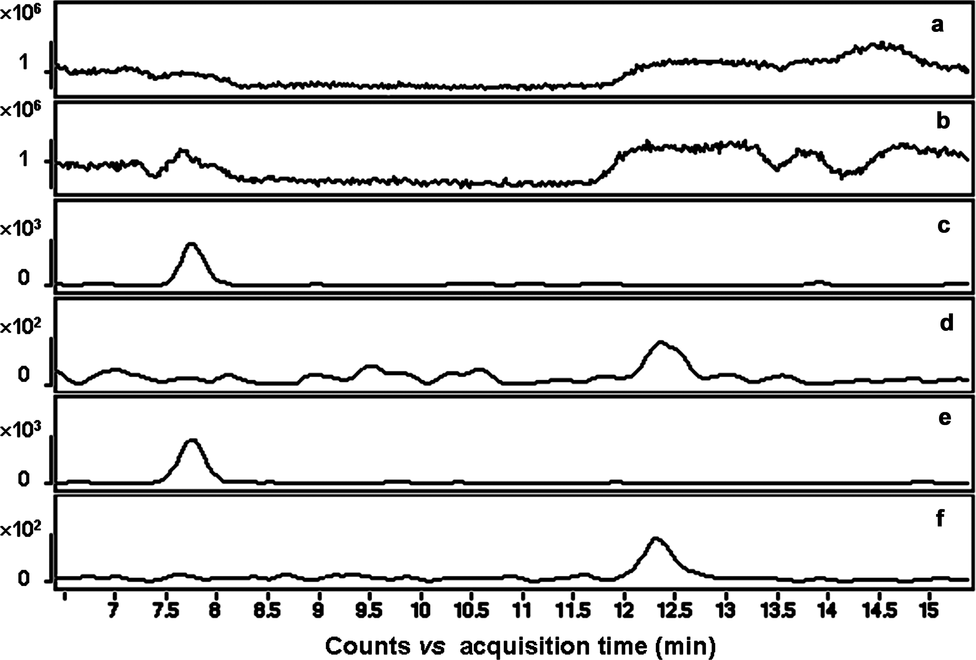
Liquid chromatography–mass spectrometry analysis of serum and kidney in cadmium-exposed rats.
Cd produces oxidative stress in different tissues by disturbing the pro-oxidant–antioxidant balance. 30 Oxidative stress appears to play a major role in chronic Cd-induced hepatic and renal toxicity because the inhibition of components of the antioxidant defense system accelerated the damage. Free radical damage to phospholipids is involved in the development of induced changes. Free radical scavengers and antioxidants are thought to be useful in protecting against Cd toxicity.
We also detected the antioxidant effects of the compounds that were observed in serum and renal tissue. NO has been implicated in physiological and pathological processes, such as vasodilation, nonspecific host defense, ischemia reperfusion injury, and chronic or acute inflammation. NO is produced by the oxidation of
Data are mean±SD values (n=6).
P<.05, ** P<.01 for difference from untreated group.
Histopathological studies showed that multiple foci of hemorrhage necrosis and cloudy swelling of tubules in the kidney were induced by Cd (Fig. 7b). According to microscopic examinations, pathological lesions induced by Cd were remarkably reduced by the administration of vitamin E (500 mg/kg) (Fig. 7c). There was little change in the kidney histology of 125 mg/kg and 250 mg/kg E. ulmoides bark extract–treated groups (Fig. 7d and e, respectively). However, pathological lesions induced by Cd were remarkably reduced by administration of 500 mg/kg E. ulmoides bark extract (Fig. 7f) compared with the control, a finding in agreement with the results of renal oxidative status.
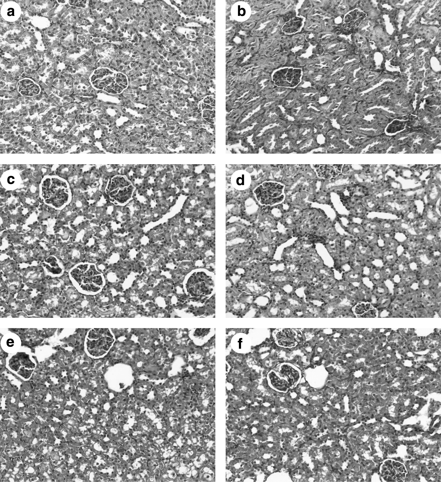
Representative photomicrographs of hematoxylin and eosin–stained histological sections of kidney from
Conclusions
In conclusion, this study demonstrates that E. ulmoides bark extract protects against Cd-induced oxidative damage in the kidney of rats. Geniposide and genipin were observed in both serum and in kidney tissue, where they inhibited NO production. The present study therefore provides biological evidence supporting the usefulness of E. ulmoides bark against Cd-induced toxic oxidative stress in rat kidney tissue.
Footnotes
Acknowledgments
This research was supported by the International Traditional Chinese Medicine Program for Corporation in Science and Technology (2008DFB30070), the Program for Changjiang Scholars and Innovative Research Team in University, the Tianjin Committee of Science and Technology, China (10SYSYJC28900), and the Important Drug Development Fund, Ministry of Science and Technology of China (2009ZX09311-002).
Author Disclosure Statement
The authors declare that there are no conflicts of interest.