
Abstract
Momordica charantia is used to treat various diseases, including inflammatory conditions. Previous reports indicated that the extract of this plant inhibits activation of nuclear transcription factor-κB (NF-κB) but activates peroxisome proliferator-activated receptor (PPAR). Additionally, cucurbitane-type triterpene glycosides are the main bioactive components of the fruit of M. charantia. Therefore, we investigated the anti-inflammatory activity of 17 cucurbitane-type triterpene glycosides
Introduction
N
Peroxisome proliferator-activated receptor (PPAR) is a member of the nuclear receptor superfamily of ligand-dependent transcription factors that is predominantly expressed in adipose tissue, 4 the adrenal glands, and the spleen. 5 Originally identified as an essential protein in glucose metabolism, PPARγ has since been shown to be important in cellular processes in endothelial cells, vascular smooth muscle cells, dendritic cells, and platelets. 5 It can dampen macrophage inflammatory responses through inhibition of pro-inflammatory molecules, such as tumor necrosis factor-α (TNFα), interleukin (IL)-6, and iNOS. Developing specific inhibitors of COX-2 and iNOS with lower toxicity and higher anti-inflammatory activity in therapeutic applications is of great interest.
Bitter melon, the fruit of Momordica charantia L. (Cucurbitaceae), a traditional herbal medicine, is used worldwide. It is widely used as a bitter stomachic, a laxative, an antidiabetic agent, an anti-inflammatory agent, and an anticancer agent and in the treatment of rheumatoid arthritis. 6 Herbal drugs have potential therapeutic application because of their effectiveness, lesser side effects, and relatively low cost. Therefore, investigation of agents from traditional medicinal plants has become more important, and researchers are competing to find new, effective, and safe therapeutic agents for the treatment of cancer, inflammatory disease, and diabetes. Bitter melon has various documented anti-inflammatory effects. It reduces prostaglandin E2, IL-1β, IL-6, IL-7, TNFα, and lipopolysaccharide levels 7 –9 and increases the secretion of transforming growth factor-β, IL-10 in RAW 264.6 macrophages, Caco-2 cells, and THP-1 cells. 8,10 Numerous cucurbitane-type triterpenes and their glycosides have been isolated from the roots, 12 fruits, 13 –23 seeds, 24,25 leaves, and vines 26 –29 of this plant. However, the anti-inflammatory activity of these cucurbitane-type triterpene glycosides has not been evaluated and reported. Recently, one compound with the same skeleton, 23,24-dihydroxycucurbitacin D from the plant Bryonia alba L., was reported to exert anti-inflammatory activity by inhibiting NO generation through blocking NF-κB activation and iNOS gene transcription. 30 Additionally, in previous studies, we isolated and identified 17 compounds from the methanol extract of M. charantia fruits, six of which were new and eleven were known. 31,32
Here we continued our previous work in screening active compounds for their effects against inflammation and type 2 diabetes. Their inhibitory effects on NF-κB activation and iNOS and COX-2 expressions as well as transactivations of a PPAR response element (PPRE) and members of the PPAR family (PPARα, PPARβ/δ, and PPARγ) were also evaluated. This study provides new insights into the ways in which bitter melon modulates metabolic functions in human HepG2 cells.
Materials and Methods
General experimental procedures
The optical rotation was determined on a Jasco (Easton, MD, USA) model DIP-370 digital polarimeter. Electrospray ionization mass spectra were obtained using an Agilent (Palo Alto, CA, USA) model 1200 LC-MSD Trap spectrometer. High-resolution electrospray ionization mass spectra were obtained using a JEOL (Tokyo, Japan) model JMS-T100LC spectrometer. The 1 H-nuclear magnetic resonance (600 MHz) and 13C-nuclear magnetic resonance (150 MHz) spectra were recorded on a JEOL model ECA 600 spectrometer, and trimethylsilane was used as an internal standard. Gas chromatography was performed on a Shidmazu (Kyoto, Japan) model 2010 instrument. Column chromatography was performed using silica gel (Kieselgel 60, 70–230 mesh and 230–400 mesh; Merck, Darmstadt, Germany) and YMC (Fujisilisa Chemical Ltd., Kasugai, Aichi, Japan) RP-18 resins.
Plant material
The fruit of M. charantia was collected in Vuthu, Thaibinh Province, Vietnam, in June 2009 and identified by Dr. Ninh Khac Ban, Institute of Marine Biochemistry, Vietnam Academy of Science and Technology, Hanoi, Vietnam. A voucher specimen (IMBC MC0609) was deposited at the herbarium of the Institute of Marine Biochemistry, Vietnam Academy of Science and Technology.
Cell culture and reagents
Human hepatocarcinoma HepG2 cells were maintained in Dulbecco's modified Eagle's medium (Invitrogen, Carlsbad, CA, USA) containing 10% heat-inactivated fetal bovine serum, 100 units/mL penicillin, and 10 μg/mL streptomycin at 37°C and 5% CO2. Human TNFα was purchased from ATgen (Seoul, Korea). Cells were counted with a hemocytometer, and the number of viable cells was determined through trypan blue dye exclusion.
Cytotoxicity assay
A 3-(4,5-dimethylthiazol-2-yl)-5-(3-carboxymethoxyphenyl)-2-(4-sulfophenyl)-2H-tetrazolium, inner salt (MTS) assay (CellTiter 96® AQueous One Solution Assay, Promega, Madison, WI, USA) was performed to analyze the effect of the different compounds on cell viability. Cells were cultured overnight in 96-well plates (1×10⁴ cells per well). Cell viability was assessed after the incubation with the compounds at a concentration of 5 μM for 24 h. The number of viable cells was determined by measuring the absorbance at 490 nm of the dissolved formazan product after addition of MTS for 30 min as described by the manufacturer.
Luciferase assay
Cells were seeded at 1.5×10⁵ cells per well in a 12-well plate and grown for 24 h. All cells were transfected using Lipofectamine™ LTX (Invitrogen) according to the manufacturer's protocol. Luciferase (Luc) activity was assayed using an LB 953 Autolumat (EG&G Berthold, Nashua, NH, USA) as described previously. 33 NF-κB-Luc and PPRE-Luc plasmids were kindly provided by Dr. Kyoon E. Kim (Chungnam National University, Daejeon, Korea). All experiments were performed in triplicate.
Reverse transcription–polymerase chain reaction
Total RNA was extracted from cells using easy-BLUE™ (iNtRON Biotechnology, Seoul). Approximately 2 μg of total RNA was reverse-transcribed using Moloney murine leukemia virus reverse transcriptase and oligo(dT) primers (Promega) for 1 h at 42°C. The resulting cDNA was polymerase chain reaction–amplified using Taq polymerase premixture (TaKaRa, Shiga, Japan). Polymerase chain reaction products were subjected to electrophoresis on 1% agarose gels and stained with ethidium bromide. Polymerase chain reaction was conducted with the following primer pairs: iNOS sense 5′-TCATCCGCTATGCTGGCTAC-3′, iNOS antisense 5′-CTCAGGGTCACGGCCATTG-3′, COX-2 sense 5′-GCCCAGCACTTCACGCATCAG-3′, COX-2 antisense 5′-GACCAGGCACCAGACCAAAGACC-3′, glyceraldehyde 3-phosphate dehydrogenase sense 5′-TGTTGCCATCAATGACCCCTT-3′, and glyceraldehyde 3-phosphate dehydrogenase antisense 5′-CTCCACGACGTACTCAGCG-3′. The specificity of the products generated using each set of primers was examined using gel electrophoresis and further confirmed using a melting curve analysis.
PPAR subtype transactivation assay
The pFA-Gal4-PPARα ligand binding domain (LBD), pFA-PPARβ/δ LBD, and pFA-PPARγ LBD expression plasmids were provided by Dr. Young Yang (Sookmyung Women's University, Seoul). HepG2 cells were seeded at 1.5×10⁵ cells in 12-well plates and grown for 24 h. All cells were transiently co-transfected with one of the expression vectors for pFA-Gal4-PPAR LBDs together with pFR-Luc using the WelFect-M™ GOLD transfection reagent (WelGENE, Seoul) as described by the manufacturer. Following a 24-h incubation, the cells were treated with various concentrations of compounds and incubated for 20 h. Luc assays were performed using a dual-Luc reporter assay system according to the instructions of the manufacturer (Promega, Sunnyvale, CA, USA), and the activity was determined in a microplate luminometer (Centro LB 960, EG&G Berthold, Bad Wildbad, Germany) by measuring light emission for 5 s.
Statistical analysis
All results are expressed as mean±SD values. Data were analyzed by one-factor analysis of variance. Quantification of polymerase chain reaction products was performed using Image Lab™ software (Bio-Rad). If a statistically significant effect was found, the Newman–Keuls test was performed to isolate the difference between the groups. P<.05 was considered to be significant.
Results
Chemistry
Seventeen compounds (

Structures of cucurbitane-type triterpene glycosides
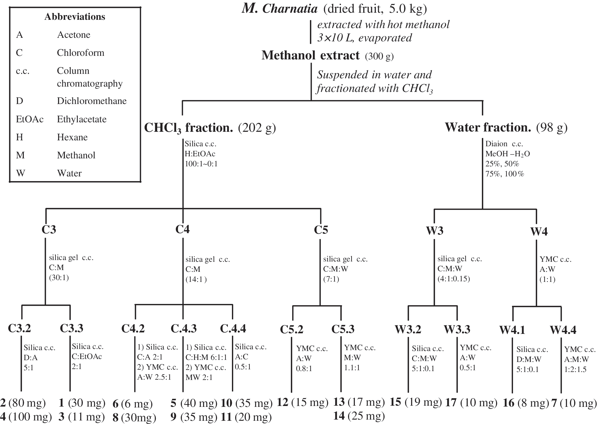
Scheme of extraction of cucurbitane-type glycosides
Biological activities
In our continuing study, the methanol extract of bitter melon has displayed the ability to markedly reduce prostaglandin E2-, IL-1β–, IL-6–, IL-7–, and lipopolysaccharide-stimulated TNFα levels and increased PPARα and PPARγ expression. 11 Thus, the 17 compounds listed above were further evaluated for their effects on NF-κB activation, iNOS and COX-2 expressions, and PPRE, PPARα, PPARβ/δ, and PPARγ transactivations.
To investigate cellular toxicity of the 17 isolated compounds, they were applied at various concentrations to HepG2 cells for 24 h, after which cell viability was measured in an MTS assay as described in Materials and Methods. None of the compounds displayed any cellular toxicity at the concentration of 5.0 μM (data not shown). They were therefore used in subsequent experiments at concentrations of 0.05, 0.5, and 5.0 μM.
Effect of compounds 1–17 on inhibition of NF-κB activation
To evaluate the anti-inflammatory activity of 17 compounds listed above, we first examined their inhibitory effects on NF-κB transcriptional activation in HepG2 cells (Fig. 3). Cells were treated with compounds at various concentrations prior to stimulation with TNFα (10 ng/mL). Compounds

Effects of compounds
Sulfasalazine was used as positive control compound.
IC50, 50% inhibition concentration; ND, not determined.
Effects on iNOS and COX-2 expression
Seven compounds (
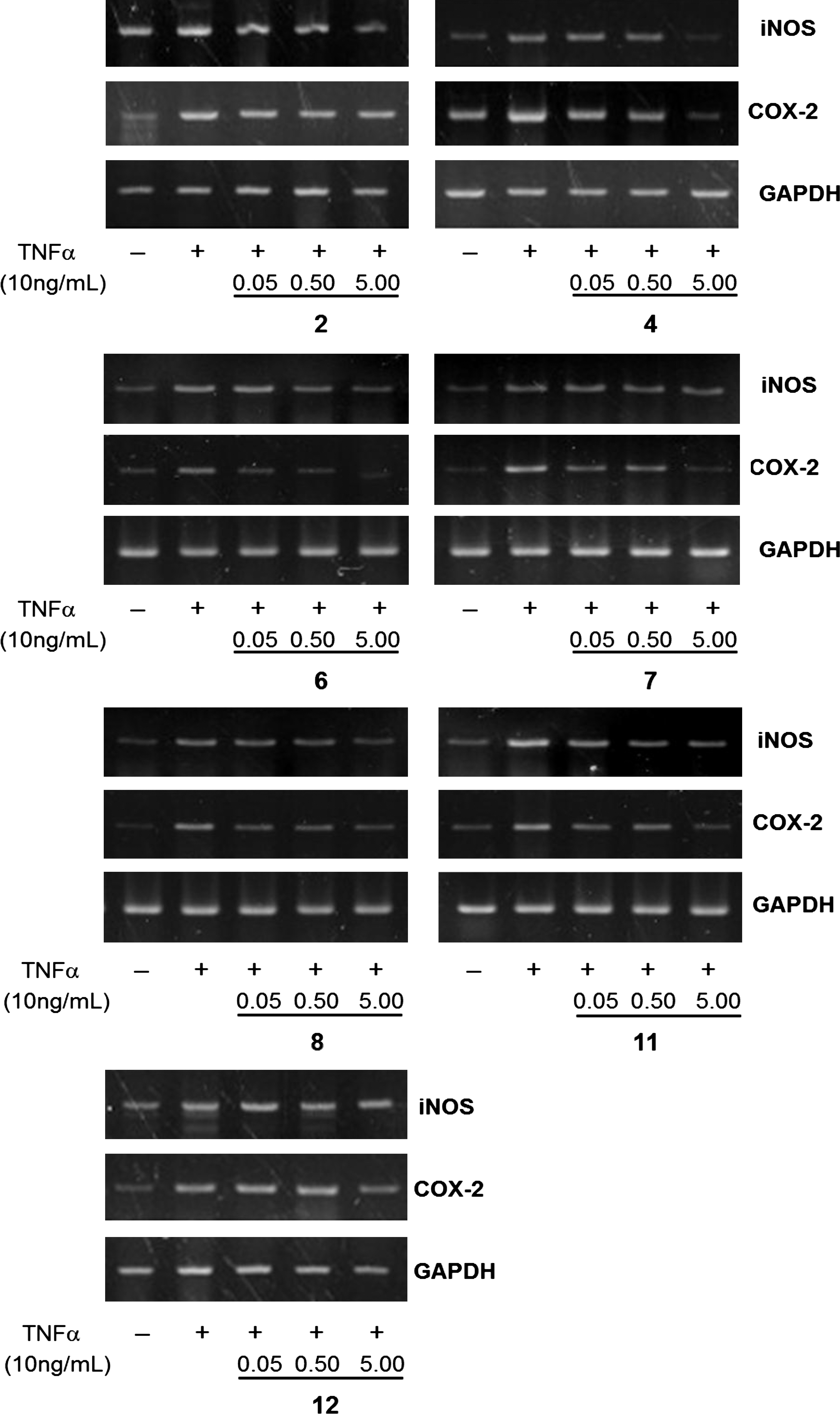
Inhibitory effects of compounds
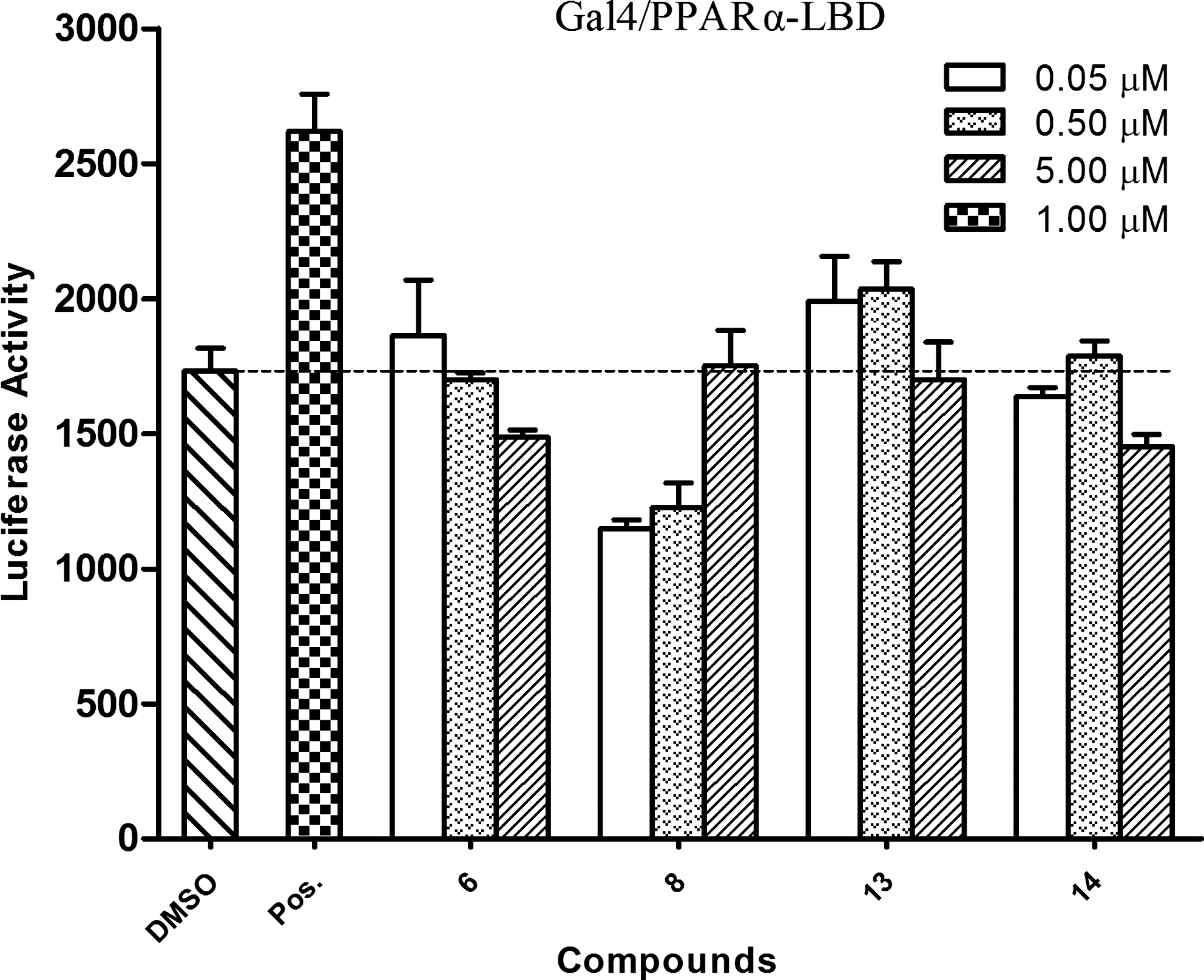
PPAR
To study the modulation of PPAR activity by compounds

Effects of compounds
Effects of compounds
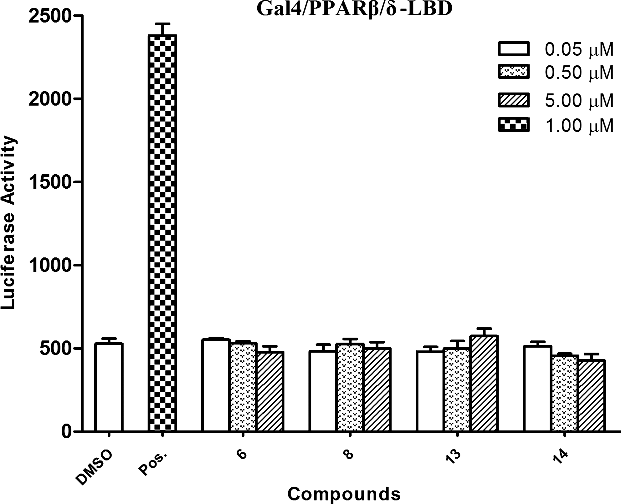
Effects of compounds
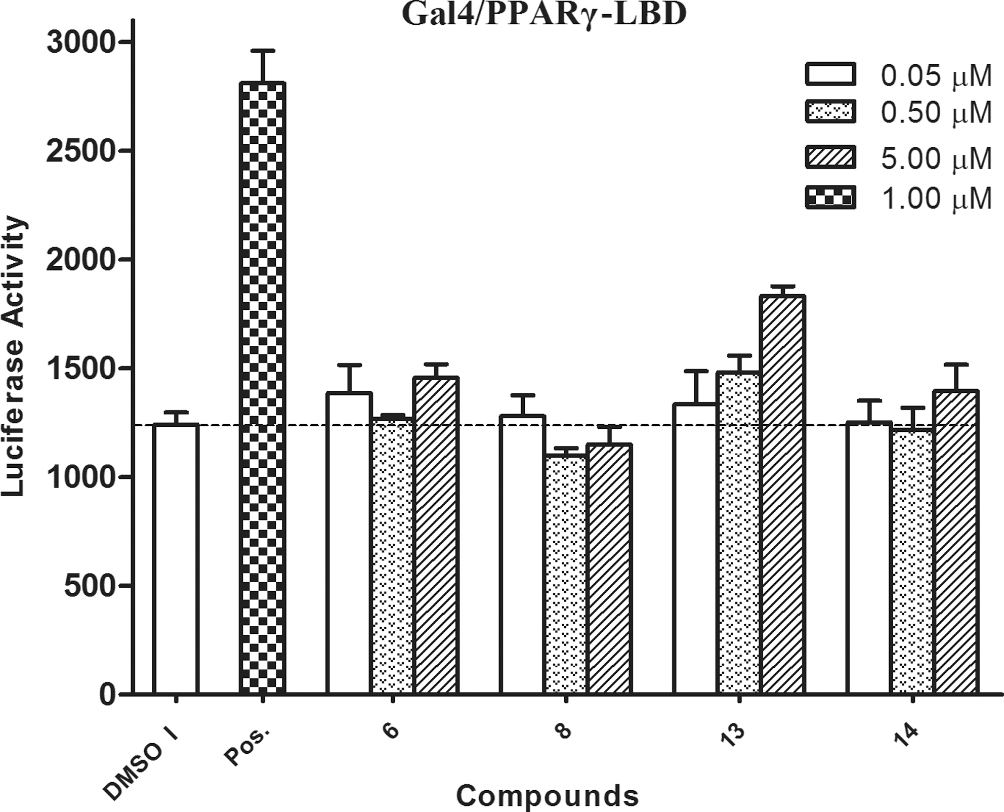
Effects of compounds
Discussion
The pharmacological properties of local food plants are an attractive area for investigation. Accumulating evidence suggests that many locally consumed foods have functional properties, notably anti-inflammatory activity.
34
The results of the present study demonstrate the inhibitory effects of the tested compounds on NF-κB transcriptional activation in HepG2 cells stimulated with TNFα. Compounds
In a previous report, the ethyl acetate extract of M. charantia was found to activate both PPARα and PPARγ.
11
However, the effects of the cucurbitane-type triterpene glycosides
In conclusion, the current study is a preliminary in vitro result of cucurbitane-type triterpene glycosides from bitter melon, which requires further investigations using in vivo and human studies to prove the efficacy.
Footnotes
Acknowledgments
This work was supported by the Priority Research Centers Program through the National Research Foundation of Korea funded by the Ministry of Education, Science and Technology (grant 2009-0093815). The authors would like to thank the Korean Basic Science Institute for performing the nuclear magnetic resonance and mass spectrometry experiments.
Author Disclosure Statement
No competing financial interests exist.