
Abstract
The present study was conducted to evaluate the effect of a fraction of macroporous resin (FMR), a bioactive component of Smilax china L., on benign prostatic hyperplasia (BPH) in castrated rats induced by testosterone propionate. Rats were randomly divided into five groups: the negative control group (sham-operated), the model group, two FMR-treated groups (at doses of 300 mg/kg and 600 mg/kg of body weight), and the positive control group (treated with finasteride at the dose of 3 mg/kg). Drugs were administered once a day for three consecutive weeks by gastric gavage. Prostates were weighed, testosterone and dihydrotestosterone (DHT) levels in serum were determined, and histopathological examinations were carried out. FMR treatment inhibited prostatic hyperplasia, reducing the DHT level in serum and improving the prostate gland morphology compared with the model group. The overall results of this study suggest that FMR is effective at inhibiting experimentally induced prostate enlargement, and it presents a valuable resource for the treatment of human BPH.
Introduction
B
The general treatment of BPH includes drug therapy and surgical treatment. By comparison, drug treatment has more advantages and has been considered as a preferable method for older men. Recently, some medicines, like α-1-adrenoceptor antagonist and 5α-reductase inhibitor, 6,7 are very popular in the treatment of BPH. However, the side effects caused by these drugs should not be disregarded. For example, finasteride, a synthesized 5α-reductase inhibitor, can cause side effects such as gynecomastia and severe myopathy because of its similar structure to that of the steroidal hormones. 8,9 In recent years, herbal therapies have become increasingly popular. The search for improved drugs from natural sources is increasing because natural products may have fewer adverse effects. 10 –15
Smilax china L., known as “Ba Qia” or “Jin Gang Teng” in China, has been long used as a traditional Chinese medicine. S. china L. was entered into the Chinese Pharmacopoeia in the 2010 version. 16 It has many pharmacological effects, such as anti-inflammatory, antinociceptive, and anticancer activities. 17,18 In China, it has been extensively used as a diuretic agent and for clinical treatment of rheumatic arthritis, acute bacillary dysentery, chronic nephritis, cancer, and inflammatory diseases. 19,20 In some areas of China, like Chongqing, it is also eaten as a food. 21
Because of the remarkable therapeutic effect of S. china L., research into its potential clinical application and pharmacological actions has increased during the past decade. A previous study has reported screening for activity against prostatitis of four fractions from S. china L. rhizome, and the fraction of macroporous resin (FMR) showed the greatest activity. 22 FMR also showed anti-inflammatory and antimicroorganism effects. 23 For the purpose of examining its pharmacological actions on other prostate-related disease, in this study, its activity in treatment of BPH in the prostatic hyperplasia rat model was investigated by using the preparation of FMR.
Materials and Methods
Chemicals and reagents
Finasteride tablets (5 mg per tablet) were purchased from Merck Sharp & Dohme Ltd., and testosterone propionate (25 mg/mL) was provided by Shanghai Tongyong Medical Co., Ltd. Enzyme-linked immunosorbent assay kits for determination of testosterone and DHT were supplied by R&D Systems.
Plant material
S. china L. rhizomes were purchased from Wuhan Humanwell Healthcare (Group) Co., Ltd., and identified by Prof. Changgong Zhang (College of Pharmacy, Tongji Medical College of Huazhong University of Science and Technology). A voucher specimen (BQ201006) was preserved and deposited for reference in our laboratory.
Preparation of FMR and quality analysis
According to the procedures described by Shu et al., 24 FMR was prepared as follows: the air-dried and powdered rhizomes of S. china L. (2 kg) were extracted with 50% ethanol (2×10 L) under reflux for 2 h, and extraction was repeated three times. The resulting solvent was eliminated under reduced pressure to obtain a dried extract, with a yield of 13.3 g/100 g of the starting crude material. The dried extract (100 g) was dissolved in 20% ethanol (1.5 L) and filtered, the pH was adjusted to about 4.5, and then NaCl was used to bring the solution to its final concentration at 1.5 mol/L. The extract solution was subjected to HPD100 macroporous resin (Cangzhou Bon Adsorber Technology Co., Ltd., Cangzhou, China) column chromatography (water→70% ethanol→95% ethanol) to give three fractions. The 70% ethanol fraction was used as FMR.
According to previous studies, total saponins are the main constituents of FMR, and their contents were determined as described by Baccou et al. 25 with some modification. In brief, 30 mg of FMR was dissolved in 5 mL of hot water, and the solution was extracted by 20 mL of butanol; the extraction was repeated three times. The extract solution was concentrated to dryness in vacuo. Then the extract was dissolved with 25 mL of methanol and diluted to 40 μL to a new test tube. The methanol was eliminated by heating at 70°C, and then 5% vanillin (0.2 mL) and perchloric acid (0.8 mL) were added to the test tube. The tube was maintained in a 70°C water bath for 15 min to develop color completely and then put in an ice bath for 3 min to stop the reaction, and lastly 5 mL of glacial acetic acid was added to the tube. The absorbance of the resulting solution was measured at 415 nm with a model 756MC spectrophotometer (Shanghai Precision & Scientific Instrument Co., Ltd., Shanghai, China). A standard curve was prepared from diosgenin using the above method. On the basis of the standard curve, the content of total saponins was calculated.
Fifty milligrams of FMR was dispensed in 30 mL of 2 M HCl solution to hydrolyze for 3 h, and then the solution was extracted three times each by 50 mL of petroleum ether. The extract solution was concentrated to dryness in vacuo, and the residue was dissolved with 20 mL of methanol for the following determination of diosgenin in FMR. The content of diosgenin in FMR was determined by high-performance liquid chromatography with ultraviolet detection. A high-performance liquid chromatography system (Hitachi, Hitachi High-Technological Corp., Japan) equipped with a model L-2130 pump, an online solvent vacuum degasser, a model L2400 ultraviolet detector, and a manual injector with a 20-μL injection loop was used. Chromatographic separations were carried out on an Amethyst C18–P chromatography column (Sepax Technologies Inc.). The column temperature was maintained at 25°C. The mobile phase was a mixture of H2O–CH3CN (10:90, vol/vol). The flow rate was 1.0 mL/min. The ultraviolet detector was set at a wavelength of 205 nm.
The methanol stock solution of diosgenin was prepared at the concentration of 1.00 mg/mL. A series of standards were prepared by diluting proper aliquots of the stock solution with methanol. The calibration curve for diosgenin was drawn by simple linear regression of its concentration versus its peak area, and the calibration curve was used to calculate the content of diosgenin in FMR.
Animals and treatments
All experiments were conducted under the European Community guidelines for the use of experimental animals and approved by the Animal Care and Use Committee of Huazhong University of Science and Technology. In total, 50 male Sprague–Dawley rats, weighing 200±20 g each, were purchased from the Research Center of Laboratory Animals, Tongji Medical Center, Huazhong University of Science and Technology, Wuhan, China. The animals were housed under controlled conditions (12-h/day controlled lighting, maintained at 25°C and 55–75% air humidity) and fed with normal rat chow and water ad libitum. All animals were allowed to acclimate for 7 days prior to the first treatment.
Forty male Sprague–Dawley rats were castrated as described by Van Coppenolle et al. 26 Chloral hydrate (350 mg/kg of body weight) was used for anesthesia. Castration was performed through the scrotum sack, cutting open the scrota and then taking out the testicles. Another 10 rats received sham operation, compared with the castrated rats: an incision was made in the same area, but without taking out the testicles, and they served as the control group. After operation, all rats were treated with penicillin for 3 days to prevent infection. After 7 days for recovery, the first drug treatment was begun.
The 10 sham-operated rats, which served as the control group, had olive oil injected subcutaneously once a day, and all the castrated rats had testosterone propionate (0.5 mg/0.1 mL per rat) dissolved in olive oil injected subcutaneously each day to create prostatic hyperplasia. 27 The 40 castrated rats were divided randomly into four groups, with 10 rats each: one model group, which was administered 0.5% sodium carboxymethylcellulose; two FMR groups, which were administered FMR at doses of 300 or 600 mg/kg of body weight; and one positive control group, which was treated with finasteride at the dose of 3 mg/kg of body weight. The drugs were administered using a stomach tube once daily for 3 weeks. All rats were weighed every 5 days during the experiment.
Blood and tissue samples
After 3 weeks of treatment, all rats were deprived of food for 18 h, weighed, and sacrificed. Blood was collected and allowed to clot, and serum was separated at 2000 g for 15 min and used for determination of testosterone and DHT by enzyme-linked immunosorbent assay kits. The prostate was removed from each rat and weighed to get the wet prostatic weight index. Then, all specimens were fixed in 10% formalin for pathological examination.
Statistical analysis
Data were expressed as mean±SD values. Results were analyzed statistically by one-way analysis of variance followed by Dunnett's post hoc test. Differences were considered statistically significant at P<.05.
Results
Quality analysis of FMR
The content of total saponins in FMR was expressed as diosgenin equivalent in milligrams per gram. The FMR was found to contain 187.2±8.67 mg/g of total saponins. The percentage content of diosgenin in FMR was determined as 1.07%.
Effect of FMR on prostate weight
FMR was well tolerated by all rats and during the entire experiment, with no side effects. As shown in Table 1, after injection of testosterone propionate for 3 weeks, the ratios of prostate weight-to-body weight were consistently higher than in control rats (P<.01), especially in the model group, where the ratio was nearly twice that of sham group rats. This result indicates that prostatic hyperplasia was successfully induced by subcutaneous injection of testosterone propionate after castration. As also can be seen, in the groups treated with finasteride (3 mg/kg of body weight) and FMR (600 or 300 mg/kg of body weight), the indices of prostate weight were reduced by 32.2%, 27.4%, and 18.6%, respectively, compared with the model group.
Data are mean±SD values (n=10). The index of prostatic weight was defined as (prostatic weight/body weight)×10−3).
P<.01 compared with control group; b P<.05, c P<.01 compared with model group.
FMR, fraction of macroprous resin.
Effect of FMR on concentrations of testosterone and DHT in serum
The effects of FMR on the concentration of testosterone and DHT in serum are presented in Table 2. DHT concentrations in all drug-treated groups were significantly decreased compared with the model group. Testosterone concentrations were not significantly different between the BPH model group and the drug-treated groups.
Data are mean±SD values (n=10).
P<.01, compared with control group; b P<.01 compared with model group.
DHT, dihydrotestosterone.
Histopathological examination
The rat prostate gland morphology in the control group had no obvious changes, but for the model group, the prostatic epithelial cells presented the typical features of glandular hypertrophy: papillary fronds protruded into the gland cavities, the prostatic epithelial height was increased notably, and the gland lumen diameter was significantly smaller (Fig. 1). Compared with the model group, the rat prostate gland morphology in the FMR-treated groups showed some improvement, especially in the FMR (600 mg/kg)-treated group (the prostatic epithelial height was reduced markedly, whereas the gland lumen volume was increased significantly).
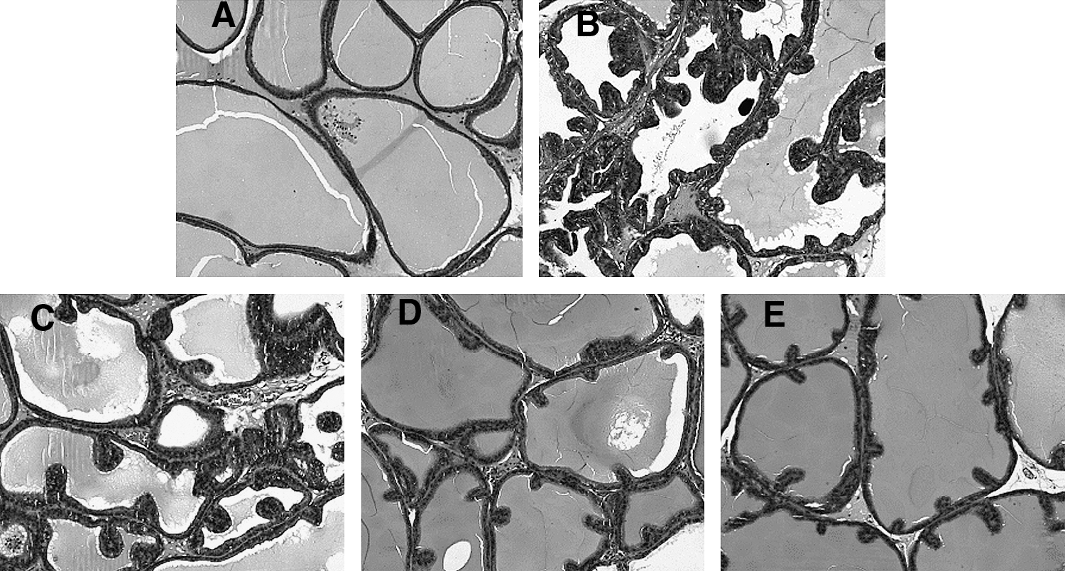
Effect of S. china L. FMR on histopathological changes in testosterone propionate–induced prostatic hyperplasia:
Discussion and Conclusions
BPH is one of the most common diseases among aged men, and its lower urinary tract symptoms reduce the quality of daily life. 28,29 Safe and effective natural interventions that reduce the symptoms and reverse or halt the progression of BPH have been the subject of considerable research interest. In this study, we demonstrated that FMR, a bioactive fraction from the rhizomes of S. china L., effectively mitigated testosterone propionate–induced BPH in a prostatic hyperplasia rat model, as evidenced by lower prostate weights and histopathological indices.
Tissue androgens stimulate prostate enlargement. Testosterone and DHT are involved in the progression of BPH. By contrast, DHT, which is converted from testosterone by steroid 5α-reductase, plays a more important role in the progress of BPH. 5α-Reductase inhibitors, like finasteride, which can block the formation of DHT, are commonly used for the treatment of BPH. Administration of FMR reduced the concentration of DHT in serum, whereas the testosterone level in serum was not changed, indicating FMR exerted its effect by inhibiting 5α-reductase.
Prostatic inflammation frequently accompanies BPH, and the inflammation can contribute to the development of BPH. 30 In BPH development, proliferation was enhanced, whereas apoptosis was suppressed. 31 Some studies that have evaluated the mechanism of FMR for treatment of prostatitis indicated that the observed anti-inflammatory and antimicroorganism effects of FMR might be due to the main active constituent of saponins in FMR. 22,23 Some literature also indicates that saponins in S. china L. showed excellent bioactivities with antitumor and anti-inflammatory effects. 32,33 Among the saponins, diosgenin, which was also found in FMR, is a safe and efficacious chemopreventive agent against a series of human cancers. Its anticancer activity involves a variety of molecular and cellular targets, including cell cycle arrest, suppression of the signal transducer and activator of transcription 3 pathway, and activation of caspase-3 and p53, and it can also reduce the expression of signal transducer and activator of transcription 3–regulated gene products, like vascular endothelial growth factor, Bcl-2, Bcl-xL, etc. 34,35 Furthermore, diosgenin can exert anti-inflammatory activity by suppressing activation of c-Jun N-terminal kinase, nuclear factor-κB, lipopolysaccharide/interferon-γ–triggered CK2, and activator protein-1. 36 It is likely that all these above activities are conducive to the protective effects on BPH.
In conclusion, a 3-week oral administration of FMR from S. china L. inhibits the prostatic enlargement of experimentally induced BPH in rats, probably by inhibiting 5α-reductase. Further ongoing studies are focused on exploring the bioactive components of FMR that mitigate BPH and elucidating their mechanisms of action.
Footnotes
Acknowledgment
Financial support from the National Nature Science Foundation of China (grant 81173065) is gratefully acknowledged.
Author Disclosure Statement
No competing financial interests exist.