




Abstract
Ceramides (Cer) and glucosylceramides (GlcCer) play an important role in moisturizing the epidermis. Dietary GlcCer has been reported to improve transepidermal water loss (TEWL). However, the effect of GlcCer on epidermal Cer and GlcCer has not been well established. Therefore, we prepared a GlcCer-rich fraction (GCFr) from rice and evaluated its effect on TEWL and epidermal Cer and GlcCer in mice. In addition, we examined the effect of GlcCer (d18:2) contained in GCFr on the changes in Cer and GlcCer in a human epidermal equivalent. Oral dosing of GCFr (3 and 10 mg/[kg·day]) improved TEWL treated with sodium dodecyl sulfate. In the skin, epidermal Cer 1 was increased, and GlcCer (esterified ω-hydroxy fatty acid and sphingosine [EOS]) and a complex mixture of GlcCer (NS), (NP), and (C24,26-AS), known as GlcCer A/B were decreased by the GCFr. These changes were accompanied with the enhancement of glucosylceramide synthase (GCSase) and glucocerebrosidase expression. On the other hand, GlcCer (d18:2) increased Cer 1, Cer 2, GlcCer (EOS), and GlcCer A/B in a human epidermal equivalent accompanied with expression of GCSase and epidermal maturation markers. These results suggest that oral dosing of rice-derived GlcCer can compensate for epidermal loss of Cer by enhancing epidermal GlcCer metabolism. Rice-derived GlcCer may improve epidermal water loss and barrier function.
Introduction
E
GlcCer are also contained in higher plants, and some of them improve skin photoageing 9 and transepidermal water loss (TEWL) 10,11 in mice. Rice gum and scum are produced as a byproduct in the purification process of rice germ oil, and are rich sources of bioactive compounds, including tocotrienols, sterols, squalane, γ-oryzanol, and GlcCer. One of the major GlcCer in rice is a compound consisting of 4,8-sphingadienine (d18:2), hydroxy fatty acids, and glucose. 12,13 The crude rice-derived GlcCer fraction (∼6%) has been reported to improve TEWL in mice. 11 However, the effects of the purified GlcCer fraction and isolated GlcCer from rice on epidermal hydration and Cer contents have not been studied. Therefore, we prepared a GlcCer-rich fraction (GCFr) from rice scum and its formulation and evaluated the effect on TEWL, Cer, and GlcCer in mice. In addition, we isolated major GlcCer in rice and examined the effect on the changes in Cer, GlcCer, and several markers of epidermal maturation in human epidermal equivalent.
Materials and Methods
Animals and cells
Male hairless mice (Hos, HR-1) aged 6 weeks were obtained from Hoshino Laboratory Animals (Kanagawa, Japan). Human epidermal equivalent cells precultured for 7 days (LabCyte EPI-MODEL [24-well]) were obtained from Japan Tissue Engineering Co. Ltd. (Gamagori, Japan).
Materials
Rice-derived GlcCer (purity: 99% by thin-layer chromatography [TLC]) was obtained from Nagara Science (Gifu, Japan). A Cer standard from a bovine brain consisting mainly of Cer 2 and Cer 5 was obtained from Larodan Fine Chemical (Malmö, Sweden). Radioimmunoprecipitation assay (RIPA) buffer and protease and phosphatase inhibitor cocktail were purchased from Thermo Scientific (Rockford, IL, USA). Mouse anti-Cer monoclonal immunoglobulin M (IgM) and rabbit anti-GlcCer IgG were obtained from Alfresa Pharma Co. (Osaka, Japan) and Glycobiotech GmbH (Kükels, Germany), respectively. Rabbit anti-GCase IgG was obtained from Sigma (St. Louis, MI, USA). Rabbit IgG for anti-actin (H-196), anti- GCSase (UGCG [H-300]), and anti-involucrin (M-116, mouse) were purchased from Santa Cruz Biotechnology, Inc. (Santa Cruz, CA, USA). Rabbit anti-transglutaminase (TGase) IgG and rabbit anti-involucrin (human) IgG were purchased from Novus Biologicals (Littleton, CO, USA) and AnaSpec, Inc. (San Jose, CA, USA), respectively. Horseradish peroxidase (HRP)-conjugated goat anti-mouse IgM was purchased from Kirkegaard & Perry Laboratories, Inc. (Gaithersburg, MD, USA). HRP-conjugated anti-rabbit IgG was purchased from Millipore (Billerica, MA, USA). The ECL Plus Blotting Detection system and Amersham Hyperfilm™ ECL were purchased from GE Healthcare (Chalfont St. Giles, Buckinghamshire, United Kingdom). Diaminobenzidine (DAB) tablet was purchased from Wako Pure Chemical Industries, Ltd. (Osaka, Japan).
Preparation of rice-derived GCFr and the formulation of rice-derived GlcCer
The GCFr and formulation of rice-derived GlcCer (FGC) were manufactured from rice gum by Oryza Oil & Fat Chemical Co., Ltd. (Ichinomiya, Japan), and the purity was confirmed using standard GlcCer by high-performance liquid chromatography (HPLC; Fig. 1). FGC is a commercial product named Oryzaceramide™-PT consisting of 25% rice extract, 1.13% dextrin, 65.6% calcium carbonate, 5% sodium caseinate, 2% pullulan, 0.5% lysolecithin, 0.5% glycerin ester of fatty acid, and 0.2% xanthan gum. HPLC was performed under the following conditions using an evaporative light-scattering detector (ELSD-LT; Shimazu, Kyoto, Japan). Silica gel column (TSK-GEL Silica-60, 4.6-mm i.d.×250 mm; Tosoh, Tokyo, Japan) and C30 column (Develosil™ C30 UG-5; Nomura Chemical Co., Ltd., Aichi, Japan) were connected in tandem, and the flow rate was fixed at 0.5 mL/min. The column temperature was kept at 25°C, and a mixture of chloroform, methanol, and water (99:11:1) was used as a solvent. The contents of GCFr and FGC were 98% and 3.5%, respectively.

High-performance liquid chromatography (HPLC) chromatogram of samples containing GlcCer. HPLC conditions were as follows: solvent: chloroform, methanol, and water (99:11:1); flow rate: 0.5 mL/min; column temperature: 25°C.
Isolation and identification of major GlcCer (GlcCer [d18:2]) of rice
GlcCer (d18:2) was repeatedly purified from the GCFr by HPLC with an attached differential reflective intensity detector according to the previously described condition. The obtained white solid was identified as 1-O-β-D-glucopyranosyl-(2S,3R,4E,8Z)-2-[2(R)-hydroxyicosanoyl)amido]-4,8-octadecadiene-1,3-diol [GlcCer (d18:2)] (Fig. 2) by comparing the 13C- and 1H-NMR spectra and [α]D with the reported values. 14 GlcCer (d18:2) exhibited a single peak on HPLC (Fig. 1).
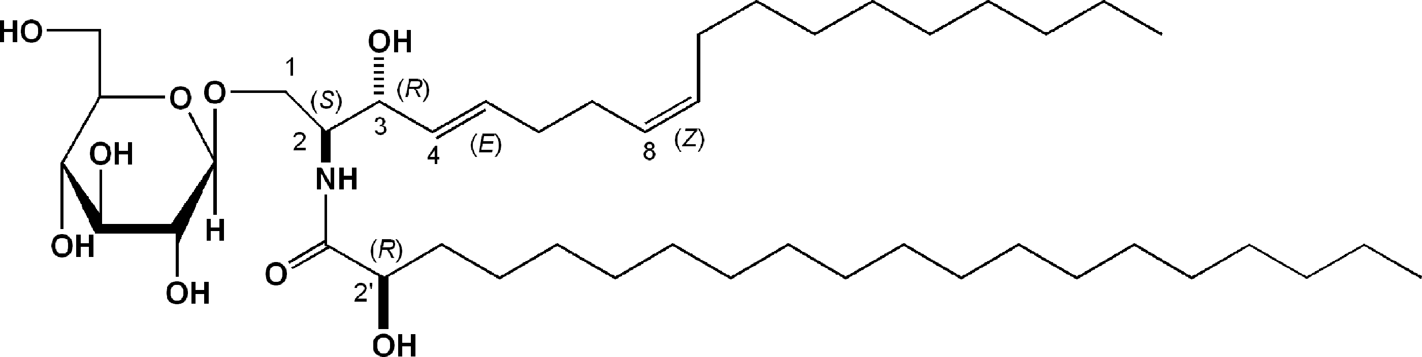
Chemical structure of GlcCer (d18:2) derived from rice GlcCer, glucosylceramides.
Animal experiment
The treatment schedule is illustrated in Figure 3A. GCFr (3 and 10 mg/[kg·day]) or FGC (85.5 and 256 mg/[kg·day]) suspended with 1% acacia solution in water was given orally to the mice for 9 days. The doses of FGC were adjusted, so that GlcCer concentrations were the same as for GCFr. An hour after the last administration, TEWL on the dorsal skin was measured using a VapoMeter (Delfin, Kuopio, Finland). Treatment with sodium dodecyl sulfate (SDS) was performed according to the method of Ideta et al. 10 Briefly, a cotton sheet that had been immersed in 10% SDS was placed onto the right side of the midline on the dorsal skin for 5 min. Then, the cotton was replaced, and the area was cleaned with warmed water. This procedure was repeated from day 10 to 15, and GCFr or FGC was given once a day. On day 12, TEWL on SDS-treated and untreated areas was measured. On day 16, the skin treated with SDS was removed. The specimen was immersed in a neutralized 4% paraformaldehyde solution for immunostaining. For Western blotting and analysis of Cer and GlcCer, the specimen was stored at −80°C.

Effects of GlcCer-rich fraction (GCFr) and formulation of rice-derived GlcCer (FGC) on transepidermal water loss (TEWL) in mice.
The experiment was performed in accordance with the Guidelines for the Proper Conduct of Animal Experiments (Special Council of Japan, June 1, 2006). The experiment was approved by the ethics committee of the research and development section in Oryza Oil & Fat Chemical Co. on September 1, 2011.
Culture of epidermal equivalent
Each cup of EPI-MODEL was placed into a 12-well plate, and the culture medium as established by Green et al. 15,16 (1.5 mL) was added to the lower well (basolateral side). After culturing (37°C, 5% CO2 atmosphere) over night, the medium was replaced with a fresh medium (1.35 mL), and then GlcCer (d18:2) dissolved in 1% dimethyl sulfoxide (150 μL) was added. After culturing for 3 days, the equivalent was immersed in a neutralized 4% paraformaldehyde solution for immunostaining. Samples were stored for Western blotting and analysis of Cer and GlcCer at −80°C.
Analysis of Cer and GlcCer
The skin tissues and epidermal equivalent were homogenized in 1 mL of a mixture of chloroform and methanol (2:1) using a homogenizer (Mini-Beadbeater-1; BioSpec Products, Bartlesville, OK, USA). After centrifugation (2800 g, 10 min) of the homogenate, the supernatant was collected in a glass tube. This procedure was repeated once, and the solvent was removed by nebulization with N2 gas. The lipid residue was weighed and dissolved in a mixture of chloroform and methanol (2:1) to a concentration of 5 mg/mL for mouse skin. For epidermal equivalent, the residue was adjusted to 1 mg/mL for Cer and 10 mg/mL for GlcCer. For TLC of Cer, the lipid solution (15 μL) and standard Cer (2 mg/mL, 5 μL) were developed on the high-performance TLC (HPTLC) plate (Silica gel 60; Merck, Whitehouse Station, NJ, USA) using a mixture of chloroform, methanol, and acetic acid (190:9:1) as a solvent. 17 The spots were visualized by immersion in 10% copper sulfate solution containing 8% phosphoric acid, followed by heating at 180°C for 4 min. For measurement of GlcCer, the lipid solution (25 μL) and GlcCer (d18:2; 2 mg/mL, 5 μL) were developed on an HPTLC plate using a mixture of chloroform, methanol, and water (40:12:1) as a solvent. 7 The spot of GlcCer was visualized by immersion in 0.1% orcinol solution containing 10% H2SO4, followed by heating at 110°C for 10 min. The spots were captured using a dual-wavelength flying-spot-scanning densitometer (CS-9300; Shimazu) at 550 nm, and the area was measured. The areas of Cer and GlcCer were corrected by the upper spot of the Cer standard (Cer 2) and GlcCer (d18:2).
Western blotting of GlcCer-metabolizing enzymes and maturation markers of epidermis
The skin specimen or epidermal equivalent was homogenized in 1 mL of RIPA buffer containing a protease and phosphatase inhibitor cocktail. After centrifugation (16,000 g, 10 min) of the homogenate, the supernatant was collected. Then, the protein concentration of the supernatant was adjusted with the RIPA buffer to 1 mg/mL. The supernatant was mixed with the same volume of the Laemmli sample buffer (62.5 mM Tris–HCl, 2% SDS, 5% 2-mercaptoethanol, 25% glycerol, and 0.01% bromophenol blue) and heated at 95°C for 5 min. The sample solution (12 μL) was separated by 10% SDS–polyacrylamide gel electrophoresis (PAGE), and the proteins were transferred to a polyvinylidene fluoride membrane. Primary antibodies were used at the following dilutions: anti-GCSase (1:1000), anti-GCase (1:1000), anti-actin (1:10,000), anti-TGase (1:4000), and anti-involucrin (1:5000). HRP-conjugated anti-rabbit IgG (1:25,000 dilution) was used as a secondary antibody. Detection was performed by the chemiluminescence method using an ECL Plus Blotting Detection system and Amersham Hyperfilm™ ECL.
Immunostaining
The mouse skin and epidermal equivalent tissues were embedded in paraffin, and the specimen was fixed on a glass slide. The deparaffinized specimen was treated with 0.3% H2O2 in methanol for 30 min and blocked with 5% skimmed milk for 1 h. The specimen was treated with a primary antibody at 4°C overnight at the following dilution ratios: anti-Cer IgM (1:100) and anti-GlcCer IgG (1:100). HRP-conjugated secondary antibodies were added at room temperature for 30 min at the following dilution ratios: anti-mouse IgM (1:200) and anti-rabbit IgG (1:500). Then, the specimen was treated with a DAB solution, followed by staining with hematoxylin solution.
Statistics
The results are expressed as means and standard error (SE). Significance of the differences was examined by the Williams method for TEWL. One-way analysis of variance followed by Dunnett's test was used for determination of Cer and GlcCer. Differences of P<.05 were considered significant.
Results
HPLC profiles of the GCFr, FGC, and GlcCer (d18:2)
Figure 1 shows an HPLC chromatogram of a commercial standard GlcCer (>99% purity; Nagara Science) and three samples evaluated in the following experiments. The chromatogram of the GCFr was quite similar to that of standard GlcCer. In the chromatogram of FGC, three peaks considered to be lipids originating from emulsifiers in the formula were observed. The isolated GlcCer (d18:2) showed a single peak.
Effect of the GCFr on TEWL and contents of Cer and GlcCer in mice
The effects of the rice-derived GCFr and FGC in mice were evaluated on the skin treated with or without SDS. Consecutive oral treatments with GCFr and FGC tended to improve TEWL by day 9 (Fig. 3). FGC (256 mg/[kg·day]) significantly suppressed increases in TEWL. On day 12, TEWL on the sites without SDS treatment were significantly suppressed by the GCFr (3 and 10 mg/[kg·day]) and FGC (85.5 and 256 mg/[kg·day]). TEWL of SDS-treated sites were also significantly improved by GCFr and FGC. The differences in TEWL between SDS-treated and normal sites were also significantly improved by the treatment with the GCFr and FGC.
Figure 4 shows TLC chromatograms of Cer and GlcCer extracted from mouse skin treated with or without SDS and GCFr. Each spot of Cer was identified by the reported rate-of-flow (Rf ) values and images. 18 –20 The spots of Cer 1 (Cer esterified ω-hydroxy fatty acid and sphingosine [EOS]) and Cer2 (Cer nonhydroxy fatty acid and sphingosine [NS]) were thinner with SDS treatment (Fig. 4, left).Treatment with GCFr enhanced expression of Cer 1 and 2. Table 1 shows the area of cholesterol, free fatty acid, Cer 1, and Cer 2. GCFr (10 mg/[kg·day]) significantly restored the expression of Cer 1 and tended to enhance Cer 2. In addition, in the immunostaining of Cer (Fig. 5A), stratum corneum was decreased by the treatment with SDS (control), but was normalized by the GCFr (3 and 10 mg/[kg·day]), and Cer in the stratum corneum and stratum lucidum was increased.
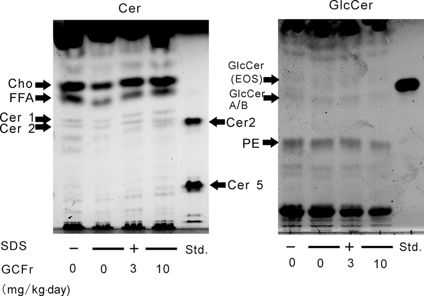
TLC chromatogram of ceramides (Cer) and GlcCer in mouse skin. For measurement of Cer and GlcCer, the lipid sample was developed on an high-performance thin-layer chromatography (HPTLC) plate using a mixture of chloroform, methanol, and acetic acid (190:9:1) and a mixture of chloroform, methanol, and acetic acid (40:12:1), respectively. The spots for Cer were visualized using 10% copper sulfate solution containing 8% phosphoric acid. The spots of GlcCer were visualized using 0.1% orcinol solution containing 10% H2SO4. Cho, cholesterol; FFA, free fatty acid; PE, phosphatidylethanolamine.
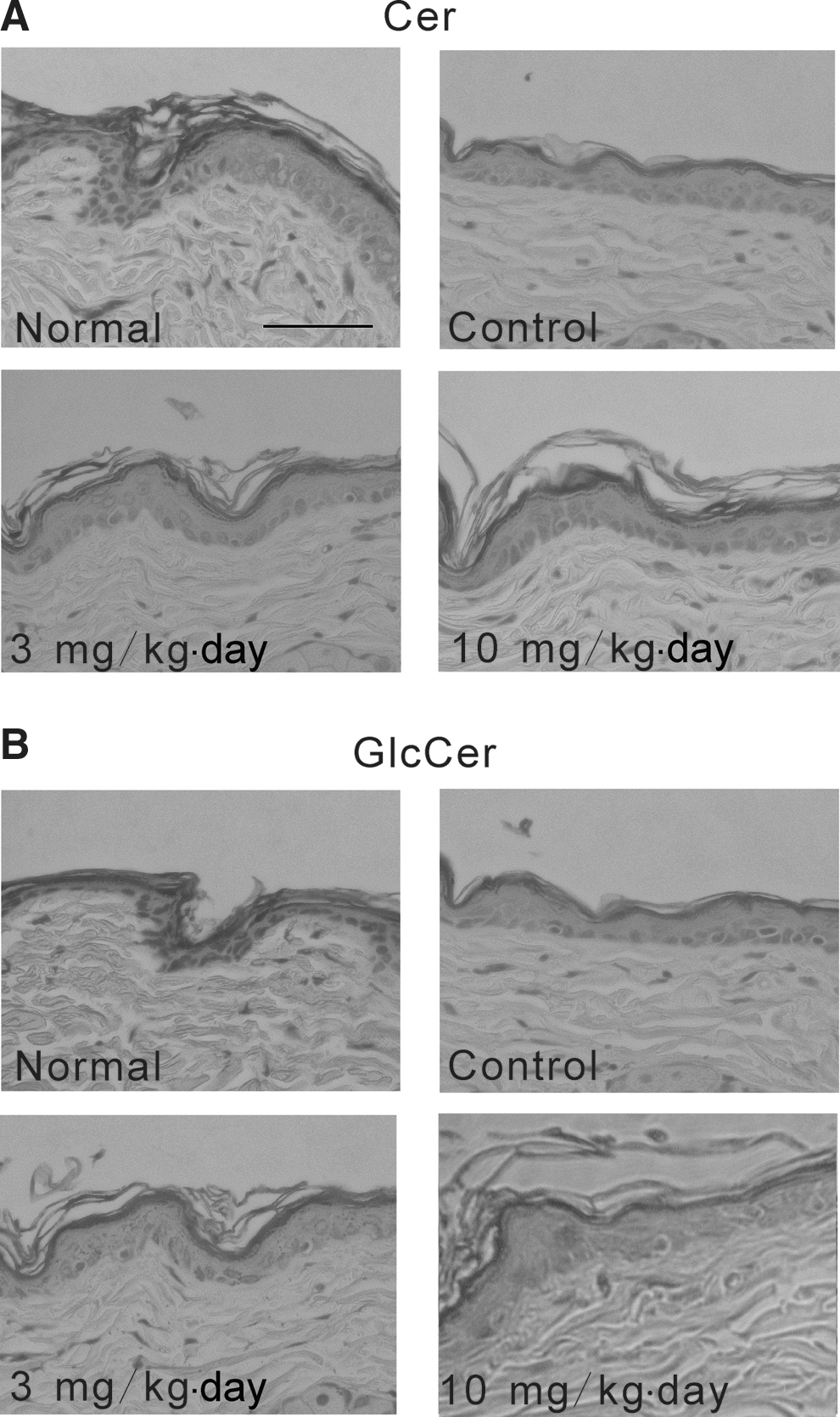
Immunostaining images of
The areas of spots were corrected by the area of standard Cer and GlcCer. Each value represents mean with the SE of five to six mice. Significance of differences was examined by one-way analysis of variance followed by Dunnett's test.
Significant difference from control (P<.05).
Cer, ceramide; GlcCer, glucosylceramide; GCFr, GlcCer-rich fraction; EOS, esterified ω-hydroxy fatty acid and sphingosine; Cho, cholesterol; FFA, free fatty acid; PE, phosphatidylethanolamine; SE, standard error.
We next investigated the expression of GlcCer. Each GlcCer was identified as GlcCer (EOS) and the complex mixture of GlcCer (NS), (NP), and (C24,26-AS) known as GlcCer A/B, as described by Doering et al. 21 SDS hardly affected the of GlcCer (EOS) and GlcCer A/B contents as revealed by the TLC analysis (Fig. 4, right). The GCFr (10 mg/[kg·day]) tended to suppress the expression of GlcCer (EOS) and GlcCer A/B. Table 1 shows the areas of GlcCer (EOS) and GlcCer A/B. GlcCer (10 mg/[kg·day]) significantly suppressed GlcCer (EOS) and GlcCer A/B expression. In the immunostaining images of GlcCer (Fig. 5B), stratum corneum and stratum lucidum were stained deeply (normal). After treatment with SDS, the amount of GlcCer was decreased (control). The GCFr (10 mg/[kg·day]) further decreased the stained areas of GlcCer on stratum corneum and stratum lucidum.
Effect of the GCFr on the expression of GlcCer-metabolizing enzymes in mice
The expression of the enzymes that regulate synthesis of GlcCer and Cer, namely GCSase and GCase, was evaluated. GCSase was expressed after the treatment with SDS (Fig. 6). The GCFr (3 and 10 mg/[kg·day]) enhanced the expression of GCSase. On the other hand, the expression of GCase was suppressed by the treatment of SDS. This decrease has been observed in the skin of patients with psoriasis. 8 The GCFr (10 mg/[kg·day]) restored the expression of GCase.
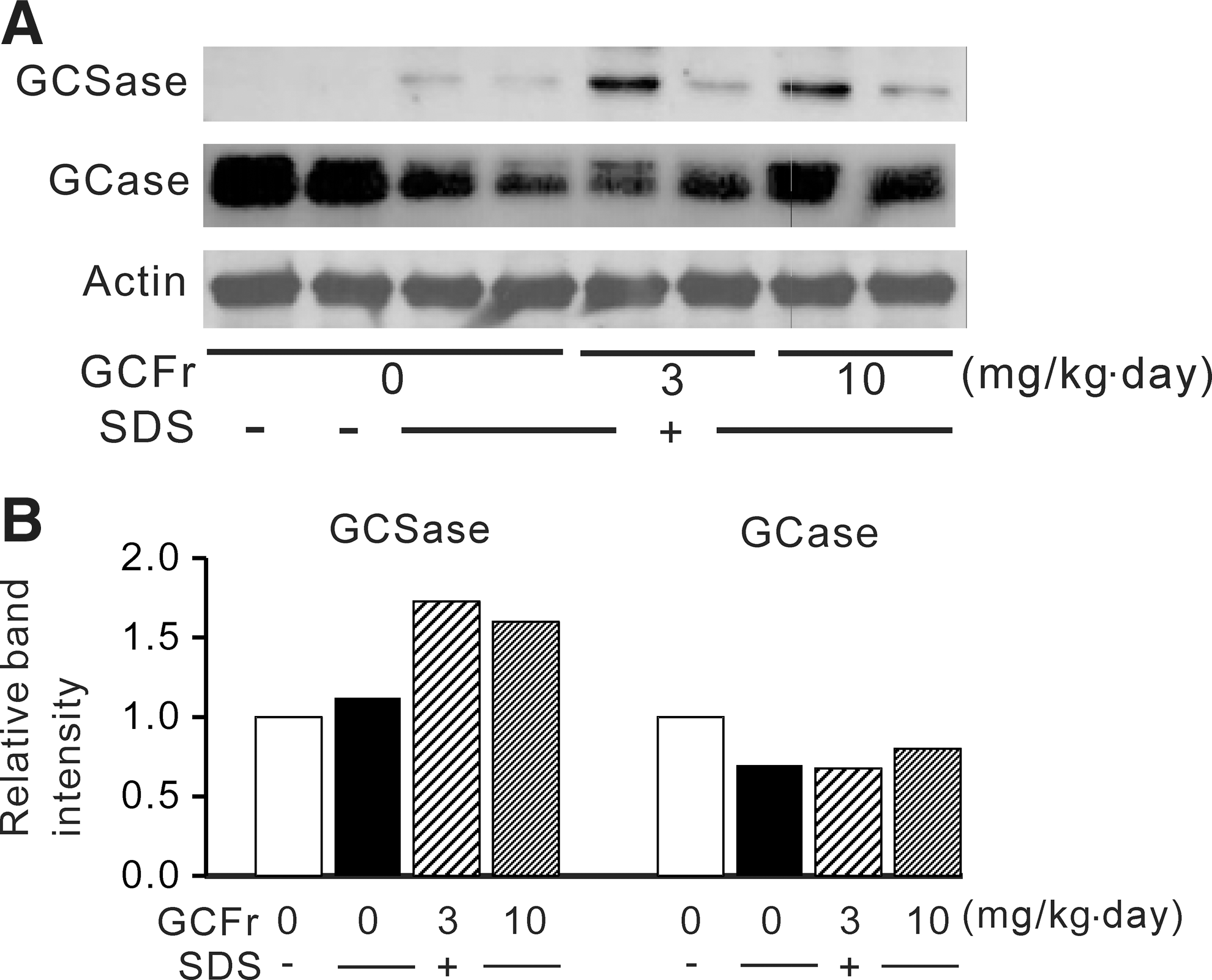
Effect of GCFr on the expression of GlcCer-metabolizing enzymes in mice.
Effect of GlcCer (d18:2) on Cer and GlcCer in epidermal equivalent
In vitro study of GlcCer (d18:2), a principal GlcCer in rice, was performed. The Cer profile of the epidermal equivalent treated with GlcCer (d18:2) was analyzed by TLC (Fig. 7, left). Cer 1 was clearly detected, and the spots of Cer 2 were thin and pale. GlcCer (d18:2) significantly enhanced the expression of Cer 1 at 1 μg/mL and Cer 2 at 10 μg/mL (Table 2). In the immunostaining images of Cer in epidermal equivalent, GlcCer (d18:2) clearly enhanced Cer expression in stratum corneum at 10 μg/mL (Fig. 8A).
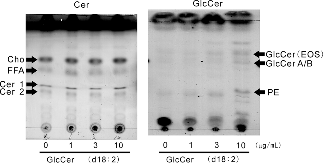
TLC chromatogram of Cer and GlcCer in epidermal equivalent treated with GlcCer (d18:2). HPTLC was performed with the same conditions as denoted for Figure 4.
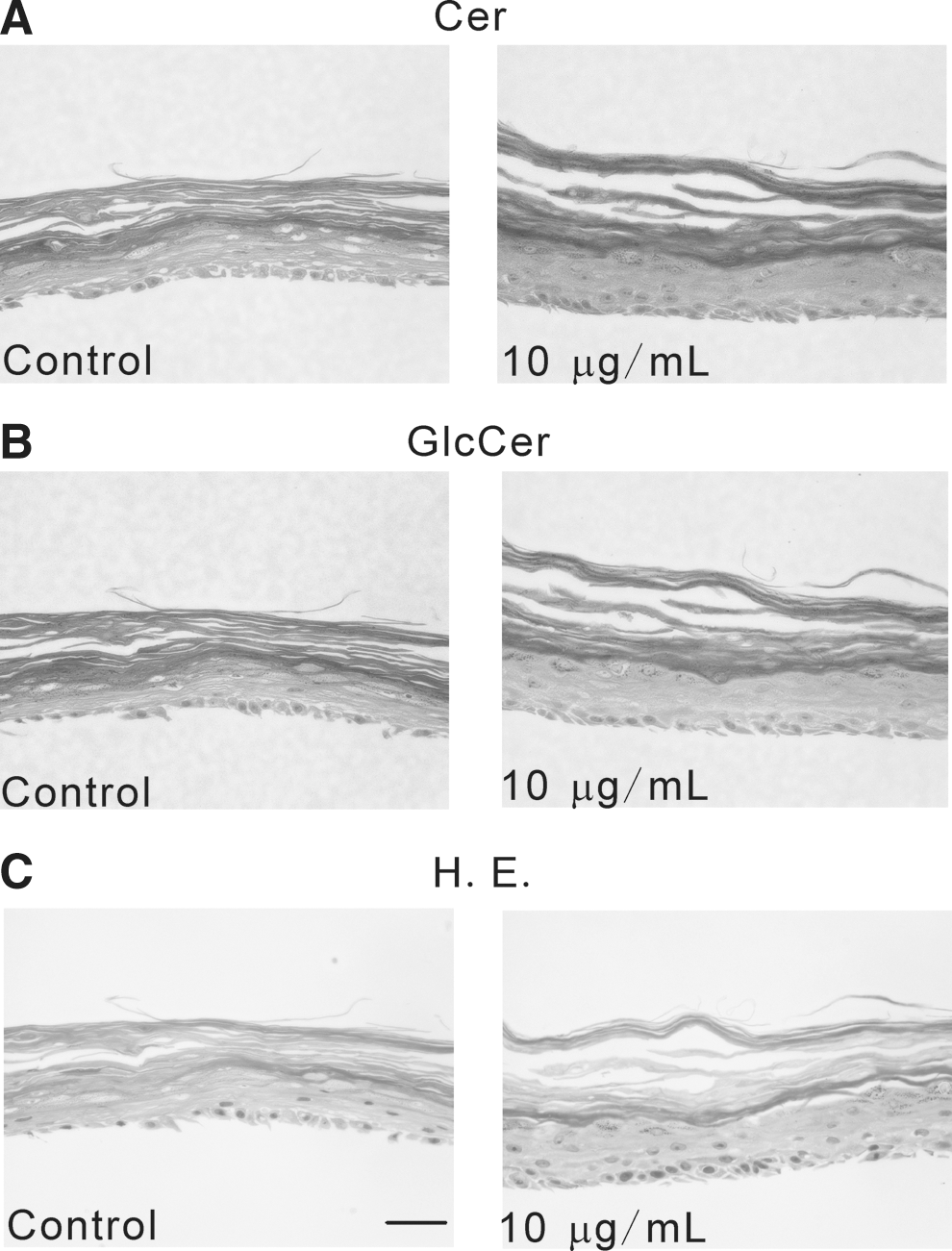
Immunostaining images of
The areas of spots were corrected by the area of standard Cer and GlcCer. Each value represents mean with the SE of three experiments. Significance of differences was examined by one-way analysis of variance followed by Dunnett's test.
Significant differences from control (* P<.05; ** P<.01).
On the other hand, in TLC analysis of GlcCer, the amount in epidermal equivalent was quite small (Fig. 7, right). After treatment with GlcCer (d18:2; 3 and 10 μg/mL), the spots of GlcCer (EOS) and GlcCer A/B clearly became thicker. As shown in Table 2, the areas of GlcCer (EOS) and GlcCer A/B were significantly increased by more than 3 μg/mL GlcCer (d18:2). Immunostaining of GlcCer (Fig. 8B) indicated that the distribution area of GlcCer was enlarged by the treatment with 10 μg/mL GlcCer (d18:2).
Effect of GlcCer (d18:2) on the expression of GlcCer-metabolizing enzymes and maturation markers of epidermis in epidermal equivalent
We evaluated the effect of GlcCer (d18:2) on the expression of synthetic and glycolytic enzymes of GlcCer (Fig. 9). GlcCer (d18:2) clearly enhanced the expression of GCSase; however, it did not affect the expression of GCase. On the other hand, GlcCer (d18:2) enhanced the expression of maturation markers of epidermis, including TGase and involucrin, at more than 1 μg/mL.
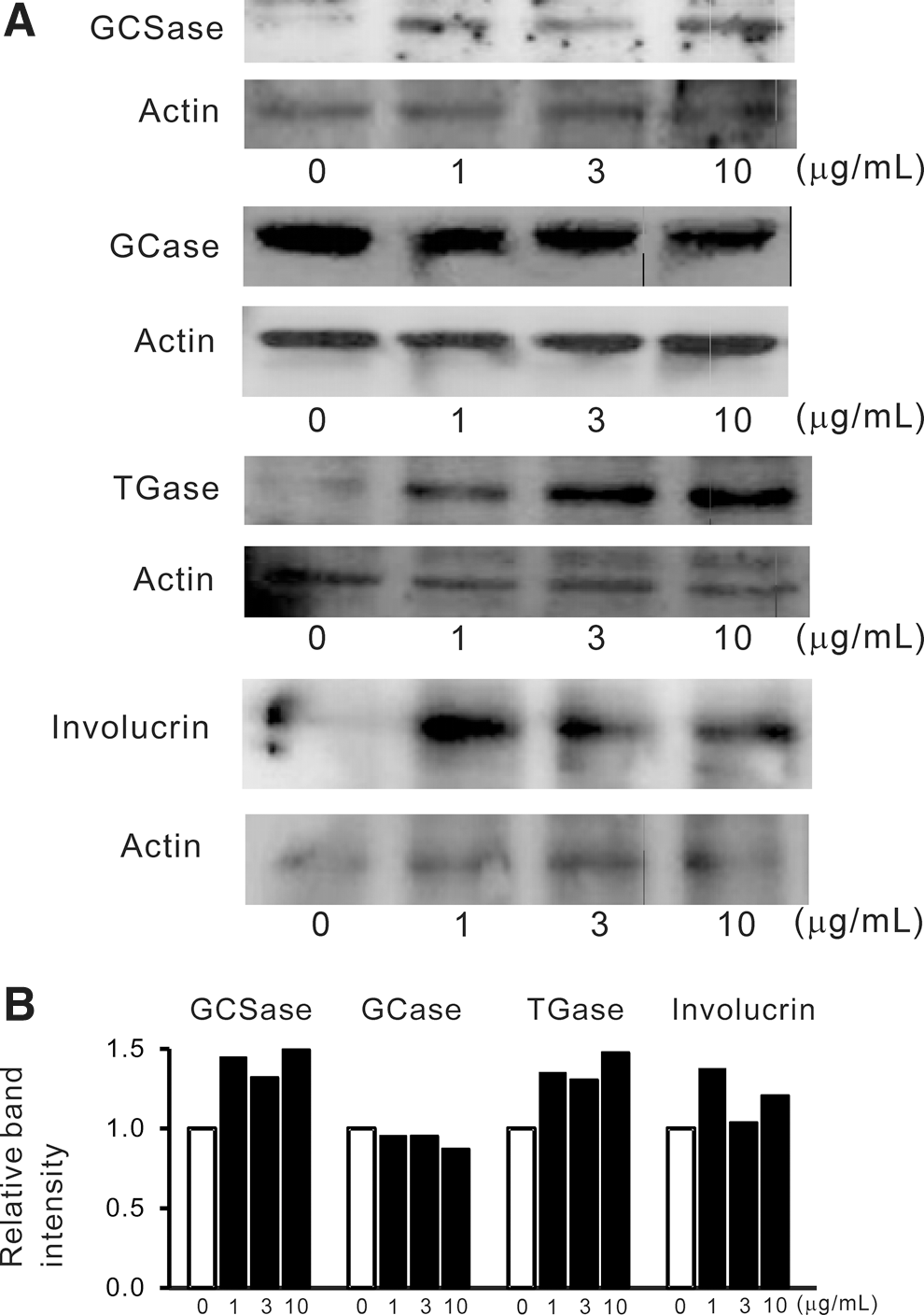
Effect of GlcCer (d18:2) on the expression of GlcCer-metabolizing enzymes and differentiation markers of epidermis in epidermal equivalent. The epidermal equivalent was treated with GlcCer (d18:2) for 3 days.
Discussion
In the present study, we investigated the effects of a rice-derived GCFr and its formulation (FGC) on TEWL and epidermal Cer in mice. The TEWL on SDS-treated and untreated sites and those differences were improved by oral dosing with the GCFr and FGC for 12 days. Thus, the GCFr and FGC were found to improve epidermal water loss by oral treatment.
The absorption of dietary GlcCer has been investigated in an ex vivo experiment in rats. 22 Maize-derived GlcCer, including GlcCer (d18:2), was absorbed from the gut and was detected in lymph fluid as Cer consisting of 4t,8c-sphingadienine. Dietary GlcCer is thought to be deglycosylated in the intestine and absorbed as the Cer form. Besides, dietary Cer (not GlcCer) has been reported to be distributed in the skin of rats. By oral dosing of radiolabeled Cer (C18-d18:0), the radioactivity was detected in plasma, and it moved to epidermis from 24 to 168 h after dosing. 23 They also found that sphingosine along with its metabolites were detected in the skin after oral dosing of radiolabeled sphingosine in mice. 24 From these reports, dietary GlcCer is thought to be absorbed as Cer and its metabolites, and they reach to the epidermis via lymph and blood flow. However, there are no reported investigations of whether rice-derived GlcCer increase Cer in epidermis, leading to moisture retention. Therefore, we determined the actual contents of Cer and GlcCer in SDS-treated skin. As a result, the GCFr (10 mg/[kg·day]) was found to increase the content of Cer 1. In addition, the increase in Cer expression in stratum corneum and stratum lucidum was also confirmed in histochemical analysis. Cer 1 contains a linoleic acid ester linked to the ω-hydroxyl group of a very long chain amide-linked ω-hydroxyacid 25 and is closely related to skin barrier function. 1,26 Acylceramides, including Cer 1, have been reported to provide superior barrier function among Cer. 27 These results suggest that oral treatment with the GCFr and FGC improves TEWL by enhancement of Cer expression in epidermis.
On the other hand, the GCFr decreased the amount of GlcCer, including GlcCer (EOS) and GlcCer A/B. GlcCer (EOS), corresponding to Cer 1, exists in lamellar granules and plays a role as a molecular rivet of the lipid bilayer. 28 GlcCer (A/B) is a mixture of GlcCer (NS), GlcCer nonhydroxy fatty acid and phytosphingosine (NP), and GlcCer (C24,26-AS). 21 These are thought to be precursors of Cer 3, Cer Y, and Cer 6, respectively. 7 GlcCer are synthesized from Cer by GCSase and hydrolyzed to Cer by GCase. 7 Immunostaining after administration of 10 mg/[kg·day] GCFr revealed suppression of GlcCer expression. From Western blotting analysis of these enzymes, both expressions were enhanced by the GCFr. Thus, decreases in GlcCer (EOS) and GlcCer A/B were suggested to be based on the enhancement of Cer production by GlcCer-metabolizing enzymes, including GCSase and GCase. AcylGlcCer, including GlcCer (EOS), are synthesized from Cer by three steps. 29 In the first step, Cer is glycosylated by GCSase and then converted to ω-hydroxyglucosylceramide by P-450 enzyme. Finally, acyl GlcCer is formed from ω-hydroxyglucosylceramide by ω-acyltransferase. Thus, the GCFr is thought to enhance GlcCer (EOS) formation in the early stage of the biosynthetic pathway from Cer. Increases in Cer accompanied by decreases in GlcCer are observed in the formation process of the coenocyte lipid envelope in mouse embryo. 30 GCFr may enhance supplementation of Cer from the GlcCer pool diminished by SDS.
We next investigated the effect of purified GlcCer (d18:2) on the Cer in human epidermal equivalent. GlcCer (d18:2) increased Cer 1 and Cer 2 at 1 and 10 μg/mL, and immunostaining image of Cer revealed clear enhancement of Cer expression. On the other hand, GlcCer (d18:2) significantly enhanced the contents of both GlcCer (EOS) and GlcCer A/B. The increase in GlcCer is opposite to the result of in vivo study. Therefore, we examined the expression of GCSase and GCase. As a result, GlcCer (d18:2) enhanced only GCSase expression and did not affect the GCase expression. Hence, the action of GlcCer (d18:2) on GCase expression was found to be different in SDS-treated mouse and human epidermal equivalent. Further investigation is required to clarify the difference of the effect of GCFr and GlcCer (d18:2) on the expression of GlcCer and GCase in normal mice and SDS-treated epidermal equivalent. Moreover, evaluation of the effects on the other minor GlcCer in the GCFr on epidermal equivalent is also needed. At least, GlcCer is thought to increase Cer by enhancement of GlcCer synthesis in vitro.
TGase and involucrin are known to be maturation markers of epidermis. 31 Involucrin is a substrate of TGase and attaches to ω-hydroxyceramide by TGase to provide the barrier function of the epidermis. 32 Recently, GlcCer derived from Amorphophallus konjac were reported to enhance TGase expression in mice leading to cornified envelope formation. 10,33 In the present in vitro study, we confirmed that GlcCer (d18:2) enhanced the expression of both markers. GlcCer (d18:2) may contribute to epidermal hydration by enhancement of involucrin content and the molecular structure of cornified envelope.
In conclusion, oral dosing of a rice-derived GlcCer (GCFr) was found to prevent epidermal water loss by increasing Cer content via acceleration of GlcCer metabolism. GlcCer (d18:2) in GCFr possesses the ability to increase Cer and GlcCer and enhances maturation of epidermis in human epidermal equivalent. Rice-derived GlcCer is suggested to be useful for improving epidermal water retention and barrier function.
Footnotes
Author Disclosure Statement
No competing financial interests exist.