
Abstract
The metabolic syndrome is a major worldwide health care issue and a dominant risk factor for cardiovascular disease. The liver manifestations of this syndrome include nonalcoholic fatty liver disease (NAFLD) and its progressive variant nonalcoholic steatohepatitis (NASH). Although significant research has been performed, the basic pathogenesis of NAFLD/NASH remains controversial and effective treatments are still unavailable. We have previously reported on a murine model of NASH induced by the neonatal injection of monosodium glutamate (MSG), which includes the clinical manifestations of central obesity, diabetes, hyperlipidemia, and ultimately liver inflammation, fibrosis, and cancer. Although MSG is considered a safe food additive, its administration to pregnant rats increases the voracity and growth hormone levels in the offspring. To further understand the biology of this model, we have investigated the influence of the calorie intake on these clinical manifestations by feeding animals a restrictive diet. MSG-treated animals fed a restrictive diet continue to manifest obesity and early stage NASH but have improvements in serum lipid profiles. At 12 months of age, mice had manifestations of obesity, whether animals were fed a restricted or control diet, but animals fed a restrictive diet had a reduction in the progression of NASH. In conclusion, MSG appears to be a critical factor in the initiation of obesity, whereas calorie intake may modulate the progression of disease.
Introduction
T
Our group and others have recently suggested that the use of monosodium glutamate (MSG) as a dietary additive may be correlated with obesity and other signs of the metabolic syndrome, despite conflicting reports on different outcomes. 23,24 Accordingly, we have recently developed a murine model in which the subcutaneous injection of MSG at birth leads to central obesity and type 2 diabetes onset by 4 to 6 months. 25,26 Furthermore, NASH appears at liver histology after six months, followed by fibrosis and neoplastic transformation by 12 months. We thus investigated whether a light diet restriction (75% volume of full stomach) changes the phenotype of mice in terms of body weight, liver and visceral fat weight, serum lipid profile, and liver pathology. Our data suggest that NASH appearance is independent from the dietary intake, which, on the other hand, modifies the disease progression.
Materials and Methods
Experimental design
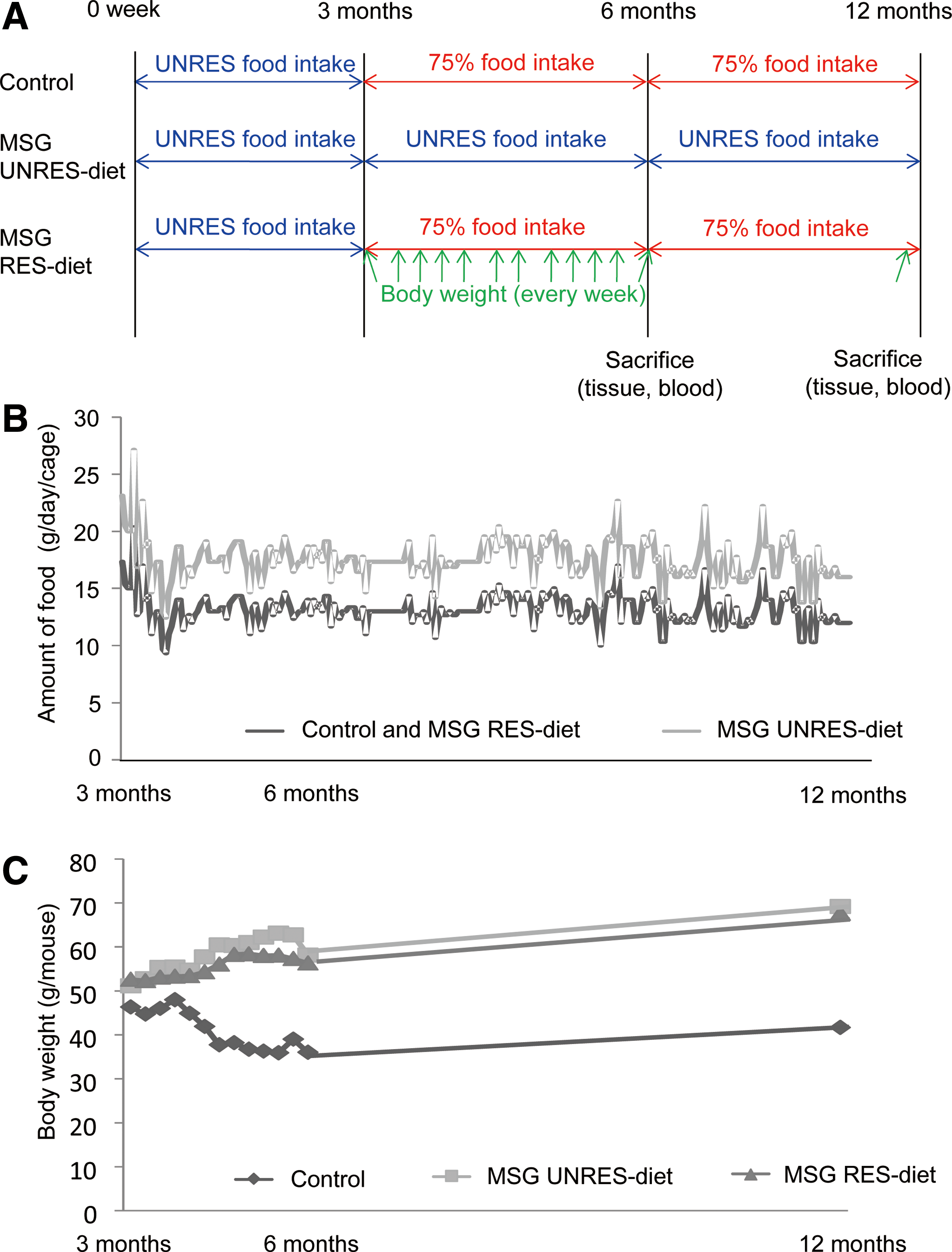
The study protocol is illustrated in Figure 1A. Fifty-four 12-week-old monosodium glutamate (MSG) male mice (n=36) and control male mice (n=18) were purchased from the Institute for Animal Reproduction. MSG mice include Crj:CD-1 (ICR) animals administered 2 mg/g of MSG subcutaneously from birth to 5 days of age. 26 For control purposes, ICR male mice were administered 2 mg/g of physiological saline at the same time points. All mice had an ad libitum chlorinated water intake until 12 weeks of age, and were housed in TPX cages (3 mice per cage) (Okazaki Sangyo Co., Tokyo, Japan) according to their treatment group in a temperature-controlled room with a 12-hour light/dark illumination cycle. All mice were fed a standard diet (MF; Oriental Yeast Co. Ltd., Tokyo, Japan). MSG mice were randomly allocated into two groups with unrestricted or restricted dietary regimens, the daily food consumption of the MSG-unrestricted group was recorded. On the following day, MSG-restricted and control group were fed 75% of the previous day average intake of the MSG-unrestricted group. 27
Outline of the experimental protocol
All animals were offered free access to water. The overall composition of the diet is shown in Table 1. Animals were sacrificed at 6 (50%) or 12 (50%) months of age, in all cases following a 12-hour fasting before the collection of liver, visceral fat, and peripheral blood samples. Measurements obtained during the study included markers of obesity such as bilateral epididymal fat (as an index of visceral fat), visceral fat weight, weekly body weight until 6 months of age, and body weight at 12 months of age. Liver specimens were obtained and fixed with 10% buffered formalin solution, paraffin-embedded for histological analysis. All animal use procedures were in accordance with the Guide for the Care and Use of Laboratory Animals and approved by the Committee on Animal Experimentation of the University of Toyama.
Liver pathology
Serial 4 mm thick sections were cut from formalin-fixed paraffin-embedded liver tissues and stained with hematoxylin and eosin (HE) and Azan. Three representative areas were analyzed in each section and the average value was used to calculate the NAFLD activity score (NAS). Liver disease activity was scored using the NAS system proposed by the NASH Clinical Research Network based on semi-quantitative items, e.g., steatosis (0–3), hepatocellular ballooning (0–2), lobular inflammation (0–2). 28 NAS values equal or greater than 5 were consistent with a diagnosis of NASH while NAS<3 ruled out the diagnosis. Although NAS has originally been established and validated in human adult and pediatric patients, recent studies have used this score to assess animal NASH. 29 –31 Furthermore, we performed immunohistochemistry to evaluate liver tumor at 12 months of age. Liver tumors were stained using a rabbit polyclonal anti-glutamine synthase antibody (LifeSpan BioSciences, Inc., Seattle, WA, USA), as glutamate synthase represents a helpful marker for the diagnosis of hepatocellular carcinoma (HCC). 32,33 The degree of liver fibrosis was evaluated only at 12 months of age, as supported in our previous study. 26 The presence of liver nodules was also recognized.
Serum analyses
An external supplier (LipoSearch, Tokyo, Japan) measured serum lipid profile included total cholesterol (T-chol), LDL cholesterol (LDL-chol), and triglycerides (TG) of all mice, using HPLC as reported elsewhere. 34 Serum non-esterified fatty acid (NEFA), alanine aminotransferase (ALT), glucose, and insulin were measured using the Determiner NEFA755 (Kyowa Medix Co., Ltd., Tokyo), Transaminase CII Test Wako (Wako Pure Chemical Ind., Ltd., Osaka, Japan), Glucose CII-Test Wako (Wako Pure Chemical), and Ultrasensitive Insulin Assay Kit (Morinaga Bio-Science Laboratories, Inc., Yokohama, Japan), respectively.
Statistical analysis
All continuous variables were expressed as mean±standard error of measurement (SEM). Pairwise comparisons between groups were performed by one-way analysis of variance models and corrected for multiple comparisons according to Bonferroni. All analyses were two-tailed and corrected P (Pc) values <.0167 were considered as statistically significant. Statistical analyses were performed using Stat View version 5.0 (Abacus Concept, Berkley, CA, USA).
Results
Body weight, liver/body weight ratio, visceral fat weight, and visceral fat/body weight ratio
At 6 months of age, the calorie intake mean of MSG-unrestricted and MSG-restricted group was 20.3 kcal/day/mouse and 15.3 kcal/day/mouse, respectively, while at 12 months of age, the calorie intake mean of MSG-unrestricted and MSG-restricted group was 20.9 kcal/day/mouse and 15.8 kcal/day/mouse, respectively. Figure 1B illustrates the progression of the amount of food intake and Figure 1C the body weight with different dietary regimens. The amount of food intake and body weight were recorded starting at 3 months of age, since mice were purchased at that time. At 3 months of age, body weight did not differ between MSG mice subject to restricted (56.3±1.1 g) or unrestricted (58.1±1.6 g) diet while, as expected, control mice had significantly lower body weight (37.0±2.4 g). MSG mice increased their body weight regardless of their dietary regimen (Fig. 1C) and the two dietary regimens did not differ in body weight at 6 and 12 months of age in MSG mice (Fig. 2). Control mice continued to decrease their body weight until 6 months of age and the endpoint body weight of mice in the control group was significantly lower compared to MSG mice at all time points. Among these mice the dietary regimens did not change body weight significantly.
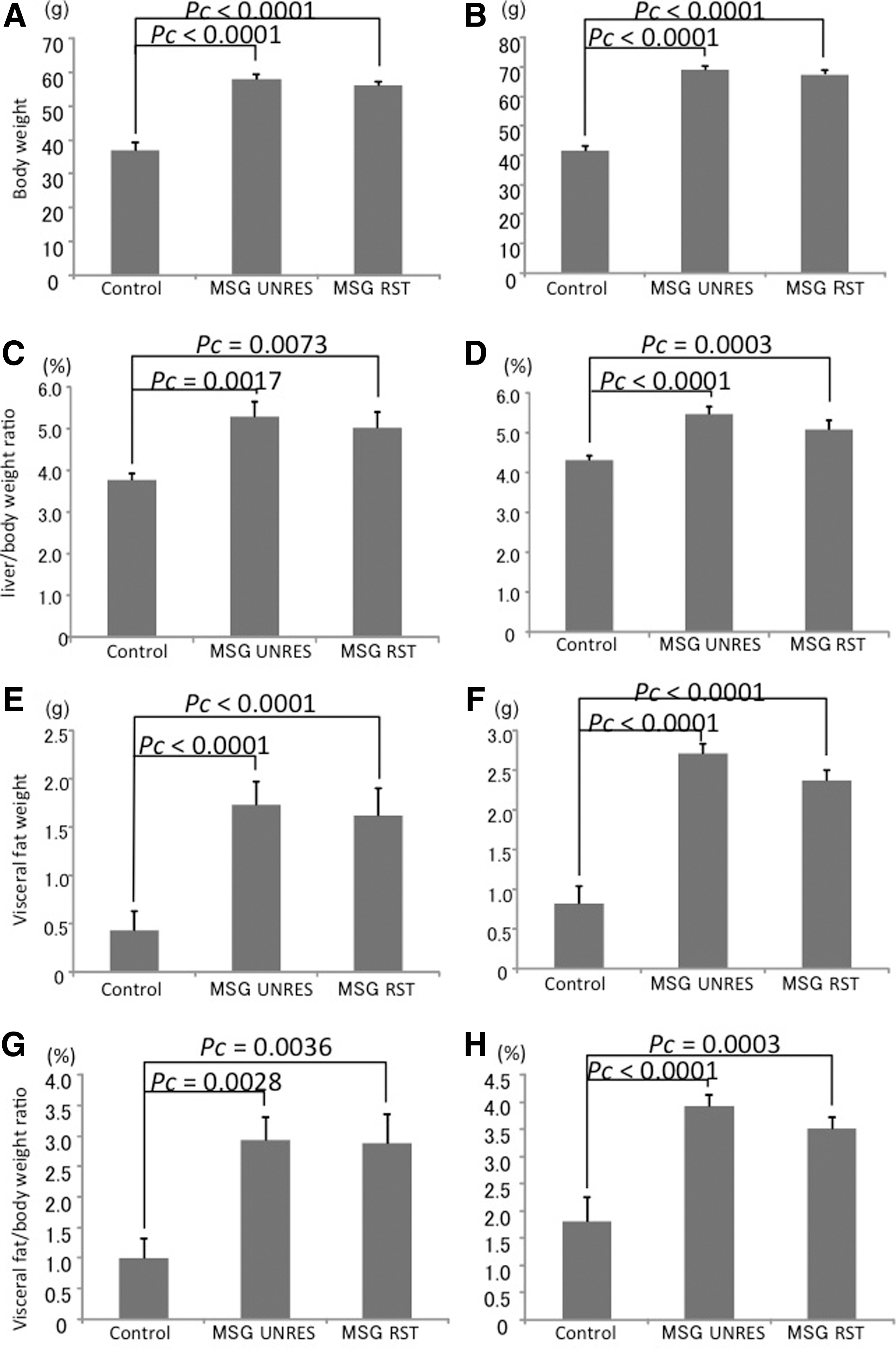
Body weight, liver/body weight ratio, visceral fat weight, and visceral fat/body weight ratio in the three study groups (MSG with restricted diet, MSG with unrestricted diet, and controls) at 6
Similarly, the visceral fat weight, liver/body weight ratio, and the visceral fat/body weight ratio at 6 and 12 months of age did not differ with the dietary regimen in the MSG groups while being significantly higher compared to the control group (Fig. 2).
Serum analyses
By 6 and 12 months of age, MSG mice manifested higher levels of serum T-chol, LDL-chol, TG, and NEFA compared to the control group, regardless of the dietary restriction. When 6-month old MSG mice allocated to a specific diet were compared, T-chol and LDL levels were lower in the restricted diet group (Fig. 3). Similarly, 12-month-old mice had significantly lower levels of T-chol, LDL-chol, and NEFA compared to mice with an unrestricted diet (Fig. 3). The MSG unrestricted group manifested significantly higher serum ALT level compared to other groups at 12 months of age (Table 2) while no difference in serum ALT levels was observed among groups at 6 months of age. The MSG-unrestricted group had significantly higher serum glucose levels than the control group, but no significant difference between MSG groups at 6 months of age. Furthermore, the MSG-unrestricted group had significantly higher serum glucose level compared to other groups at 12 months of age (Table 2). Both MSG-treated groups manifested significantly higher serum insulin levels. The MSG-unrestricted group showed significantly higher serum insulin levels than those of MSG-restricted group at 6 months of age. Furthermore, it showed significantly higher serum insulin levels compared than those of the other groups at 12 months of age.
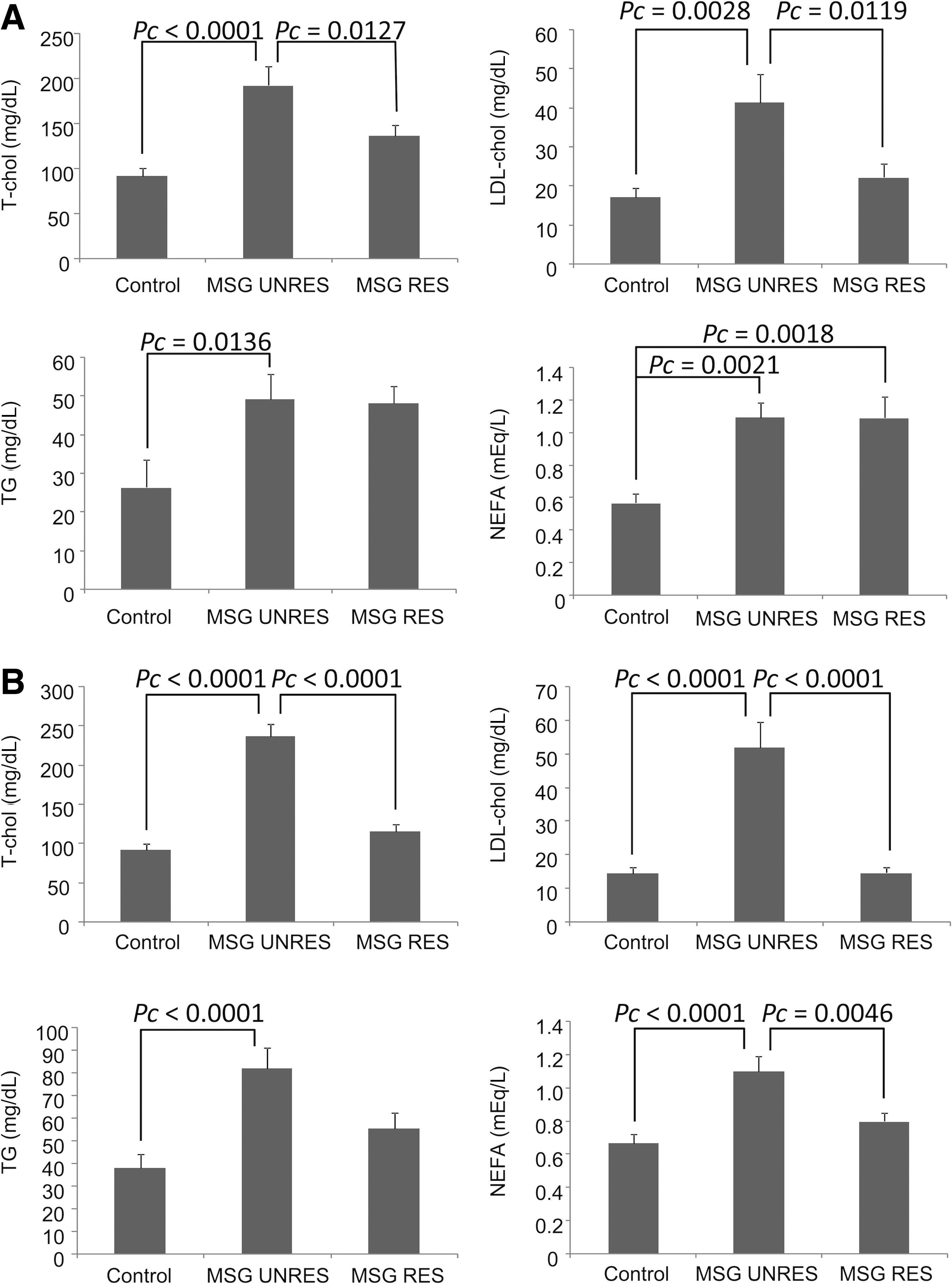
Serum lipid profiles in the three study groups (MSG with restricted diet, MSG with unrestricted diet, and controls) at 6
n=9 for each group.
*P<.0167, ** P<.0033 vs. Control at 6 months of age.
†P<.0033 vs. MSG-unrestricted at 6 months of age.
‡P<.0033 vs. Control at 12 months of age.
§P<.0167, §§ P<.0033 vs. MSG-unrestricted at 12 months of age.
ALT, alanine aminotransferase; MSG, monosodium glutamate.
Liver histology
Both MSG groups manifested marked fatty changes in the liver by 6 months, with major microvesicular and macrovesicular steatosis in the perivenular areas and, in some cases, diffuse throughout the parenchyma (Fig. 4A). Hepatocellular ballooning, inflammatory infiltrate with neutrophil aggregation, and Mallory body formation but no fibrosis were also observed. By 12 months of age, the MSG-unrestricted mice had mild perivenular to pericellular fibrosis with clear signs of steatohepatitis and numerous liver nodules (Fig. 4B). These nodules had no capsule and manifested an increased cellularity with irregular hepatocellular arrangements. Further cellular atypia, such as nuclear swelling and higher nucleus/cytoplasm ratio, were frequently observed along with thick trabecular arrangements. Immunohistochemistry is very useful in distinguishing between dysplastic and early malignant hepatocellular nodules 32 and, in our study, the immunostaining of tumors had strong intensity (Fig. 4C). Taken all together, these histological characteristics suggested a dysplastic/neoplastic nature of the nodules. The restricted diet, conversely, was associated with neither fibrosis nor liver nodules. The pathology grading features are illustrated in Table 3 for 6-month-old and 12-month-old mice. No significant liver abnormalities were observed in control mice at 6 (Fig. 4A) and 12 months (data not shown) of age.
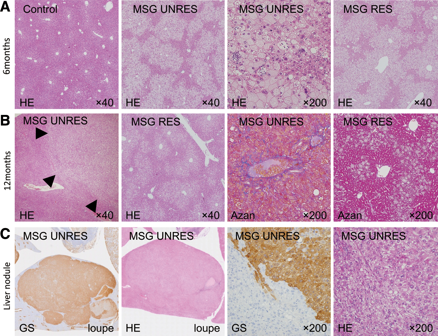
Liver histology following hematoxylin/eosin (HE) staining in the three study groups (MSG with restricted diet, MSG with unrestricted diet, and controls) at 6
n=9 for each group.
P<.0167, ** P<.0033 vs. Control at 6 months of age.
P<.0033 vs. MSG-unrestricted at 6 months of age.
P<.0033 vs. Control at 12 months of age.
P<.0167, §§ P<.0033 vs. MSG-unrestricted at 12 months of age.
NAS, nonalcoholic fatty liver disease activity score.
Discussion
The etiology of the metabolic syndrome and its liver phenotype NAFLD/NASH remains largely unknown based on major issues remaining in terms of individual susceptibility and disease penetrance 35 as well represented by discordant monozygotic twins. 36 Weight loss appears beneficial in NAFLD, but few studies showed liver fibrosis improvement. 37 –40 A recent Cochrane review analyzed the effects of massive weight loss induced by bariatric surgery on liver histology, but the lack of randomized clinical trials does not allow a scientifically sustained conclusion. 41 Healthcare resource allocation data suggest that NASH is rapidly becoming the major cause of cirrhosis and hepatocellular carcinoma. 42 This is highly important if one considers that some degree of NAFLD is observed in a large part of the general population. 43,44 Accordingly, the current hypothesis states that calorie intake may be necessary but not sufficient to cause obesity 45 and we submit that MSG intake may play a significant role in this scenario. Based on our MSG-induced animal model, and in agreement with epidemiological data, we propose that MSG may play a major role in developing obesity, type 2 diabetes, hyperlipidemia, and NASH. 26 We now report that a moderate dietary restriction influences NASH progression to fibrosis and cancer while not affecting the onset of obesity.
MSG is a naturally-occurring amino acid widely used as a flavor enhancer in increasing amounts. 46 MSG as a food additive is considered safe and it is found in breast milk. The Joint FAO/WHO Expert Committee on Food Additives (JECFA) allocated an “acceptable daily intake not specified” to glutamic acid and its salts. 47 The conclusions of a subsequent review by the Federation of American Societies for Experimental Biology (FASEB) and the Federal Drug Administration (FDA) did not discount the existence of a sensitive subpopulation but otherwise agreed with the safety evaluation of JECFA and the Scientific Committee for Food (SCF). 48,49 The SCF of the European Commission also allocated an “acceptable daily intake not specified” to glutamic acid and its salts in 1991. The SCF report demonstrated that numerous reproduction and teratology studies in mice, rats, rabbits, and monkeys revealed no deleterious effects on the offspring. 50 MSG increases insulin levels without impairing glucose tolerance in fasting human subjects. 51 As with the use of salt, 52 the use of MSG is associated with obesity, type 2 diabetes, and the metabolic syndrome as well-illustrated in the Chinese population. 53 MSG injected at high doses in newborn rodents, but not adult animals, 54 causes obesity and insulin resistance 55 along with adipose tissue hypertrophy, hyperinsulinemia, hyperglycemia, and hyperleptinemia. It also causes decreased insulin-stimulated glucose transport in adipocytes and muscle cells. 54,56,57 On the other hand, Hermanussen and Colleagues reported that oral administration of MSG to pregnant rats affects the offspring birth weight, voracity, and serum growth hormone levels. 58 –60 As observed for obesity, the increasing incidence of NAFLD has been connected with the massive use of MSG (up to 10 g per kg of food) in the food industry as a flavor enhancer. MSG is an amino acid considered safe at doses as high as 16 g by the U.S. Food and Drug Administration and its consumption has been increasing worldwide from 200,000 tons in 1969 to 800,000 tons in 2001. 61 Most recently, epidemiological findings from the Chinese population suggest that the incidence of obesity correlates with the MSG intake 53 and a maternal MSG-rich diet may induce early life programming to determine adiposity and obesity 62 through different mechanisms. 63 We report herein that the liver phenotype of MSG-induced metabolic syndrome is independent from the calorie intake in our animal model.
The impact of dietary changes on NASH severity seems well established despite some paradoxical changes with patients having a more pronounced reduction of liver fat and a faster weight loss developing mild portal inflammation or fibrosis. 64 A rapid weight reduction also leads to excessive fat catabolism, and marked elevation of free fatty acids along with a lack of essential amino acids in the serum and liver, which might ultimately induce or aggravate NASH and liver fibrosis. 65 As a result of the conflicting data, the adequate degree and rate of weight loss to effectively influence NASH remains to be determined and our data support the hypothesis that calorie intake may influence the progression of NAFLD but possibly not its initial appearance.
In the present study, mice administered MSG at birth were treated with a restricted (75% of a full stomach) or ad libitum diet starting at 3 months of age. First, 6-month-old MSG mice on a restricted diet manifested the same degree of obesity, serum ALT, and liver histology changes compared to their unrestricted diet counterpart and opposite of control mice on a restricted diet. These findings suggest that MSG induces metabolic changes by decreasing metabolism rates, rather than by increasing energy intake, as a moderate diet restriction is not sufficient to halt the fatty liver development. By 12 months, MSG mice had developed obesity with visceral fat deposition regardless of the dietary regimen while a restricted diet was associated with a less-advanced liver histology stage (as represented by the absence of fibrosis or liver cancer) and a less severe dyslipidemia nor hyperglycemia. In this study, both MSG-treated groups did not show significant lower body weight regardless of diet restriction at both evaluation points. This result is congruent with the other reports using MSG-treated animal models. 66,67 It is a specific feature of MSG-induced obesity that increased adiposity occurs in the absence of hyperphagia. 68,69 MSG treatment raises low basal metabolic rate. 70 Both MSG-treated groups in this study did not show a significant difference about visceral fat weight despite calorie restriction. Although the control mice continued to decrease their body weight until 6 months of age, they gained weight at similar rates compared to both MSG-treated groups from 6 months to 12 months of age, thus suggesting that MSG in the newborn ICR male mice may decrease basal metabolism until 6 months old. One of the most frequently reported outcomes of calorie restriction is hypotriglyceridemia 71,72 and the MSG-restricted group did not have lower serum TG value than MSG-unrestricted mice. MSG-treated rats are characterized by reduced basal metabolic rate, with increase in lipogenesis and diminished lipolysis 70 and hyperinsulinemia. 73 Regardless of the diet restriction, both MSG-treated groups presented hyperinsulinemia. Hyperinsulinemia induced hypertriglyceridemia on both MSG-treated groups. In subjects with the metabolic syndrome, the secretion of adiponectin, which plays a role in maintaining insulin sensitivity, is decreased, while the secretion of leptin, TNF-α, and IL-6 is increased, inducing hyperinsulinemia. Both these changes contribute to the progression of liver inflammation and fibrosis. 74 We also note that macrophages infiltrate visceral fat in obese mice and humans, and these cells further aggravate insulin resistance by locally producing inflammatory cytokines. 75,76 We previously reported that this mouse model presents with hypoadiponectinemia and hyperleptinemia, and that anti-IL-6 antibody-positive, anti-TNF-α antibody-positive macrophages infiltrate the visceral fat. 77 That MSG may affect the expression of genes involved in development of NASH is an obvious hypothesis but our study did not address this issue. This animal model is similar in human NAFLD/NASH histopathology in that it presents with metabolic syndrome with normal breeding. The long-term breeding was complicated by hepatic fibrosis and liver tumors and that some present with the liver pathology findings similar to primary biliary cirrhosis, so it seems to be largely preferred as an NAFLD/NASH animal model. 26
We are aware that not including an MSG control mouse group fed ad libitum is a limitation of the present study. This group could have helped to get more solid conclusions on the effects of dietary effects. Future steps will also include the measurement of liver triglycerides to evaluate possible mechanisms of MSG action on the liver, particularly in cancer predisposition. Furthermore, since MSG was not administered orally in this study, we cannot infer strict advices on an acceptable intake of MSG for humans based on our data. Nonetheless, we believe that our results and the fact that MSG administered orally may pass through the placenta should be considered in future assessments.
In conclusion, we hypothesize that MSG plays a role in the onset of obesity and NASH by decreasing metabolism rates and that the calorie intake is capable of influencing its progression without affecting the degree of obesity in our animal model. Accordingly, we can hypothesize future developments in the study of gene expression changes or fibrosis development mechanisms. Similar to our data, the Japanese tradition suggests Hara-hachibu, that is the 80% of a full stomach, as the ideal degree of satiety to pursue good health and a moderate dietary restriction could prove an effective first-line measure against NASH and liver cirrhosis.
Footnotes
Author Disclosure Statement
The authors declare that there is no conflict of interest.