
Abstract
This article presents a study of vanillin encapsulation inside multilamellar liposomes, with emphasis on the evaluation of antioxidant activity, the hemolytic effect, and the antisickling properties of these products. Egg phosphatidylcholine-cholesterol and egg phosphatidylcholine-cholesterol-1-O-decylglycerol liposomes were prepared by mechanical dispersion, all with vanillin included. Vesicles were characterized by determination of encapsulation efficiency and vanillin retention capacity. Antioxidant activity was determined by the 2,2-diphenyl-1-picrylhydrazyl (DPPH) method. The hemolytic effect of liposomes was also evaluated by spectrophotometry, as well as the antisickling activity by the Huck test using optical microscopy. Results showed that the lipid composition of liposomes did not significantly affect the encapsulation efficiency. Stable vesicles were obtained with a high retention percentage of vanillin. Liposomes exhibited a high capture of the DPPH radical compared to free vanillin and 1-O-decylglycerol (C10) in solution. Vesicles caused no significant hemolisys in normal erythrocytes, nor in those coming from patients with sickle cell anemia. Vanillin encapsulated in liposomes retained its antisickling activity, with a greater effect for C10-containing vesicles. Our results show that vanillin encapsulation in liposomes is a way to enhance the pharmacologic properties of this molecule using a suitable vehicle.
Introduction
V
Although vanillin has these medicinal effects, its biological activity in animal models has been limited because it has low oral bioavailability, probably due to its degradation in the gastrointestinal tract. 10 The half-life of this molecule is very short, fast disappearing from peripheral blood. 11 Its low solubility in water raises the problem of administering it by nonaqueous vehicles, which are often toxic. 12 All of these factors make it necessary to have a suitable vehicle for the efficient administration of vanillin.
Recently developed drug delivery systems such as liposomes serve as efficient vehicles, and there are several FDA-approved formulations. 13 These vesicles can be modified to influence the process of transporting the encapsulated drug. Among the changes that have been made in lipid membranes is the inclusion of 1-O-alkylglycerols. 14 These lipid ethers, derivatives of shark liver oil, have antitumor, antimicrobial, and anti-inflammatory activities. 15 –17 They are also absorption promoters and enhance the passage of several molecules through the blood–brain barrier, the gut, and cell membranes. 18,19 The lipidic ether 1-O-decylglyerol is an example of this family of compounds. 15 –19 The 1-O-alkylglycerol ethers may be used to modify liposomes and improve the capacity of these systems to transport molecules through the barriers mentioned above. The 1-O-alkylglycerols, and specifically 1-O-decylglycerol (C10), can also synergize the effects of drugs encapsulated in the liposomes, due to its pharmacological properties.
In this work, we performed the encapsulation of vanillin in liposomal vesicles, some of them modified with the synthetic ether C10, and then studied the influence of encapsulation on some pharmacological properties of vanillin. Specifically, we studied the antioxidant activity by evaluation of 2,2-diphenyl-1-picrylhydrazyl (DPPH) capture. Since vanillin can be used in sickle cell anemia and hemolysis is frequent in this pathology, 8 we also assayed the hemolytic activity of liposomes to determine if those vesicles may aggravate this problem. Finally, we assayed the capacity of vesicles to inhibit the sickle deformation.
Materials And Methods
Reagents
To obtain the liposomal vesicles, egg yolk phosphatidylcholine (ePC) obtained by the method of Pangborn et al. 20 was used. The C10 was provided by the Food and Pharmacy Institute, Havana University. Cholesterol (Cho) was purchased from Sigma®, vanillin was provided by Panreac®, and organic solvents (chloroform and methanol) by UNI-chem®.
Preparation of liposomes
Organic solutions were prepared containing ePC, Cho, vanillin, and C10 in a chloroform–methanol 2:1 mixture. Two preparations were developed, one containing ePC and Cho at the molar ratio 1:0.5 and another containing ePC–Cho–C10 at the molar ratio 1:0.5:0.1. In all solutions, 3 mg of vanillin were added to ensure a molar 1:1 ratio with ePC. Each preparation was performed in triplicate. The organic solutions were evaporated in a rotary evaporator (IKA®), until a thin lipid film formed on the walls of the vessel containing the mixture. One milliliter of saline was then added to the reaction vessel and subjected to vortexing (IKA), thereby obtaining a suspension containing liposomes. Subsequently, the vesicles were purified by centrifugation 60 min at 10,000 g in a refrigerated centrifuge (Heal Force®) and washed three times to remove the unencapsulated solute, then resuspended in 1 mL of saline and stored at 4°C. Encapsulation efficiency (EE%) of vanillin in the liposomal vesicles was determined indirectly as follows:
where C vi is the concentration of vanillin added to the initial mixture and C vs is the concentration of vanillin in the supernatants after purification.
The vanillin concentration was determined spectrophotometrically at 610 nm using the method reported for the determination of this flavoring agent in foods, 21 by reaction with Folin Ciocalteu's reagent. The retention percent was determined by quantification of vanillin released from the liposomes after 7 days of storage and subsequent centrifugation.
Free radical scavenging capacity
To determine the scavenging activity of the compounds, a 0.5 mL ethanol solution of DPPH at 0.1 mM and 1 mL of different preparations were mixed in a vial. These reaction mixtures were prepared in triplicate for each liposomal preparation and incubated at 25°C for 1 h. Then, they were subjected to centrifugation at 10,000 g for 60 min, and the supernatants were collected. DPPH was also mixed with ethanolic solutions of vanillin to the concentration of 1.5 mg/mL. Reactivity was also evaluated using solutions of C10 (10 μmol/mL) and ePC (20 μmol/mL) with the same concentration into liposomes, proceeding in a similar manner. In the supernatants of the preparations, as well as in the solutions of vanillin, C10, and ePC, the absorbance was determined at 517 nm. A solution of DPPH itself was used as the negative control, while the positive control was an ascorbic acid solution.
The equation to determine the percentage of DPPH radical capture is
where I p is capture percent, A is the absorbance, and the subscripts m and p represent the sample and the DPPH or white, respectively.
Hemolytic effect
The study of the hemolytic activity was performed using the spectrophotometric method of Stanley. 22 Five tests were performed in whole blood from patients with sickle cell disease (SCD) from the special hematology consultation at Children's South Hospital of Santiago de Cuba, with previous informed consent and in accordance with the ethical standards of the institution. Five tests were also conducted with blood from volunteer donors, provided by the Provincial Blood Bank of Santiago de Cuba. The exclusion criteria were infants born prematurely, those who had received transfusions within two months before the taking of samples, and pregnant women. Patients with low hemoglobin values and those who were receiving any type of treatment were also excluded.
Heparinized whole blood was centrifuged at 1400 g for 10 min and the cell pellet was subjected to three successive washes with phosphate buffer solution (PBS), pH=7.4. Thereafter, the pellet was diluted with PBS to a final volume of 10 mL. The hemolytic effect was studied in triplicate for each preparation. Positive control was a solution of erythrocytes treated with Na2CO3 at 0.1%, and negative control was erythrocytes with PBS (Table 1). After adding, compounds were stirred and mixtures were allowed to stand 30 min and subsequently centrifuged. Supernatants were collected, and the absorbance was determined at 545 nm to detect the release of hemoglobin. Using the absorbance values, the percentage of hemolysis caused was calculated, according to the formula:
PBS, phosphate-buffered saline.
where the subscripts cn and cp represent the negative and positive controls, respectively.
Effects on SS erythrocytes
To study the effect of liposomal preparations on SS erythrocytes from patient with SCD, five tests were performed with whole blood from patients with SCD. Reaction mixtures were obtained by mixing 200 μL of blood with 200 μL of liposomal preparations. This ensures sufficient amount of vanillin to be able for reacting with S hemoglobin (HbS) as has been reported previously by our group. 23 Each experiment was performed with six replicates. Liposomal preparations and free vanillin were evaluated. The mixtures were incubated at 36°C, and then a 10-μL aliquot of each one was deposited in a slide for running the Huck test. 24 This involves placing a cover slip over deposited blood, and then sealing it with paraffin to subject the samples to anoxygenic conditions. The mixtures were prepared in triplicate. Slides were observed under an optic microscope (Novel) with a digital camera (Canon) at the initial time (t 0) and at 24 and 48 h. In each case, a sickle cell counting was performed. The observation field was fixed to 200 cells.
Statistical analysis
Analysis of variance was performed using simple classification, coupled with the Duncan's test with a significance level of P<.05. All data processing was performed using Statistica for Windows version 8.1.
Results And Discussion
Characterization of liposomes
Characteristics of vesicles are shown in Table 2. The encapsulation efficiency showed a mean value of 47.5%±2.6% in the preparation containing C10 and 50.3%±1.3% in the one composed by ePC–Cho alone, without statistically significant differences between the two results (P>.05). We obtained high retention values of vanillin, 98.48%±0.37% for ePC–Cho and 94.63%±0.64% for ePC–Cho–C10, with no significant differences between the two preparations (P>.05). The absence of statistically significant differences between the encapsulation efficiencies and the retention capacity of the tested liposomes suggests that the presence of C10 does not affect these parameters at the molar ratio used in the study. The high retention percentage of vanillin in storage conditions affirms the stability of the obtained liposomal vesicles.
Values are given as the arithmetic mean±standard deviation of three replicates. No significant differences were found with analysis of variance (P>.05).
ePC, egg yolk phosphatidylcholine; Cho, cholesterol; C10, 1-O-decylglycerol.
Free radical scavenging capacity
Differences were statistically significant (P<.05) in the capture of DPPH for tested liposomes of different composition. As shown in Figure 1, C10 vesicles showed a capture of 98.56%, a percentage that is significantly (P<.05) higher than the rest of the preparations. The preparation ePC–Cho showed a scavenging activity value of 76.44%, which is significantly higher than C10 (23.81%), ePC (45.01%), and the unencapsulated vanillin (43.90%). These results demonstrate that the incorporation of C10 into the liposomal structure increases the ability to scavenge the DPPH radical. Vesicles appeared to be stable after centrifugation in the retention percent determination step, with very low liberation of entrapped material to the medium. Therefore, this is evidence that the antioxidant potential was not due to vanillin being released from the liposomes as it was being centrifuged in this assay.

2,2-diphenyl-1-picrylhydrazyl (DPPH) scavenging percent of different tested preparations. All values are given as the arithmetic mean±standard deviation of three replicates. Different letters indicate statistically significant differences in ANOVA coupled with Duncan's test (P<.05). Control (positive), ascorbic acid; Cho, cholesterol; ePC–Cho–C10, liposomes with 1-O-decylglycerol; ePC–Cho, liposomes without 1-O-decylglycerol; C10, 1-O-decylglycerol; ePC, egg yolk phosphatidylcholine.
The fact that the liposomal preparation without C10 showed a greater reactivity with DPPH than C10, ePC, and vanillin can be explained by assuming a synergism between the antioxidant action of vanillin and the ePC vesicle structure. It has been shown that vanillin has antioxidant capacity, and the antioxidant properties of ePC have also been confirmed, through studies of its interaction with singlet oxygen. 25 It can be assumed that in the vesicles under study, the capacity of immobilized vanillin to capture free radicals is added to the free radical scavenging effect of ePC. ePC contains double bonds and is able to react with oxygen- and nitrogen-reactive species. It is noteworthy that the preparation with C10 is the one that has the best scavenging activity. This result cannot be explained by merely taking into account the direct capture of DPPH by C10, since its structure has no double bonds or other groups capable of interacting significantly with free radicals. C10 is likely to form curvature regions in the lipid bilayers, which modify lipid packaging. These regions can be considered areas where the membrane lipid density decreases, favoring the inward diffusion of substances such as DPPH. In previous works, it has been demonstrated that lipids which do not form bilayers, such as stearylamine (SA) and phosphatidylglycerol (PG), are inducers of curvature regions in liposomes and this allows proteins like epidermal growth factor (EGF) to pass through them. 26,27 C10, like SA and PG, does not form bilayers, 28 so it is very likely that curvatures formed by this ether are of sufficient size to let large molecules pass through. For many substances, phospholipids with their electric charges and external groups may constitute barriers to movement through the membranes. In the areas of lower density and packing of the polar heads of phospholipids, molecules like DPPH and other reactive species may diffuse into the bilayers and have a greater probability of interaction with the immobilized vanillin within. This would increase the capacity of DPPH scavenging, as observed in the experiment under analysis. A fact that supports this hypothesis is the property of alkylglycerols to act as absorption promoters. 29
Hemolysis
The polymerization of HbS causes the sickle deformation in SS erythrocytes and hemolysis; 24 therefore, any compound with the potential to treat SCD needs to have a low hemolytic effect to prevent increase of hemolysis due to the illness. This fact makes it necessary to test the compounds for hemolytic effect.
Figure 2 shows the hemolysis percent of the compounds on erythrocytes from apparently healthy individuals (AA) and patients with SCD (SS erythrocytes). For the ePC–Cho–C10 preparation, higher values of hemolysis were detected: 2.04% on AA erythrocytes and 2.56% on SS erythrocytes. Hemolysis caused by ePC–Cho liposomes showed a lower average, 1.43% for AA and 1.78% for SS erythrocytes. Comparing the effect of both types of liposomes on SS erythrocytes, the differences were not statistically significant (P>.05). Similar results were observed in experiments with blood from voluntary donors. The action of each kind of liposome preparation was also compared for both types of erythrocytes. In this case, there were no significant increases of hemolysis with any of the two preparations (P>.05).
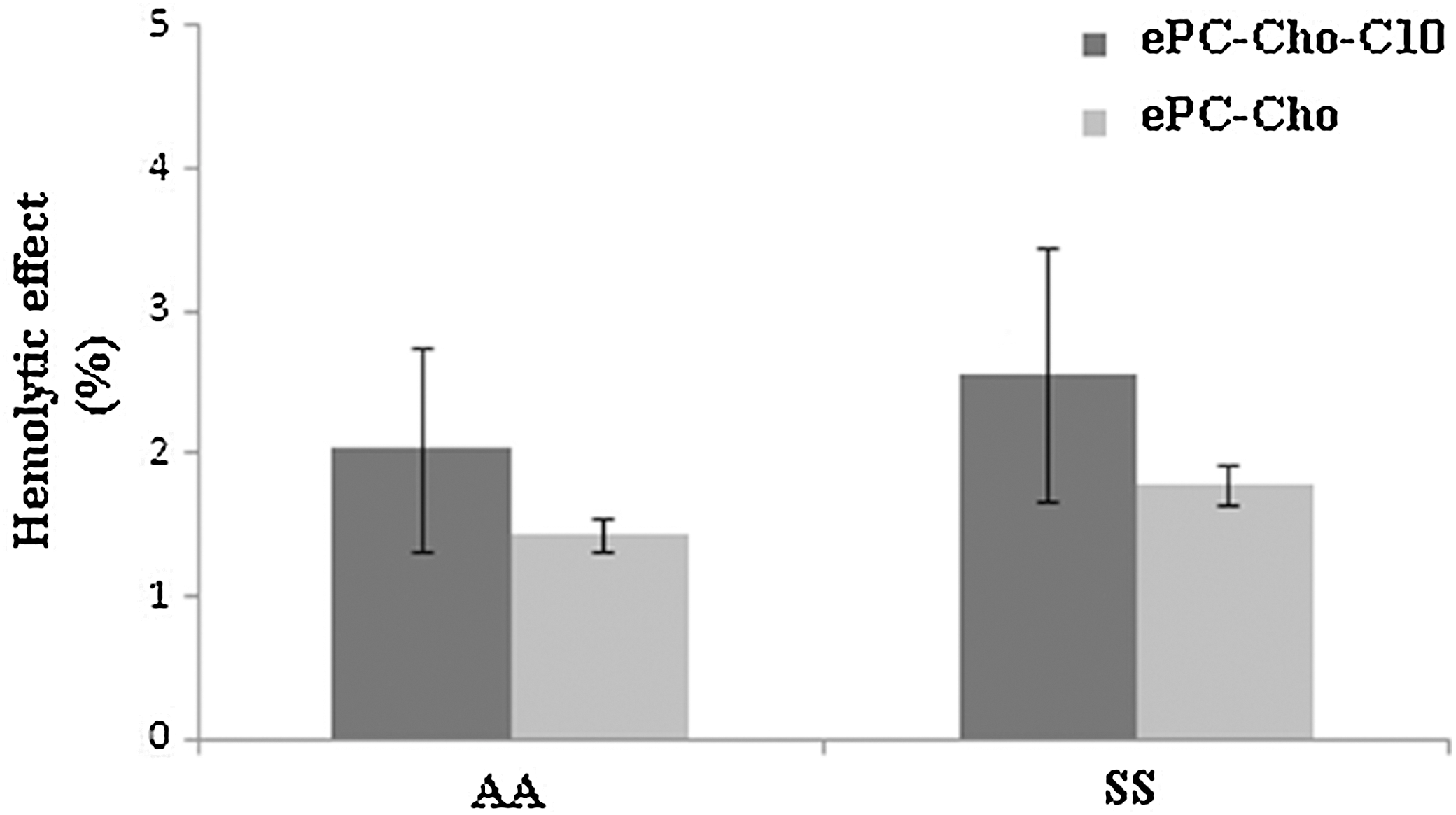
Hemolytic effect calculated in vitro by the spectrophotometric method for C10-modified and unmodified liposomes on erythrocytes of sickle cell disease patients (SS) and volunteer donors (AA). No statistically significant differences were observed between treatments when analysis of variance was applied (P>.05). All values are given as the arithmetic mean±standard deviation of three replicates. SS, erythrocytes from patient with SCD; AA, erythrocytes from healthy individuals.
Although not significant, there was a trend toward increased hemolysis in preparations containing C10. This is consistent with previous studies where hemolytic events in SS erythrocytes were increased by the presence of this lipidic ether. 30 These results may be caused by the ability of C10 to interact with cell membranes. Since polymerization of HbS causes intracellular membrane damage and hemoglobin release with values of up to 40% of hemolysis in patients with SCD, 24 it can be considered that C10 acts to increase the hemolytic effect in the case of SS erythrocytes. In general, the hemolytic effect of the tested preparations is very low compared with the maximum hemolysis reported as permissible in the literature, which is 10%. This is the maximal hemolysis that a compound may cause without toxicity in the in vitro test developed by Stanley et al. 22
Effect on erythrocytes SS
All tested preparations showed a marked decrease in the number of sickle cells at 24 h of incubation, compared to positive control (Table 3). For the positive control (Fig. 3a, b), a significant increase (63%±4.2%) in the number of sickle cells at 48 h was observed, compared with the number of cells at 24 h of incubation, 25%±3.2% (P<.05).
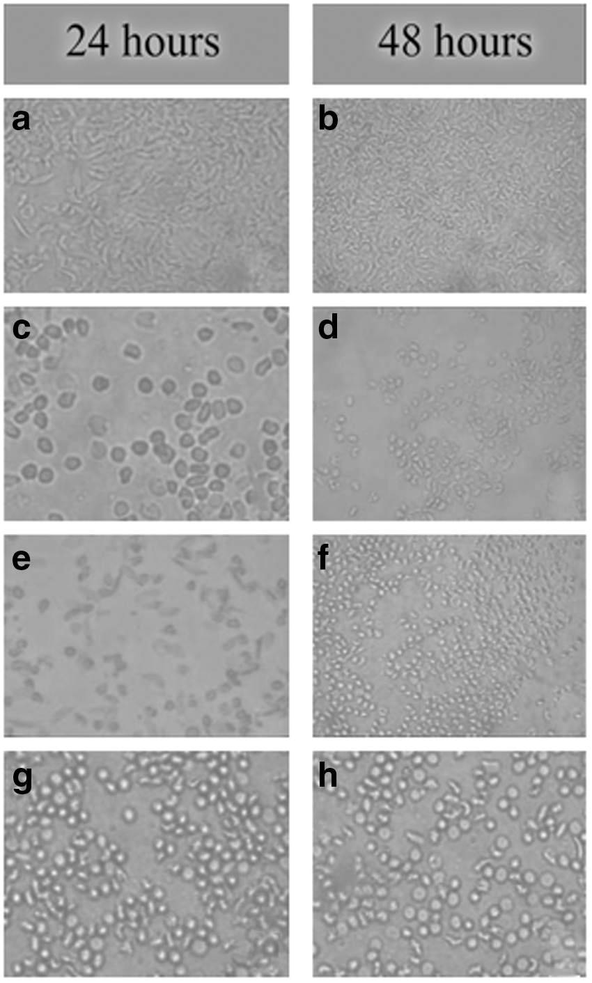
Effects of liposomes and unencapsulated vanillin on SS erythrocytes (magnification, ×40). Pictures were taken at 24 and 48 h after treatment in oxygen-lacking conditions:
Sickle cells were counted at different times after subjecting the erythrocytes with each compound to anoxygenic conditions by the Huck test. Values are given as the arithmetic mean±standard deviation of six replicates.
Different letters indicate significant differences using analysis of variance coupled with Duncan's test (P<.05).
The liposomal preparation containing C10 showed the lowest values in sickle cell formation, with values of 6.0%±1.1% at 24 h and 5.0%±2.8% at 48 h, with erythrocytes appearing similar to echinocytes (Fig. 3c, d), which are red cells with prolongations regularly distributed in the surface and are more flexible than sickle cells. These averages differ significantly (P<.05) for the positive control and the other preparations for each incubation time. No significant differences between the two incubation times (P>.05) were observed. The change of the red cells into echinocytes is reversible, and the erythrocytes may recover their normal shape. 31
Compared with the positive control, the liposomal preparation without C10 (Fig. 3e, f) showed a significant difference (P<.05) in the number of sickle cells, with the average value of 15%±1.7% at 24 h and 13.5%±2.1% at 48 h; no significant differences between stages (P>.05) were observed.
Unencapsulated vanillin (Fig. 3g, h) showed significant differences (P<.05) with the positive control and did not differ significantly (P>.05) from the preparation without C10. The average number of sickle cells for vanillin was 13.3%±0.5% at 24 h and 12.5%±3.6% at 48 h and no significant differences were found for these values (P>.05).
Previous works 8,30 have shown the potential of these compounds to inhibit sickle deformation. Investigations showed that vanillin and C10 exert their effects by different routes. Vanillin is covalently attached to HbS, forming Schiff bases with the free terminal amino groups and inhibiting the polymerization process of this molecule. 8,9 C10 appears to exert its actions by modifying the critical micelle concentration of the cell membrane. 30 In SCD, there is a reduction in the mechanical resistance of the erythrocyte membranes due to dehydration and the polymerization of HbS. 24 Membrane becomes more rigid and less flexible due to tight junctions between the phospholipids, leading to irreversible sickle deformation and hemolysis. 24,32 There are also alterations in the cytoskeleton. Incorporation of C10 makes the membrane more flexible by intercalation between tight lipids, and thus modifies the lipid composition and concentration of cellular bilayers. 33
Considering these routes, the synergistic effect observed in C10-containing preparations can be attributed to an inhibition of HbS polymerization by vanillin and an influence of C10 in the micellar state of the cell membrane. Furthermore, C10 may promote the entry of vanillin into erythrocytes. In vivo it is likely that the previously postulated mechanisms act together. An important consideration is the fact that experiments were performed with whole blood from patients with SCD. In the blood, there are multiple components besides erythrocytes, including plasma proteins, lipids, and electrolytes. Encapsulation of vanillin could prevent the interaction of this molecule with such components, which could potentially decrease its interaction with erythrocytes.
Regarding the administration route of liposomal vanillin, some considerations must be made. During oral administration, the liposome stability may be affected. Mechanical agitation due to peristaltism in the gastrointestinal tract may cause breakdown of membranes and liberation of the entrapped material. 34 The changes in pH through the medium lead to changes in the superficial charge of vesicles, which may promote aggregation and changes in membrane permeability. 35 The bile salts can destroy the membrane structure and promote the active principal releasing before it reaches the blood. 36 The enzymes present in the gut and the liver may also affect membrane integrity. 36,37 For that reason, oral administration of liposomes is not always successful and some alternatives, such as the polymer coats, 38 have been developed to improve the oral stability of vesicles. Parenteral administration is preferred for liposomes, and this may be an alternative to oral administration of vanillin. Nevertheless, both administration methods should be tested.
The fact that modification of multilamellar liposomes with C10 enhances the pharmacological properties of a solute such as vanillin without increasing hemolytic events is evidence that this type of vehicle may improve the pharmacokinetics and pharmacodynamics of an active ingredient. This suggests the possibility of using these compounds for the future treatment of diseases such as sickle cell anemia. Considering that the synergism observed in the present study was prompted by a structural component of the liposomal vesicles (C10), the result can be considered as an attempt to incorporate the liposomal vesicles to the pharmacological activity.
In summary, liposomal vesicles are valuable tools as vehicles for vanillin administration. The fact that the encapsulation of this molecule does not affect their pharmacological properties supports the use of liposomes in future pharmacokinetic and pharmacodynamic studies, both in vivo and in vitro.
Footnotes
Acknowledgment
The authors thank the Biochemistry Group, Department of Biology, Faculty of Natural Sciences, University of Oriente.
Author Disclosure Statement
No competing financial interests exist