
Abstract
North American ginseng (NAG) has received increasing attention as an alternative medicine for the treatment of diabetes. Extract of the NAG root has been reported to possess antidiabetic properties, but the underlying mechanisms for such effects have not been identified. Here we investigated the effects of NAG root extract on type 1 and 2 diabetes and the underlying mechanisms involved for such effects. Type 1 [C57BL/6 mice with streptozotocin (STZ)-induction] and type 2 (db/db) diabetic models were examined. Groups of diabetic mice (both type 1 and 2) were treated with alcoholic extract of the NAG root (200 mg/kg BW/day, oral gavage) for 1 or 2 months following onset of diabetes. Ginseng treatment significantly increased the body weight in type 1 diabetic animals in contrast to the type 2 model, where it caused diminution of body weight. Blood glucose and glycated hemoglobin levels diminished in the diabetic groups of both models with NAG treatment. Interestingly, plasma insulin and C-peptide levels were significantly increased in the STZ-diabetic mice, whereas they were reduced in the db/db mice following NAG treatment. Histological and morphometric analyses (islet/pancreas ratio) of the pancreas revealed an increase in the islet area following the treatment compared to both the untreated diabetic groups. These data indicate that NAG possibly causes regeneration of β-cells resulting in enhanced insulin secretion. On the other hand, in type 2 diabetes, the additional effects of NAG on body weight might have also resulted in improved glucose control.
Introduction
D
Previously, we and others have demonstrated that North American ginseng (NAG) root extract prevents chronic diabetic complications. 16 –19 It has been also reported by us and other groups that the high-performance liquid chromatography (HPLC) profile of NAG root extract contains a higher content of total ginsenosides with a distinct ginsenoside profile. 17 –21 In this study, we investigated the pharmacological effects of NAG root extract on insulin secretion and pancreatic islet using mouse models of type 1 and type 2 diabetes.
Materials and Methods
Reagents
All reagents were obtained from Sigma-Aldrich (Oakville, ON, Canada) unless otherwise specified.
Preparation of ginseng root extract
Four-year-old NAG roots (collected in 2007 from five different farms in Ontario, Canada) were provided by the Ontario Ginseng Growers Association. Dried ginseng root samples were shipped to Naturex (South Hackensack, NJ, USA) for extraction. A detailed methodology for alcoholic extraction and characterization has been previously published. 18,21 Briefly, dried ginseng root samples (4 kg) were ground and soaked thrice in a 75% ethanol solution (16 L) at 40°C for 5 h. The resultant solution was filtered and the excess solvent was removed by a rotary evaporator under vacuum at 45°C. The three pools were combined and concentrated until the total solid on dry basis is ∼60%. The concentrated was further lyophilized and the final yield of the powdered alcoholic extract was approximately 35.3% of the initial ground root. After that, HPLC of the alcoholic extract (100 mg/mL methanol) was performed by using the reversed-phase Inspire C-18 column. The gradient program consisted of water and acetonitrile at a flow rate of 1.3 mL/min and elutes were monitored at 203 nm. The extract in powdered form was analyzed to ensure no excessive contamination with pesticides and heavy metals were present.
Animals
All animals were cared following the Guiding Principle of the Care and Use of Animals. All experiments were approved by the University of Western Ontario Council on Animal Care Committee. Male C57BL/6 mice weighing 23–36 g were obtained from Charles River (Montreal, QC, Canada). To create a model of type 1 diabetes, groups of mice received three intraperitoneal injections of streptozotocin (STZ, 50 mg/kg in the citrate buffer, pH 5.6) on alternate days. Controls (C) were injected with the same volume of the citrate buffer. After three STZ injections, animals with stable blood glucose levels at ≥25 mM on three consecutive days were considered diabetic. Randomly selected diabetic animals (D) were monitored for 4 and 8 weeks. Furthermore, a group of the diabetic mice were subjected to NAG treatment for 4 and 8 weeks (D+NAG) at the onset of diabetes. Alcoholic ginseng extract was dissolved in Dulbecco's phosphate-buffered saline, pH 7.4 and administered by daily gavage (200 mg/kg BW). For the type 2 diabetic model, male db/db (Leprdb) mice and controls (CO, C57BL/6) were purchased from Jackson Laboratories (Bar Harbor, ME, USA. Randomly selected diabetic animals (db) were monitored for 4 and 8 weeks. Furthermore, a group of the diabetic mice were subjected to NAG treatment for 4 and 8 weeks (db+NAG) following the onset of diabetes. In each group (controls, diabetic, and diabetic treated with NAG), five animals were included.
Methods
Body weights of all mice were monitored before and every 7 days after the onset of treatment with NAG. Blood (heparinized/nonheparinized) was collected from the tail vein and blood glucose was measured before and every 7 days after onset of treatment with NAG by a Glucometer (Free Style Freedom Lite, Inc., Saint-Laurent, PQ, Canada). Glycated hemoglobin (GlycoHb) levels were measured every 4 weeks after initiation of NAG administration by using the GlycoHb estimation kit (Stanbio Laboratories, Boerne, TX, USA). The mouse insulin ELISA kit (DRG Diagnostics, Marburg, Germany) was used to measure the plasma insulin concentration in all the groups after 4 and 8 weeks of treatment with NAG. Serum C-peptide levels were measured using the Rat/Mouse C-Peptide 2 ELISA kit (Millipore, MA, USA) after 4 and 8 weeks after induction of NAG administration.
The animals were sacrificed after the follow-up period (4 or 8 weeks) by cervical dislocation and pancreas was collected. Pancreatic tissues were fixed in 10% neutral-buffered formalin, dehydrated in a series of graded ethanol, and were embedded in paraffin. The tissues were sectioned at 5 μm and were stained with hematoxylin and eosin. The islet/pancreas area ratio was analyzed by SPOT Basic 5.0 software (Diagnostic Instruments, Sterling, MI, USA).
Statistical analysis
Data are expressed as mean±standard error of the mean. Statistical significance was determined by analysis of variance followed by the Bonferroni–Dunn test. Differences were considered to be statistically significant at values of P<.05.
Results
Phytochemical characteristics of NAG root extract
NAG root extract used in this study had a total ginsenoside content of 28.25%. With respect to the ginsenoside content, 1 g of alcoholic NAG root extract contained 2.9 mg of Rg1, 89.6 mg of Re, 164.5 mg of Rb1, 12.6 mg of Rc, 1.6 mg of Rb2, and 11.1 mg of Rd. The Rb1 and Re were two predominant ginsenosides in the extract with the Rb1/Re ratio of 1.8. The Rb1/Rg1 ratio was 56.74.
Effect of NAG on body weight
In the type 1 diabetic mice (D), significant diminution of body weight was observed compared to respective controls (Fig. 1A). Treatment with NAG resulted in gradual improvement of body weight of the diabetic mice (D+NAG; Fig. 1A). It should be mentioned here that body weight improvement in the (D+NAG) mice was initiated from the end of 2 weeks of treatment regime. On the other hand, in type 2 diabetic mice (db), significant elevation of body weight was observed in comparison to the controls (Fig. 1B). NAG treatment resulted in normalization of the body weight gain in (db+NAG) mice treated for 8 weeks (Fig. 1B).

Effects of North American ginseng (NAG) root extract on body weights in
Effects of NAG on blood glucose and GlycoHb levels
Both in type 1 and type 2 diabetic mice (D and db), significant elevation of blood glucose levels occurred compared to respective controls (C and CO; Fig. 2A, B) monitored for 8 weeks. NAG treatment of the diabetic mice (D+NAG and db+NAG) caused significant reduction of blood glucose levels (Fig. 2A, B). GlycoHb levels were found to be significantly increased in all the diabetic mice (both D and db) compared to the controls (C and CO; Fig. 3A–D). Treatment of type 1 diabetic mice with NAG for 4 (Fig. 3A) or 8 weeks (Fig. 3B) caused significant decrease of GlycoHb levels. Similarly, in type 2 diabetic mice, NAG treatment for 4 (Fig. 3C) or 8 (Fig. 3D) weeks caused reduction of the GlycoHb levels both after 4 and 8 weeks of follow-up.
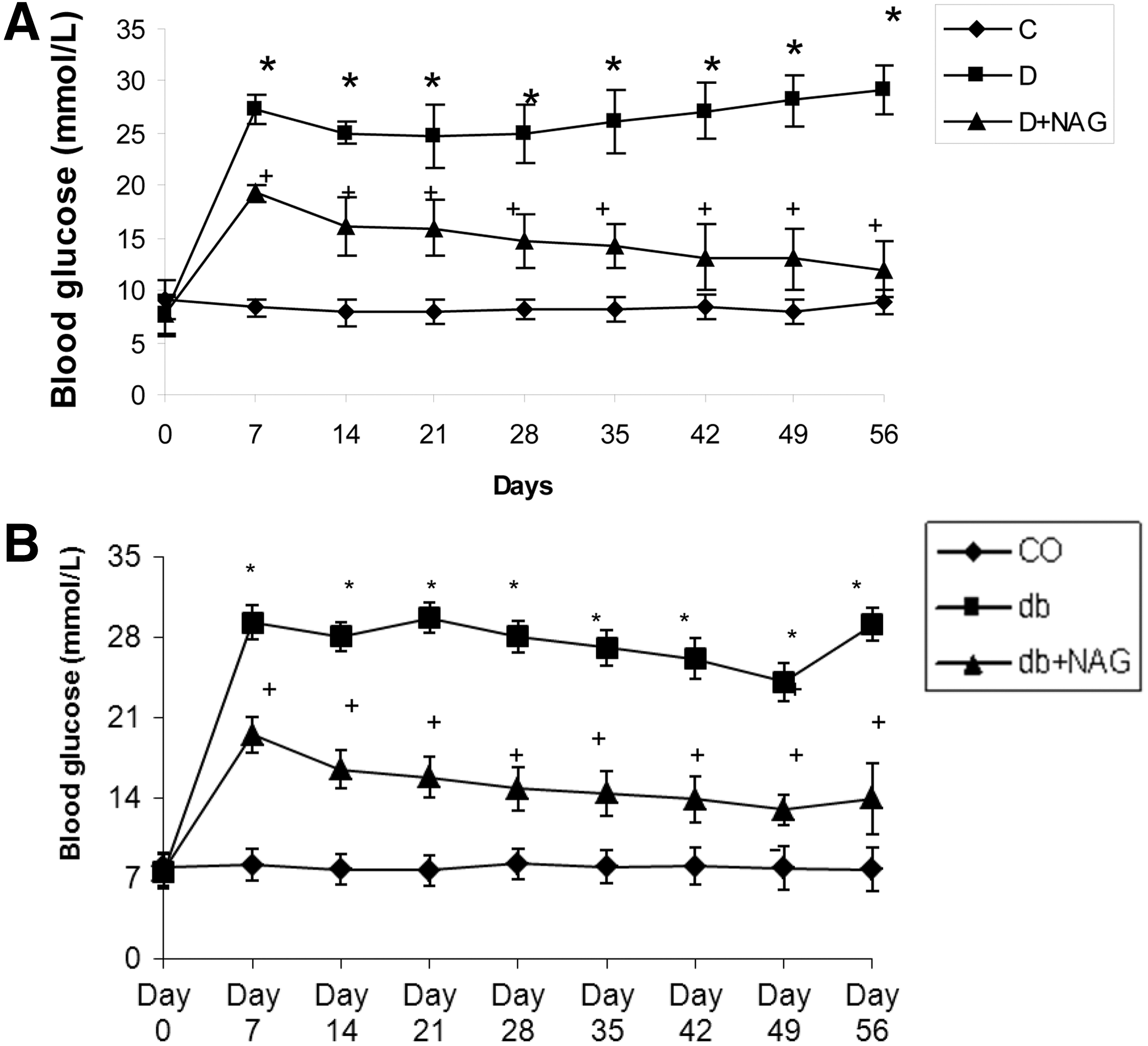
Effect of NAG root extract on blood glucose levels in
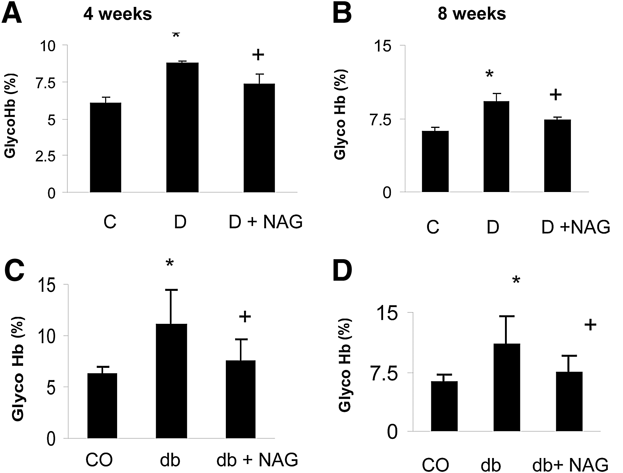
Effect of NAG root extract on glycated hemoglobin levels in
Effects of NAG on plasma insulin and C-peptide levels
Lowering of blood glucose and GlycoHb levels by NAG in diabetic mice prompted us further to investigate its effect on insulin levels. Lowering of the plasma insulin level was observed in type 1 diabetic mice (Fig. 4A, B) in comparison to the controls. NAG resulted in elevation of plasma insulin levels at 4 weeks (Fig. 4A) of treatment in the type 1 diabetic mice. Such increase was pronounced following 8 weeks of treatment (Fig. 4B). In the type 2 diabetic mice, the plasma insulin level was significantly increased (Fig. 4C, D) compared to the controls. Treatment with NAG for 4 and 8 weeks caused significant lowering of the plasma insulin level (Fig. 4C, D). To confirm that this elevation is due to secreted insulin, we measured the plasma C-peptide level. The C-peptide level is indicative of the synthetic activity of the β-cells. The C-peptide level was significantly diminished in type 1 diabetic mice (Fig. 5A, B). NAG treatment resulted in significant elevation of the plasma C-peptide level in type 1 diabetic mice (Fig. 5A, B). Interestingly, in type 2 diabetic animals, the C-peptide level significantly increased (Fig. 5C, D) and was reduced after 8 weeks of NAG treatment (Fig. 5C, D).
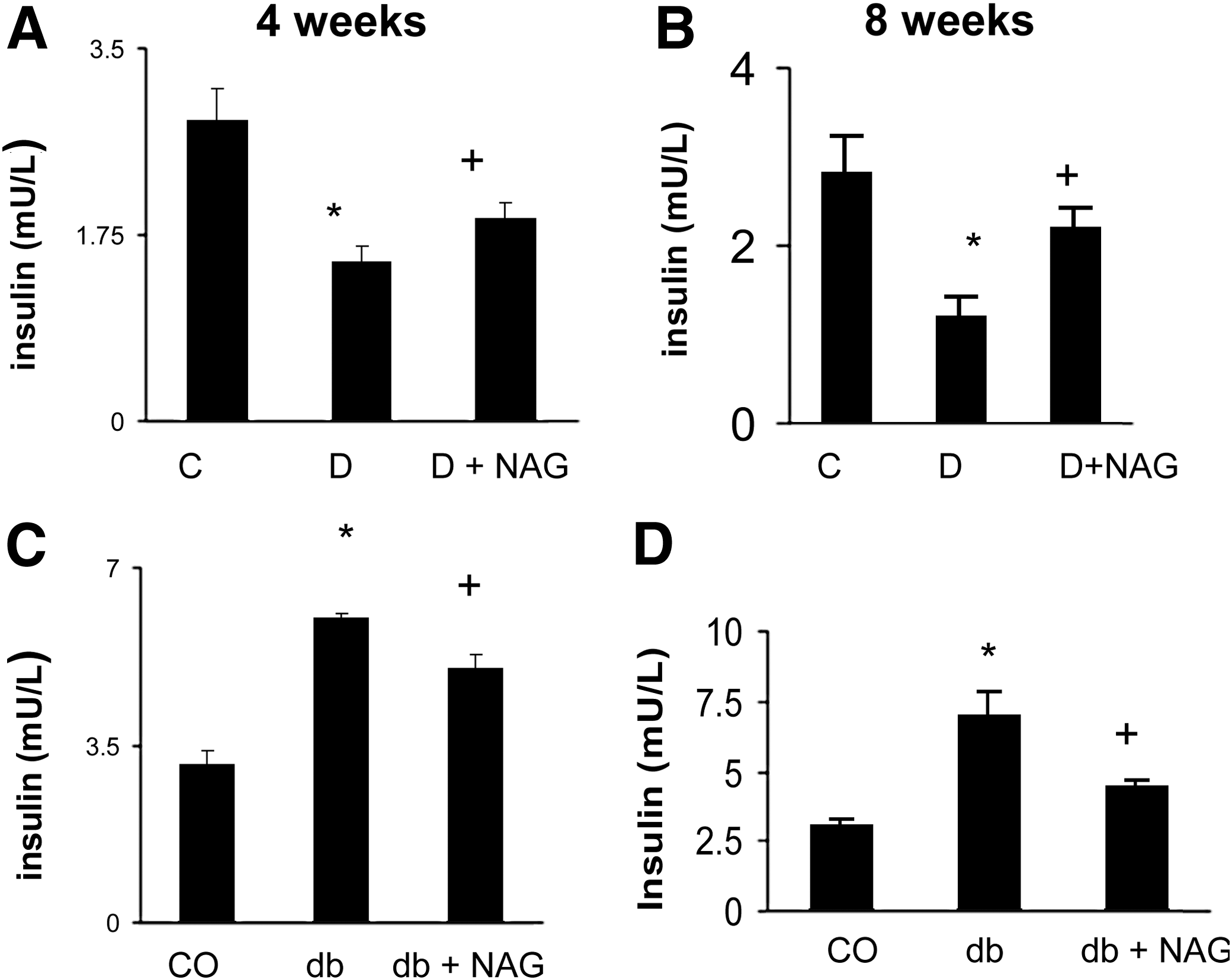
Effect of NAG root extract on plasma insulin levels in
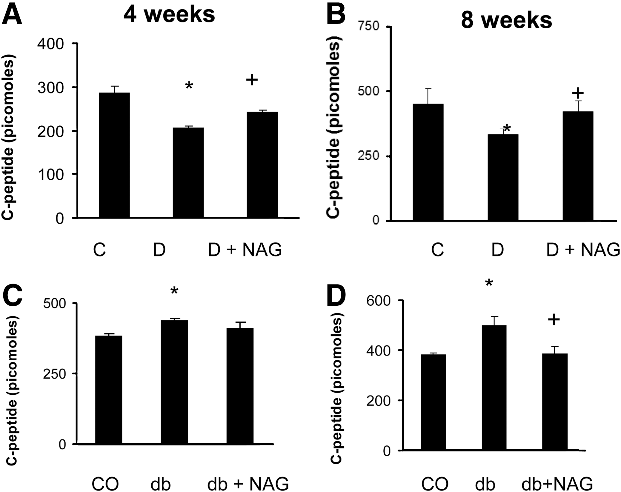
Effect of NAG root extract on plasma C-peptide levels in
Histological analysis of the pancreas
Histological examination of pancreatic sections revealed a significant decrease of the pancreatic islet area in type 1 diabetic mice (Fig. 6B) in comparison with control (Fig. 6A). The islet/pancreas area ratio was found to be diminished in type 1 diabetic mice (Fig. 6D, E). NAG-treated type 1 diabetic mice showed significant improvement of such parameters (Fig. 6C–E). On the other hand, in db/db mice, islet areas were not significantly changed compared to the controls. However, treatment with NAG increased the islet/pancreas ratio after 8 weeks of follow-up (Fig. 6F, G).

Effect of NAG root extract on the histoarchitecture of pancreas in
Discussion
The primary objective of treatment of diabetes is to maintain a normal range of the blood glucose concentration. This can be achieved by increasing the insulin secretion or augmenting insulin utilization. Here we have studied the antihyperglycemic efficacy of NAG root extract in mouse models of type 1 and type 2 diabetes and explored its effects on insulin secretion and pancreatic β-cells.
In the present study, we observed a significant antihyperglycemic effect of NAG root extract in type 1 and type 2 diabetic mice, as evidenced by decreases in the blood glucose levels after 8 weeks of treatment. Such antihyperglycemic effects were further confirmed by reduced glycated hemoglobin levels in STZ-induced and db/db mice. We also observed that NAG treatment significantly ameliorated the body weight loss in type 1 diabetes. In accordance with the previous studies, the mechanisms of action of ginseng's antidiabetic activity could be multifaceted, attributable to the modulation of gastrointestinal absorption, the regulation of insulin secretion and/or sensitivity, C-peptide levels, or a combination of these factors. 7,22 –24 Here we demonstrated that the improvement in the blood glucose levels in NAG-treated STZ-induced diabetic mice is, at least in part, associated with a significant increase in C-peptide levels and insulin secretion. Such improvements were associated with a significant increase of the islet/pancreas area ratio. Park et al. reported enhancement of β-cell mass in type 1 diabetes after treatment with Panax ginseng berry extract. They also demonstrated β-cell proliferative effects of ginseng in INS-1 cells, suggesting that an increase in cell mass is one possible mechanism explaining its antidiabetic effects. 25 In the present study, increment of the islet/pancreas area ratio in the NAG-treated type 1 diabetic mice in comparison to the untreated diabetic animals also indicates that the possible mode of action of NAG is regeneration or sensitization of pancreatic β-cells that in turn elevated the serum insulin and c-peptide levels thereby rectifying hyperglycemia and elevated glycated Hb levels.
In type 2 diabetic mice, NAG treatment reduced the body weight gain associated with lowering of glycated hemoglobin, plasma C-peptide, and insulin levels. Enhancement of the islet/pancreas area ratio was also observed in db/db mice treated with NAG for 8 weeks.
Blood glucose and glycated Hb are indicators of the diabetic dysmetabolism. The blood glucose level is a day to day indicator of the diabetic state, whereas glycated Hb levels indicate the state of long-term glucose control. In clinical and experimental studies, it has been demonstrated that lowering of blood glucose and glycated Hb levels are associated with prevention of chronic diabetic complications in both type 1 and type 2 diabetes. 26 –28 In our study, we found partial correction of both blood glucose and glycated Hb levels, indicating the beneficial effects of NAG. We believe that, at least in part, this was mediated through increased insulin synthesis by the β-cells. This is due to the fact that, ginseng treatment was associated with an increased insulin and c-peptide level, a cleavage product of insulin following its synthesis. These data are in keeping with partial morphologic restoration of the islet size. However, the additional direct glucose-lowering effects of NAG cannot be excluded. These findings are further in keeping with our previous report that NAG causes reduction of body weight, plasma insulin, and HOMA-index in db/db mice. 18 Xiong et al. 29 also proposed about the antiobesity and anti-hyperglycemic activities of the ginsenoside, Rb1 in high fat-fed obese rats. Rb1 has also been demonstrated to stimulate basal and insulin-mediated glucose uptake in a time- and dose-dependent manner in both 3T3-L1 adipocytes and C2C12 myotubes. 30 Our HPLC analysis indicated that NAG alcoholic root extract contained a very high concentration of Rb1, which is also consistent with other groups. 17 –21,31,32 Thus, along with enhancement of islet mass, the additional body weight-lowering effect of NAG in type 2 diabetic mice may also be partially responsible for improved control of hyperglycemia and better utilization of glucose. This was further confirmed by reduced glycated Hb levels in these mice. Several other groups have also reported pancreatic β-cell regenerative properties of other plant extracts and their active ingredients, namely, Gymnema sylvestre, 33 glycyrrhizin (the active ingredient of Glycyrrhiza glabra), 34,35 etc.
Data from this study indicate that NAG has a protective action against diabetes possibly through regeneration of β-cells. In type 2 diabetes, the additional effect of NAG on body weight might have also resulted in improved glucose homeostasis and uptake.
Footnotes
Acknowledgment
This research was supported by the Ontario Research Fund from the Ministry of Research and Innovation, Government of Ontario.
Author Disclosure Statement
No competing financial interests exist.