
Abstract
Porphyromonas gingivalis is a key etiologic agent of chronic periodontitis. This Gram-negative anaerobic bacterium produces several virulence factors and can induce a host inflammatory response that contributes to periodontal disease. In the present study, we investigated green tea, white tea, oolong tea, and black tea extracts with a high polyphenol content for their effects on (i) the growth and adherence of P. gingivalis, (ii) the activity of host and bacterial proteases, and (iii) cytokine secretion by oral epithelial cells. All the tea extracts inhibited the growth of P. gingivalis (minimal inhibitory concentrations ranging from 200 to 500 μg/mL; minimal bactericidal concentrations=500 μg/mL). In addition, they dose dependently reduced the adherence of P. gingivalis to oral epithelial cells. Tea extracts also inhibited the catalytic activity of matrix metalloproteinase (MMP)-9, neutrophil elastase, and P. gingivalis collagenase. Lastly, the tea extracts dose dependently inhibited the secretion of interleukin (IL)-6, IL-8, and chemokine (C-C motif) ligand 5 (CCL-5) by P. gingivalis–stimulated oral epithelial cells. No marked differences in the various effects were observed among the four tea extracts. Extracts from green tea, white tea, oolong tea, and black tea show promise for controlling periodontal disease by their capacity to interfere with P. gingivalis growth and virulence properties, host destructive enzymes, and inflammatory mediator secretion. Such extracts may be incorporated to oral hygiene products or locally delivered into diseased periodontal sites.
Introduction
O
The gingival epithelium, which covers the periodontal tissues, plays a crucial protective role as a mechanical barrier that prevents the invasion of the periodontium by periodontopathogens. 7 Gingival epithelial cells react to bacterial challenges by signaling host responses and integrating innate and acquired immune responses. 7 P. gingivalis has developed different strategies to perturb the structural and functional integrity of the gingival epithelium. 8 P. gingivalis adheres to, penetrates, and replicates inside gingival epithelial cells. 8,9 In addition, proteolytic enzymes produced by P. gingivalis can interfere with both the cell–matrix and cell–cell adhesion of epithelial cells, which may cause the junctional epithelium to detach from the root surface and regenerating tissues and promote the formation of gingival pockets. 8 Interactions between P. gingivalis and epithelial cells lead to the activation of several complex signaling cascades that ultimately regulate the transcription of target genes that encode effectors and regulators of the immune response. 8,9 More specifically, effectors of the innate immune system, including proinflammatory cytokines, chemokines, and matrix metalloproteinases (MMPs), are upregulated and may have a direct impact on periodontal disease progression and inflammation processes. 8,9
Tea, an aqueous aromatic infusion of cured leaves of the plant Camellia sinensis, is the most popular beverage in the world after water. It contains numerous components, including catechins, caffeine, amino acids, carbohydrates, proteins, chlorophyll, volatile compounds, fluoride, minerals, and other undefined compounds. 10 Teas can be classified as nonfermented (green and white teas), semifermented (oolong tea), and fermented (black tea). Green tea differs from white tea by the fact that the latter is produced only from the buds or first leaves. The chemical composition of teas depends on how they are processed. For example, green tea has a high catechin content; black tea has a high bisflavanol, theaflavin, and thearubigin content; 11 white tea has a high epigallocatechin-3-gallate (EGCG), epicatechin, and methylxanthine content. 12 Traditional Chinese medicine has considered tea as a medicine and healthful beverage since ancient times. Several biological properties have been associated to tea polyphenols, including antioxidant, anticarcinogenic, and antimicrobial activities. 11 Many studies have shown that the constituents of tea may contribute to reducing the risk of cardiovascular disease and cancer and have a variety of other beneficial effects on human health. 11 –13 Epidemiological and clinical studies have provided evidence that green tea consumption may have potential oral health benefits. 14 –17 However, few studies have compared the beneficial biological properties of different types of tea with respect to periodontal disease. The aim of the present study was to investigate the effects of major types of tea on (i) the growth and adherence of P. gingivalis, (ii) the activity of host and bacterial proteases, and (iii) cytokine secretion by oral epithelial cells.
Materials And Methods
Tea extracts
Extracts from green tea, white tea, oolong tea, and black tea were purchased from Organic Herb, Inc. (Changsha, China). Information provided by the company indicated that these extracts (water/ethanol) were prepared from tea leaves and all have a polyphenol content ≥92%. Stock solutions were prepared by dissolving 20 mg of powder in 1 mL of sterile distilled water and filtering the solution through a 0.45-μm-pore-size membrane filter.
Bacteria and culture conditions
P. gingivalis ATCC 33277 was grown in the Todd-Hewitt broth (THB; BBL Microbiology Systems, Cockeysville, MD, USA) supplemented with hemin (10 μg/mL) and vitamin K (1 μg/mL). Bacterial cultures were incubated for 24 h at 37°C under anaerobic conditions (80% N2, 10% H2, 10% CO2).
Determination of minimal inhibitory and minimal bactericidal concentrations
A 24-h culture of P. gingivalis was diluted in a fresh broth medium to obtain an optical density of 0.02 at 655 nm (OD655). Tea extracts (100 μL) diluted in a culture medium (20–2000 μg/mL) were placed in the wells of flat-bottomed 96-well microplates (Sarstedt, Newton, NC) to which an equal volume of P. gingivalis suspension was added. Wells containing only bacteria or tea extract were used as controls. After a 24-h incubation at 37°C under anaerobic conditions, bacterial growth was monitored by measuring the OD655 using a microplate reader (BioTek Instruments, Winooski, VT, USA). Minimal inhibitory concentration (MIC) values (μg/mL) were defined as the lowest concentration of the extract at which no growth occurred. To determine minimal bactericidal concentration (MBC) values (μg/mL), aliquots (10 μL) from each well with no visible growth were spread on culture plates, which were incubated for 5 days at 37°C under anaerobic conditions. MBC values were defined as the lowest concentration at which no colonies grew. The MIC and MBC assays were performed in triplicate and were repeated three times to ensure reproducibility.
P. gingivalis adherence to human oral epithelial cells
The immortalized human oral epithelial cell line GMSM-K, which was kindly provided by Dr. Valerie Murrah (Department of Diagnostics Sciences and General Dentistry, the University of North Carolina, Chapel Hill, NC, USA), was cultured in the Dulbecco's modified Eagle's medium (DMEM) supplemented with 10% heat-inactivated fetal bovine serum (FBS) and 100 μg/mL of penicillin G/streptomycin at 37°C in a 5% CO2 atmosphere. The epithelial cells were harvested by gentle trypsinization (0.05% trypsin-EDTA) (Gibco-BRL, Grand Island, NY, USA), seeded (100 μL, 1.5×10⁶ cells/mL) in sterile 96-well clear bottom black microplates (Greiner Bio One, Frickenhausen, Germany), and incubated until they reached confluence. The wells were then washed three times with 50 mM phosphate-buffered saline (PBS) pH 7.2, blocked with 1% bovine serum albumin (BSA) for 30 min to prevent nonspecific bacterial attachment, and treated with the tea extracts diluted in the DMEM at final concentrations ranging from 10 to 100 μg/mL for 30 min in a 5% CO2 atmosphere at 37°C. P. gingivalis cells from an overnight culture were suspended (109/mL) in a bicarbonate buffer (0.15 M NaCl/0.1 M Na2CO3, pH 9.0), incubated for 30 min with continuous shaking in the presence of 15 μg/mL of fluorescein isothiocyanate isomer I (FITC; Sigma-Aldrich Canada, Oakville, ON, Canada) in the dark, washed three times with PBS containing 0.05% Tween 20, resuspended in PBS in the original volume, applied at a multiplicity of infection of 200 (200 bacteria per epithelial cell) to treated or control epithelial cells, and incubated for 2 h at 37°C under anaerobic conditions. The incubation and washing steps were carried out in the dark. Following the incubation, unbound P. gingivalis cells were removed by aspiration, and the wells were washed three times with PBS. P. gingivalis cells that had adhered to the epithelial cell monolayer were quantified by monitoring fluorescence using a Synergy 2 Multi-Mode Microplate Reader (BioTek Instruments). The excitation and emission wavelengths were set at 488 and 522 nm, respectively. The assays were performed in triplicate and were repeated three times.
MMP-9, elastase, and P. gingivalis collagenase activities
Human active recombinant MMP-9 and neutrophil elastase were purchased from Calbiochem (San Diego, CA, USA). MMP-9 (1 μg/mL) diluted in the TCNB buffer (50 mM Tris-HCl, 10 mM NaCl, and 0.05% Brij 35, pH 7.5) was incubated with tea extracts (10 to 100 μg/mL) and gelatin DQ™ (150 μg/mL). A 48-h P. gingivalis culture supernatant was incubated with tea extracts (10–100 μg/mL) and type I collagen DQ™ (150 μg/mL). Elastase (50 μg/mL) was mixed with the substrate I (Calbiochem) (4 mM) and reaction buffer (100 mM Tris-HCl, 500 mM NaCl, pH 7.5), and then incubated with tea extracts (10–100 μg/mL). The assay mixtures were incubated for 4 h at 37°C for MMP-9 and elastase, and for 4 h at room temperature for P. gingivalis collagenase. Mixtures with no substrate or enzyme were used as controls. Fluorescence was measured using the excitation and emission wavelengths set at 490 and 525 nm, respectively. Hydrolysis of the elastase substrate was assayed by measuring the absorbance at 415 nm. The assay was performed in triplicate and was repeated three times.
Preparation of P. gingivalis extract
The P. gingivalis extract was prepared using the method described by Shenker and Slots 18 with some modifications. Briefly, P. gingivalis was grown in THB-HK for 48 h at 37°C under anaerobic conditions. Bacterial cells from a 1-liter culture were harvested by centrifugation (10,000 g for 20 min at 4°C) and were washed with cold PBS. The bacterial cells were sonicated for 10 min on ice using an ultrasonic disruptor. The supernatant was collected by centrifugation at 10,000 g for 20 min and sterilized using a 0.45-μm filter. The protein concentration was evaluated using a protein assay kit (DC protein assay, Bio-Rad Laboratories, Mississauga, ON, Canada), with BSA as a standard. The P. gingivalis sonic extract was kept at −80°C and boiled for 15 min before use.
Cytotoxicity of tea extracts and P. gingivalis sonic extract
A 3-[4,5-diethylthiazol-2-yl]-2,5-diphenyltetrazolium bro-mide (MTT) assay performed according to the manufacturer's protocol (Roche Diagnostics, Mannheim, Germany) was used to determine the effect of the tea extracts (10–100 μg/mL) and the P. gingivalis extract (10 μg/mL) on the viability of GMSM-K oral epithelial cells.
Cytokine secretion by P. gingivalis–stimulated oral epithelial cells
GMSM-K human oral epithelial cells were grown and harvested as described above. The epithelial cells were suspended (4×10⁵ cells/mL) in the DMEM containing 1% heat-inactivated FBS and were seeded (1 mL) in the wells of a 12-well plate. The plate was incubated overnight at 37°C in a 5% CO2 atmosphere to allow cell adhesion before use. The epithelial cells were pretreated with tea extracts (10–100 μg/mL) at 37°C in 5% CO2 for 2 h before adding the P. gingivalis extract (10 μg/mL). After a 24-h incubation, the supernatants were collected and stored at −20°C until used. Epithelial cells incubated with tea extract, but no P. gingivalis extract, or with P. gingivalis extract, but no tea extract were used as controls. Each experiment was performed in triplicate. Commercial enzyme-linked immunosorbent assay (ELISA) kits (R&D Systems, Minneapolis, MN, USA) were used to quantify interleukin-6 (IL-6), interleukin-8 (IL-8), and chemokine (C-C motif) ligand 5 (CCL5) concentrations in the cell-free supernatants according to the manufacturer's protocols. The absorbance at 450 nm was read using a microplate reader, with the wavelength correction set at 550 nm. The rated sensitivities of the kits were 9.3 pg/mL for IL-6, 31.2 pg/mL for IL-8, and 15.6 pg/mL for CCL-5.
Statistical analysis
Values are expressed as the means±standard deviations of at least three assays. The differences between the means were analyzed for statistical significance using the Student's t-test with Bonferroni corrections, and an analysis of variance was performed using the Dunnett's post hoc multiple comparison test. A P value<.05 was considered statistically significant.
Results
The different tea extracts (green, white, oolong, and black) exhibited a comparable dose-dependent antibacterial activity against P. gingivalis (Fig. 1). The MICs of the tea extracts were 200 μg/mL, except for the black tea extract, which had an MIC of 500 μg/mL. The MBCs of the tea extracts were 500 μg/mL. At 50 μg/mL, green tea, white tea, oolong tea, and black tea extracts reduced P. gingivalis growth by 21%±2%, 16%±5%, 19%±4%, and 22%±5%, respectively (Fig. 1).
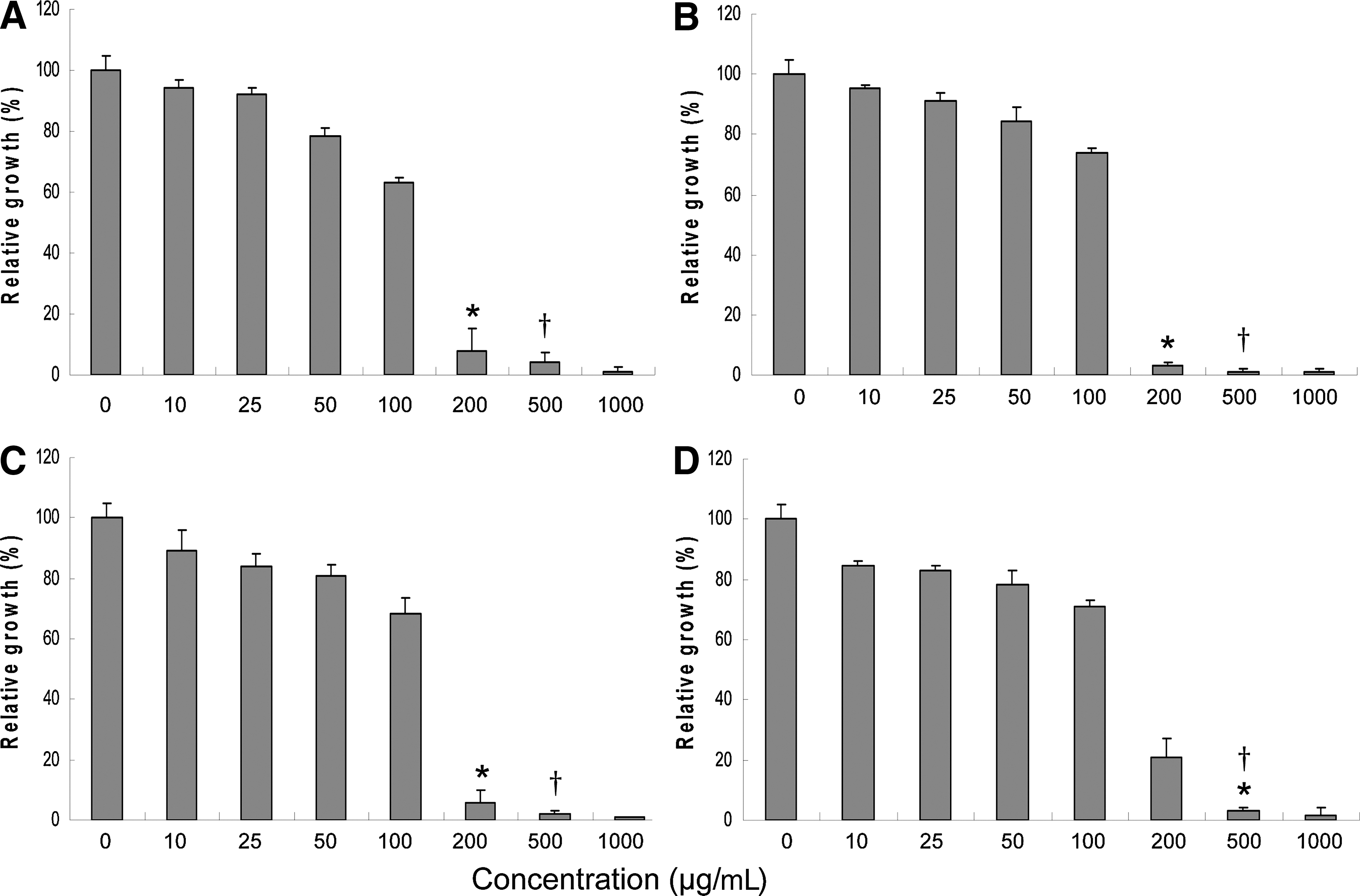
Effect of tea extracts on the growth of P. gingivalis:
The tea extracts all exhibited a marked capacity to dose dependently inhibit the adhesion of P. gingivalis to oral epithelial cells (Fig. 2). The inhibitory effect of the black tea extract was the most pronounced, while the effects of the other tea extracts were similar. The green tea, white tea, oolong tea, and black tea extracts (25 μg/mL) decreased the adherence of P. gingivalis to oral epithelial cells by 63%±3%, 59%±2%, 61%±7%, and 84%±3%, respectively (Fig. 2).
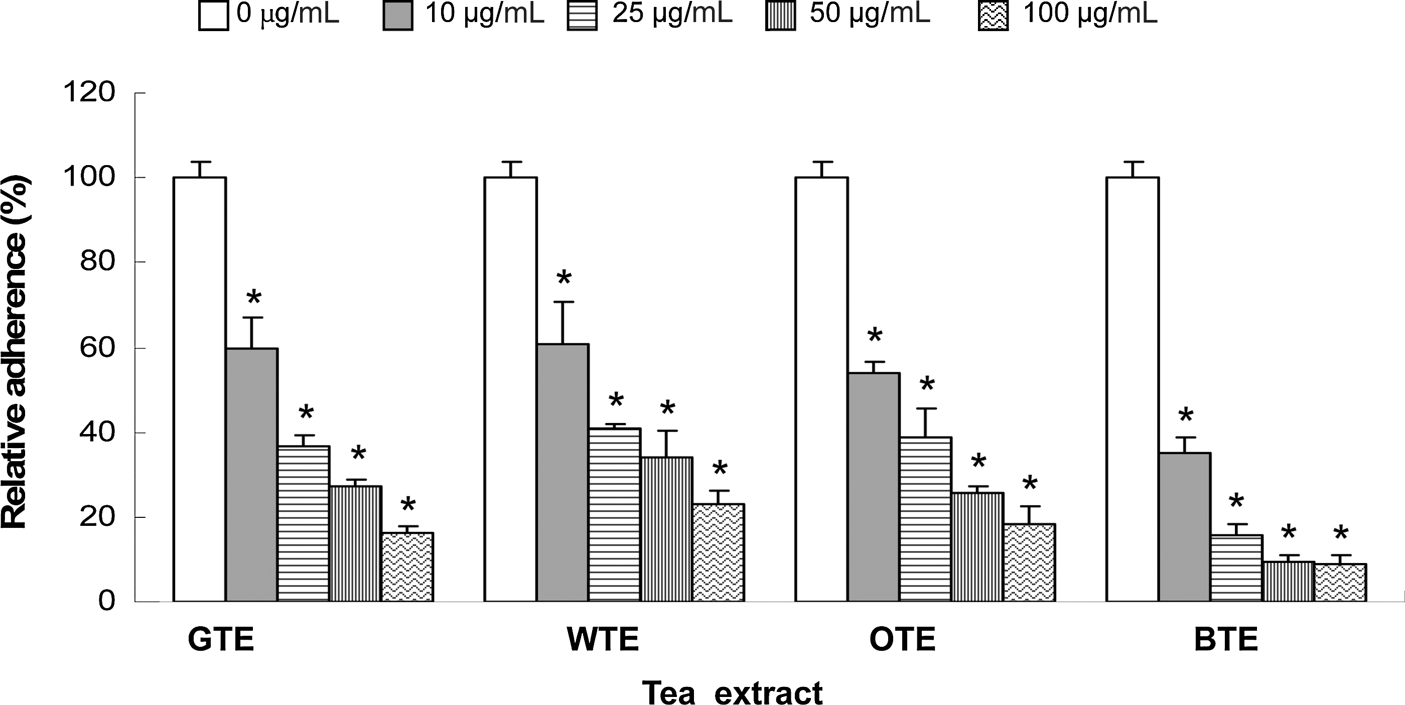
Effect of tea extracts on the adherence of P. gingivalis to human oral epithelial cells. A value of 100% was assigned to fluorescence values obtained in the absence of tea extracts. Results are expressed as the means±SD of triplicate assays for two independent experiments. *Significantly lower than the value for the untreated control (P<.05). GTE, green tea extract; WTE, white tea extract; OTE, oolong tea extract; BTE, black tea extract.
While the tea extracts all dose dependently inhibited P. gingivalis collagenase, MMP-9, and neutrophil elastase activities, the effect on the collagenase activity was more pronounced (Fig. 3). At the lowest concentration tested (10 μg/mL), the green tea, white tea, oolong tea, and black tea extracts significantly (P<.05) inhibited the P. gingivalis collagenase activity by 57%±2%, 51%±1%, 51%±2%, and 38%±1%, respectively (Fig. 3A). The tea extracts (10 μg/mL) also inhibited the MMP-9 activity by 20%±2% to 33%±1% (Fig. 3B), and the neutrophil elastase activity by 11%±2% to 38%±3% (Fig. 3C).

Effect of tea extracts on P. gingivalis collagenase
No obvious cytotoxic effects were observed following a 24-h treatment with up to 100 μg/mL of tea extract (Fig. 4). The tea extracts did, however, appear to stimulate epithelial cell proliferation.

Effect of tea extracts on oral epithelial cell viability as determined using a 3-[4,5-diethylthiazol-2-yl]-2,5-diphenyltetra-zolium bromide (MTT) assay. A value of 100% was assigned to the viability observed in the absence of tea extracts. Results are expressed as the means±SD of triplicate assays for two independent experiments.
The ability of tea extracts to inhibit the secretion of IL-6, IL-8, and CCL-5 by oral epithelial cells stimulated with a P. gingivalis extract (10 μg/mL) was then tested. This treatment of epithelial cells with P. gingivalis did not result in a loss of cell viability (data not shown). However, treating epithelial cells with the P. gingivalis extract significantly (P<.05) increased IL-6, IL-8, and CCL-5 secretion by 3.1-, 1.6-, and 2.7-fold compared to control cells. The most pronounced inhibition in cytokine secretion by tea extracts was observed for IL-6 and CCL-5 (Fig. 5A, C). In several cases, cytokine secretion by epithelial cells was below the basal levels observed for nonstimulated cells. As shown in Figure 5A, extracts from white tea and black tea were the most efficient in decreasing the secretion of IL-6. Regarding CCL-5 secretion, the results presented in Figure 5C show a more pronounced inhibitory effect caused by green tea and oolong tea extracts since the amounts secreted were below the basal levels. Lastly, while 10 μg/mL of the tea extracts had no effect on IL-8 secretion, 25, 50, and 100 μg/mL of all the extracts caused significant (P<.05) inhibition (Fig. 5B).
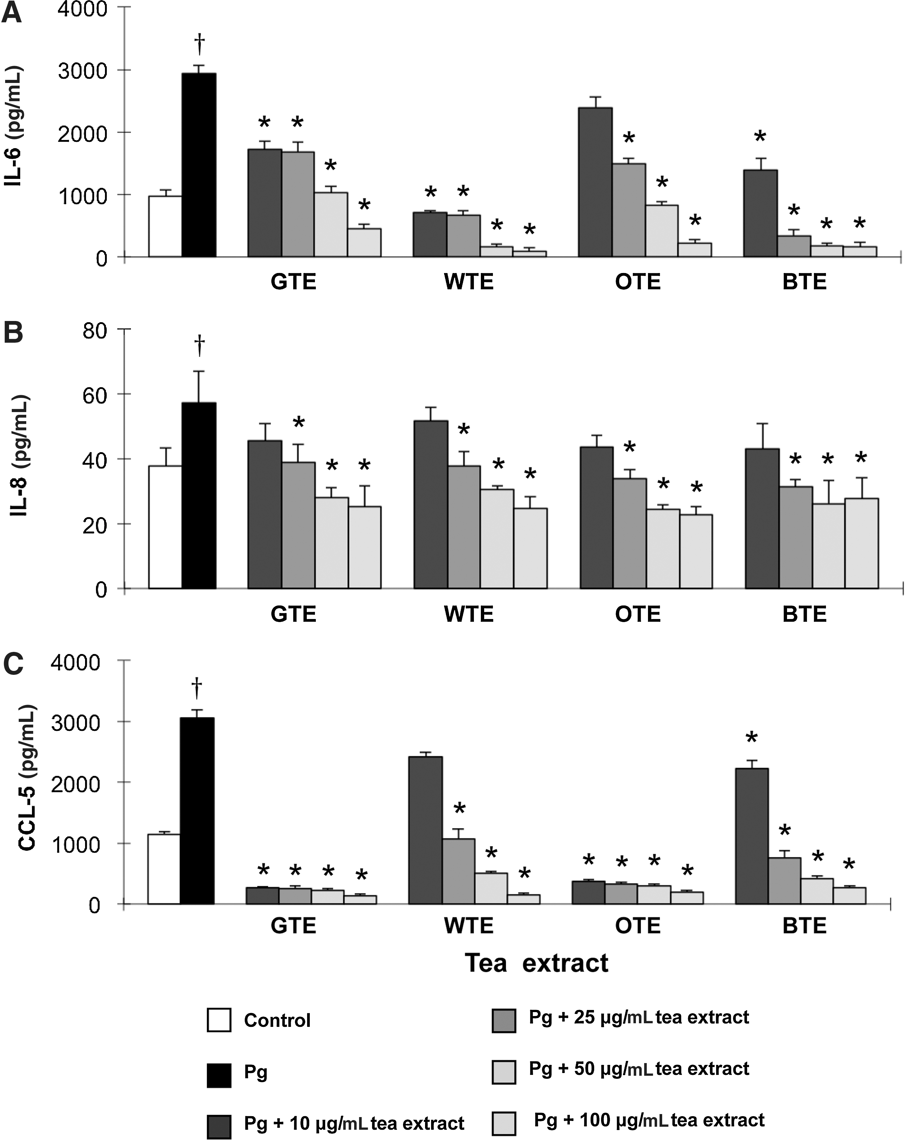
Effect of tea extracts on the secretion of interleukin (IL)-6
Discussion
There is increasing evidence suggesting that tea has a number of beneficial effects on health. 11,13,14,19,20 The pharmacological benefits of tea have been associated with various substances, including flavonoids, theaflavin, theanine, alkaloids, and polysaccharides. 10 The composition of tea is mainly determined by how the tea leaves are processed. Unfermented teas (green and white) are rich in methylxanthines and several kinds of catechins, including epigallocatechin, catechin gallate, epicatechin gallate, and EGCG, 12,21 while oolong and black teas are rich in oxidized phenolic compounds such as gallic acid, theaflavin, and thearubigin, but are poor in catechins. 22 In addition, the fluoride content of black tea is reported to be five times higher compared with green tea. 23 Anecdotal evidence suggests that green tea has a more pronounced effect on promoting health and preventing or treating chronic diseases, including periodontitis, due to its high catechin content. 11,13,14,19,20 It has been reported that green tea drinkers have healthier gums and teeth, 15 and that green tea consumption is associated with a decreased probability of tooth loss. 16 In addition, a clinical study has shown that a slow-release buccal delivery system containing green tea catechins significantly reduces the periodontal pocket depth. 17 Other compounds in tea also exhibit antibacterial, anti-inflammatory, and antioxidant activities. For example, theaflavin, one of the major oxidized polyphenols in black tea, inhibits LPS-induced ICAM-1 and VCAM-1 expression in epithelial cells, 24 suppresses oncostatin M-induced CXCL10 production in human fibroblasts, 25 and possesses the antioxidant activity comparable to that of green tea catechins. 26 Theasinensin A from oolong tea has been shown to dose dependently inhibit the mRNA, protein, and promoter activity of cyclooxygenase-2 in LPS-activated macrophages in addition to inhibiting the MMP activities of human fibrosarcoma HT1080 cells. 27 White tea has also been reported to have stronger antielastase, anticollagenase, and antioxidative activities than green tea. 28 In the present study, we compared four types of tea extracts (green tea, white tea, oolong tea, and black tea) for their effects on different aspects involved in the initiation and progression of periodontal diseases.
Since P. gingivalis is widely considered as the key etiologic agent of periodontitis, more specifically the chronic form, 4,6 the inhibition of this bacterium may be a potentially valuable strategy for interfering with the initiation of periodontitis. A previous study found that the MICs of green tea polyphenols, including epigallocatechin and EGCG, against the P. gingivalis range from 250 to 1000 μg/mL. 29 The different types of tea used in the present study also inhibited P. gingivalis growth, with MICs ranging from 200 to 500 μg/mL. While the exact mechanism by which tea inhibits bacterial growth remains obscure, some studies have provided interesting clues. The tea components, theaflavins and catechins, have been reported to irreversibly damage the bacterial cytoplasmic membrane. 30 –32 For example, EGCG generates hydrogen peroxide in the lipid bilayer of the bacterial cytoplasmic membrane, resulting in leakage of intracellular materials. 31 Membrane damage may also facilitate the diffusion of bioactive molecules into the cells. In addition, Navarro-Martínez et al. 33 provided evidence that the antibacterial action of catechins against Stenotrophomonas maltophila, a Gram-negative opportunistic pathogen, is due to its ability to inhibit cytoplasmic dihydrofolate reductase. Dihydrofolate reductase reduces dihydrofolic acid to tetrahydrofolic acid, which is required by bacteria to synthetize purine, thymidylate, and nucleic acid precursors, which are very important for cell proliferation and growth.
All the tea extracts significantly inhibited the adherence of P. gingivalis to oral epithelial cells, even at the lowest concentration tested (10 μg/mL). Gallate-type tea polyphenols have already been claimed to possess an inhibitory effect on the adherence of P. gingivalis to human oral epithelial cells. 29 In addition, some catechin derivatives inhibit Rgp and Kgp gingipains, which are involved in the adherence of P. gingivalis to host cells. 34 Matsumoto et al. 35 reported that high and low molecular weight oolong tea fractions can bind to bacterial surface proteins, decreasing cell surface hydrophobicity of Streptococcus mutans. The inhibition of adherence observed in the present study may thus result from the binding of tea components to P. gingivalis cell surface proteins.
In their natural subgingival environment, periodontopathogens degrade tissue proteins into low molecular weight peptides and amino acids, which can be used as carbon and energy sources to support bacterial growth. 36,37 Bacterial proteinases play a pivotal role in this process by direct degradation of host proteins and the activation of latent host enzymes. 36,37 Since type I collagen is the predominant protein of periodontal tissue, this constituent of the gingival matrix may be a major source of nutrients for P. gingivalis, which possesses a complex proteolytic system that can degrade type I collagen into small fragments. 38 In the present study, we showed that tea extracts strongly inhibit the collagenase activity of P. gingivalis. In addition to reducing tissue destruction, this inhibition may affect bacterial growth.
The MMP-9 activity has been strongly associated with periodontitis progression. 39 Ding et al. 40 showed that P. gingivalis can concomitantly trigger the release and activation of MMP-9 from polymorphonuclear leukocytes. In addition, it has been shown that infections of an engineered human oral mucosa model with P. gingivalis result in a significant increase in the MMP-9 protein and mRNA levels. 41 Mounting evidence also points to an important role for the elastase released from polymorphonuclear leukocytes in periodontal destruction. 42 –44 This neutrophil enzyme can degrade several matrix proteins, including elastin, collagen, and fibronectin, and its activity is significantly correlated with probing depth, attachment loss, and the gingival index of periodontal patients. 45 The present study showed that tea extracts significantly inhibit MMP-9 and elastase activities and may thus contribute to reducing periodontal tissue destruction. These results are in agreement with those reported by Demeule et al. 46 indicating that green tea polyphenols, especially EGCG, are potent inhibitors of MMP-9 and MMP-12 (also known as macrophage elastase). The inhibition of the MMP activity by green tea catechins has been associated with conformational changes. 47 In a recent study 28 comparing the anticollagenase and antielastase activities of plant extracts, white tea was shown to inhibit elastase and collagenase more than green tea. In the present study, we observed no marked differences among the four tea extracts tested.
The mechanisms underlying the destructive processes associated with periodontitis are not only related to the direct tissue damage caused by bacterial and host-derived proteinases, but also involve indirect damage mediated by host immune and inflammatory responses elicited by periodontal pathogens. P. gingivalis cells and components can induce a strong proinflammatory cytokine response in gingival epithelial cells. 8 In the present study, an extract of P. gingivalis upregulated the secretion of IL-6, IL-8, and CCL-5 by oral epithelial cells. Since growing evidence 11,13,19,48 –50 suggests that tea polyphenols have anti-inflammatory properties, we evaluated the ability of green tea, white tea, oolong tea, and black tea extracts to inhibit IL-6, IL-8, and CCL-5 secretion by epithelial cells stimulated with the P. gingivalis extract. Our results showed that all four tea extracts attenuated the P. gingivalis-induced inflammatory response to various degrees. More specifically, the expression of CCL-5, a chemokine that enhances the recruitment and infiltration of immune cells to diseased periodontal sites, was significantly suppressed by all four tea extracts. Preliminary results indicated that the four tea extracts also inhibit Aggregatibacter actinomycetemcomitans LPS-induced inflammatory cytokines production by oral epithelial cells (data not shown). Previous in vivo and in vitro studies 24 –27,49,50 have shown that tea components such as catechins, theaflavin, and thearubigin have anti-inflammatory activities. The molecular mechanisms may involve interference with signaling pathways such as NF-κB and AP-1. A recent study 50 using a macrophage genome-wide DNA microassay to investigate the anti-inflammatory genes targeted by theasinensin A showed that the activities of 63.8% of the genes upregulated in LPS-activated macrophages are attenuated by theasinensin A and that the activities of 65.7% of the downregulated genes are restored by theasinensin A. The genes suppressed by theasinensin A include those coding for tumor necrosis factor (TNF), IL-1β, and IL-6. Interestingly, the genes coding for anti-inflammatory cytokines, which were decreased in LPS-treated macrophages were restored by theasinensin A.
To summarize, the present study showed that green tea, white tea, oolong tea, and black tea extracts possess a number of properties (antibacterial, antiadherence, antiprotease, and anti-inflammatory) that may contribute to maintaining periodontal health. None of the properties were specifically associated with a particular type of tea, suggesting that all teas may have beneficial effects. Although the different tea extracts showed similar properties in regard to antibacterial, antiadherence, antiprotease, and anti-inflammatory activities, the bioactive ingredients of the various extracts may differ considering their chemical composition.
In conclusion, the present study compared the potential impacts of four tea extracts on periodontal disease therapeutic targets. They all exhibited comparable activities, including the ability to inhibit (i) the growth of P. gingivalis and its adherence to oral epithelial cells, (ii) the activity of host and bacterial proteases, and (iii) the secretion of proinflammatory mediators by oral epithelial cells. Bioactive molecules in tea thus hold promise as preventive or therapeutic agents for treating periodontal diseases.
Footnotes
Acknowledgments
This study was supported by the Laboratoire de Contrôle Microbiologique of Université Laval. We wish to thank V. Murrah (University of North Carolina, Chapel Hill, NC, USA) and J.M. Dirienzo (University of Pennsylvania, Philadelphia, PA, USA) for providing the GMSM-K epithelial cell line.
Authors' Contributions
All authors contributed equally in data acquisition and in writing of the manuscript. All of the authors read and approved the final version of the manuscript.
Author Disclosure Statement
The authors have no conflicts of interest related to this study.