




Abstract
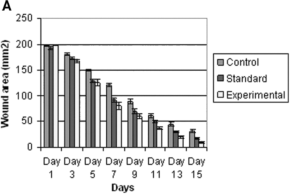
The skin of the fruit and the bark of Punica granatum are used as a traditional remedy against diarrhea, dysentery, and intestinal parasites. The fruit skin extract of P. granatum was tested for its wound healing activity in rats using an excision wound model. The animals were divided into three groups of six each. The experimental group of animals was topically treated with P. granatum at a dose of 100 mg/kg every day for 15 days, while the controls and standard group animals were treated with petroleum jelly and mupirocin ointment, respectively. Phytochemical analysis of the extract revealed the presence of saponins, triterpenes, tannins, alkaloids, flavonoids, and cardiac glycosides. Extract-treated animals exhibited 95% reduction in the wound area when compared with controls (84%), which was statistically significant (P<.01). The extract-treated wounds were found to epithelize faster compared with controls. The hydroxyproline content of extract-treated animals was significantly higher than controls (P<.05). The fruit skin extract did not show any antimicrobial activity against the microrganisms tested. P. granatum promotes significant wound healing in rats and further evaluation of this activity in humans is suggested.
Introduction
W
P. granatum L. (Lathyraceae) is a fruit-bearing deciduous shrub or small tree growing between five and eight meters tall. It is commonly known as pomegranate and is an important fruit of tropical and subtropical regions. It originated in the Middle East and India and has been used for centuries in ancient cultures for its medicinal purposes. It is widely reported that pomegranates exhibit antivirus, antioxidant, anticancer, and antiproliferative activities. 4 –6 The antioxidant activity of pomegranates is typically higher in commercial juices extracted from whole pomegranates than in experimental juices obtained from the arils only. This can be attributed to its high content of polyphenols in the peel, such as condensed tannins and anthocyanins. It has been reported that the peel in particular possesses relatively higher antioxidant activity than seeds or pulp and therefore might be a rich source of natural antioxidants. 7,8 The astringent qualities of the flower juice, skin, and tree bark are considered valuable for a variety of purposes, such as stopping nose and gum bleeds, toning skin, firming-up sagging breasts, and treating hemorrhoids. 9
All of the above medicinal uses either from fruit, seed, or leaves of P. granatum prompted us to investigate the wound healing activity of the P. granatum fruit skin extract.
Materials and Methods
Preparation of extract
The skin of P. granatum fruit was carefully peeled and shade dried. The dried fruit skin (200 g) was blended using a blender. The fine powder obtained (190 g) was used for our study.
Animals
The study was approved by the Ethics Committee for Animal Experimentation (AHC06/07/1), Faculty of Medical Sciences, University of the West Indies. Healthy inbred Sprague–Dawley male rats weighing 180–200 g were individually housed and maintained on normal food and water ad libitum. Animals were periodically weighed before and after the experiment. The excision wound model was used to evaluate the wound healing activity of P. granatum extract. The male rats were randomly distributed into three groups of six each.
Excision wound model
Rats were inflicted with excision wounds according to the method of Morton and Malone. 10 Animals were anesthetized with 1 mL of intravenous ketamine hydrochloride (120 mg/kg body weight) and shaved on both sides of the back with an electric clipper. The area of the wound to be created was outlined on the back of the animals with methylene blue using a circular stainless steel stencil. A full thickness excision wound of 200 mm2 circular area and 2 mm in depth was created along the markings. The wound was left open. Animals were closely observed for any infection, and those that showed signs of infection were separated, excluded from the study, and replaced. Animals were divided into three groups of six each. Group 1 was treated with petroleum jelly (control), group 2 was treated with fruit skin extract of P. granatum mixed with petroleum jelly (1:1 ratio, experimental, 100 mg/kg body weight), and group 3 was treated with mupirocin ointment (standard, 100 mg/kg body weight). The treatment was topically applied in all cases for 15 days. Wound areas were measured on alternate days (1, 3, 5, 7, 9, 11, 13, and 15) using a transparency sheet and a permanent marker. Recordings of the wound areas were measured using graph paper. The day of eschar falling after wounding, without any residual raw wound, was considered as the period of epithelialization.
Phytochemical screening methods
On day 15, all animals were sacrificed using ketamine and pieces of wet granulation tissue were excised from the healed area for hydroxyproline and histological studies.
Saponins
The extract (2 g) was boiled with 20 mL water for about 4 min. This was mixed vigorously and left for a few minutes. The formation of frothing indicates the presence of saponins. 11
Test for tannins
To an aliquot of the extract (dissolved in water), 2 mL of 1% ferric chloride was added. Color development from red-brown to blue-black indicates the presence of tannins. 11
Triterpenes
The extract (1 g) was mixed with 10 mL chloroform and warmed at 55°C for 30 min. A few drops (1–2 mL) of concentrated sulphuric acid were added and mixed well. The appearance of a reddish brown color indicates the presence of triterpenes. 12
Alkaloids
The extract (1 g) was boiled with 50 mL methanol for 20 min in a water bath and the cooled filtrate was tested separately with Mayer's, Wagner's, Hager's, and ammonium reineckate reagents. Cloudy precipitate of the alcoholic layer indicates the presence of alkaloids. 12
Flavonoids
About 1 g of extract was boiled with 10 mL ethyl acetate over a steam bath for 3 min. The filtrate of ∼4 mL was mixed with 1 mL of dilute ammonia solution and a yellow precipitate indicates the presence of flavonoids. 11
Antimicrobial activity
Staphylococcus aureus (ATCC 4827) Escherichia coli (ATCC 25922), Pseudomonas aeruginosa (ATCC 25853), and Klebsiella pneumoniae (ATTCC (494272) were the bacterial strains obtained from fresh colonies grown on Mac Conkey blood agar plates. Sensitivity testing was done using Muller Hinton agar plates. A known volume of bacterial suspension was transferred to each microplate well. Ten microliters of ethanol extract of P. granatum was added to the microplate wells and incubated at 35–37°C for 18–20 h. Results were determined by visual inspection of zones of growth inhibition.
Histological study
The granulation tissue samples obtained on day 15 from all three groups were fixed in 10% buffered formal saline and processed for routine histological evaluation. Sections of 7.0 μm were cut from the tissues and stained with van Gieson's stain.
Statistical analysis
The means of wound area measurements between groups at different time intervals were compared using one-way ANOVA, followed by Tukey's post-hoc tests. Data were analyzed using SPSS (Version 12.0, Chicago, USA) and P value significance was set <.05 for all analyses.
Results
The phytochemical analysis of the extract showed the presence of saponins, triterpenes, tannins, alkaloids, flavonoids, and cardiac glycosides. The P. granatum did not exhibit any antimicrobial activity when tested against S. aureus, E. coli, P. aeruginosa, and K. pneumoniae.
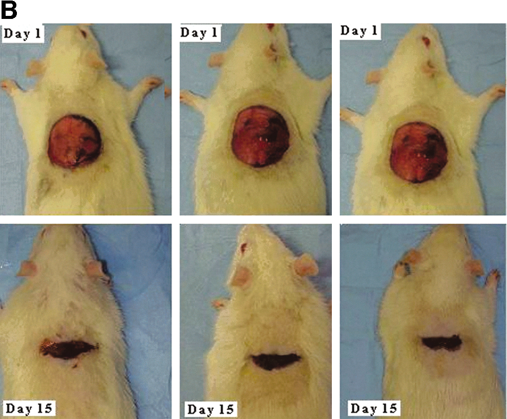
A significant increase in wound healing activity was observed in the P. granatum–treated rats (experimental) compared with the petroleum jelly– (control) and mupirocin-treated rats (standard). In the excision wound model, the experimental animals showed wound contraction of 95% (P<.01) by day 15, while those of control and standard group of rats showed wound contraction of 84% and 91%, respectively (Table 1). Experimental and standard group animals showed similar wound contraction by day 15 (Fig. 1). Animals treated with fruit skin extract of P. granatum showed higher hydroxyproline content (168 mg) compared with control (75 mg) and standard groups (114 mg) of animals (Fig. 2). A significant increase in hydroxyproline content was noted for the granulation tissues obtained from the experimental group of animals (t=3.42; P<.05).
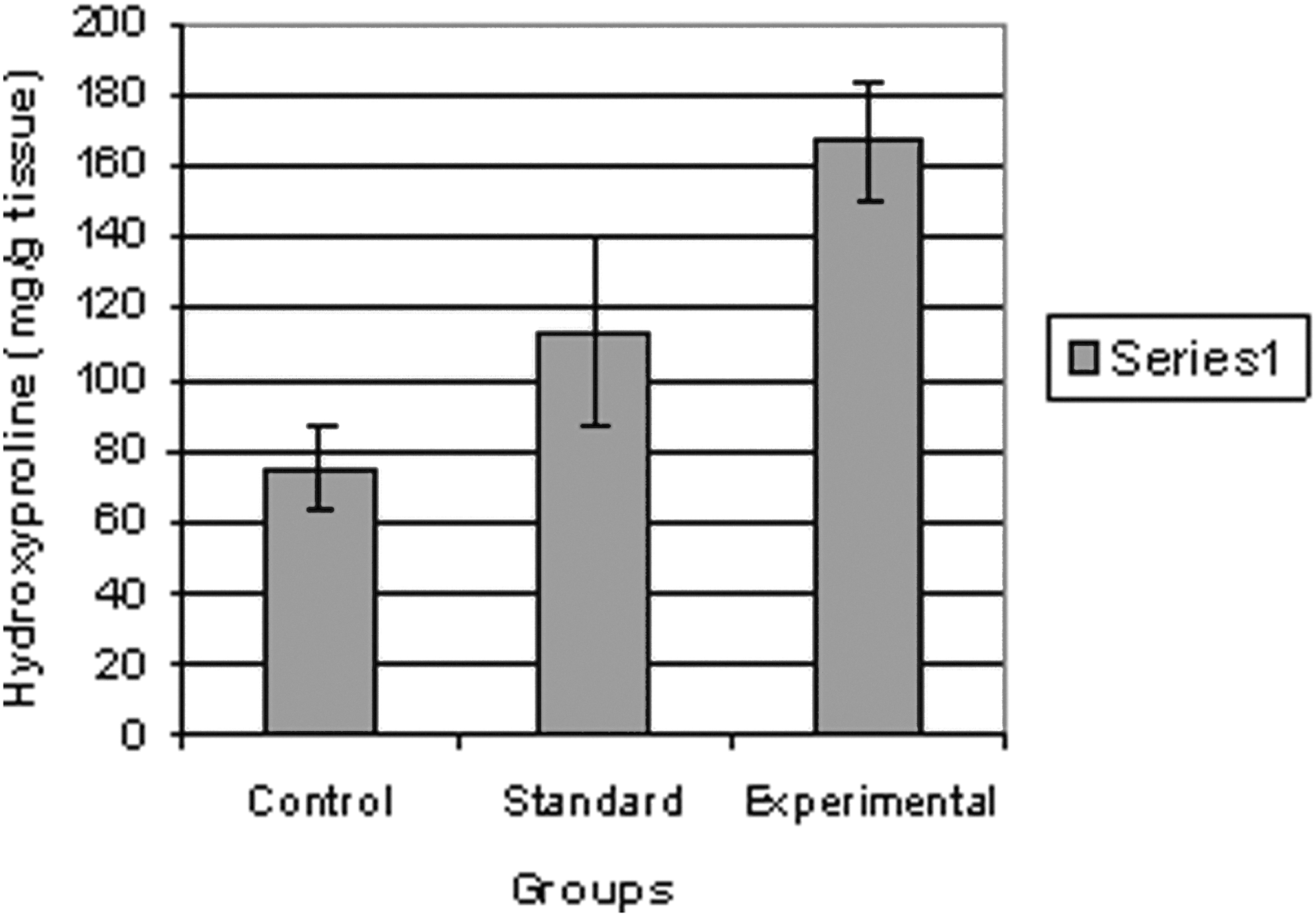
Hydroxyproline content of granulation tissue from control, standard, and experimental groups of animals.
Values are mean±SE from six animals in each group.
Statistically significant differences are indicated: * P <.05, *** P <.001 vs. both control and standard groups.
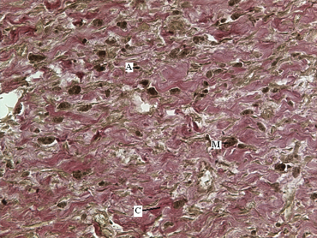
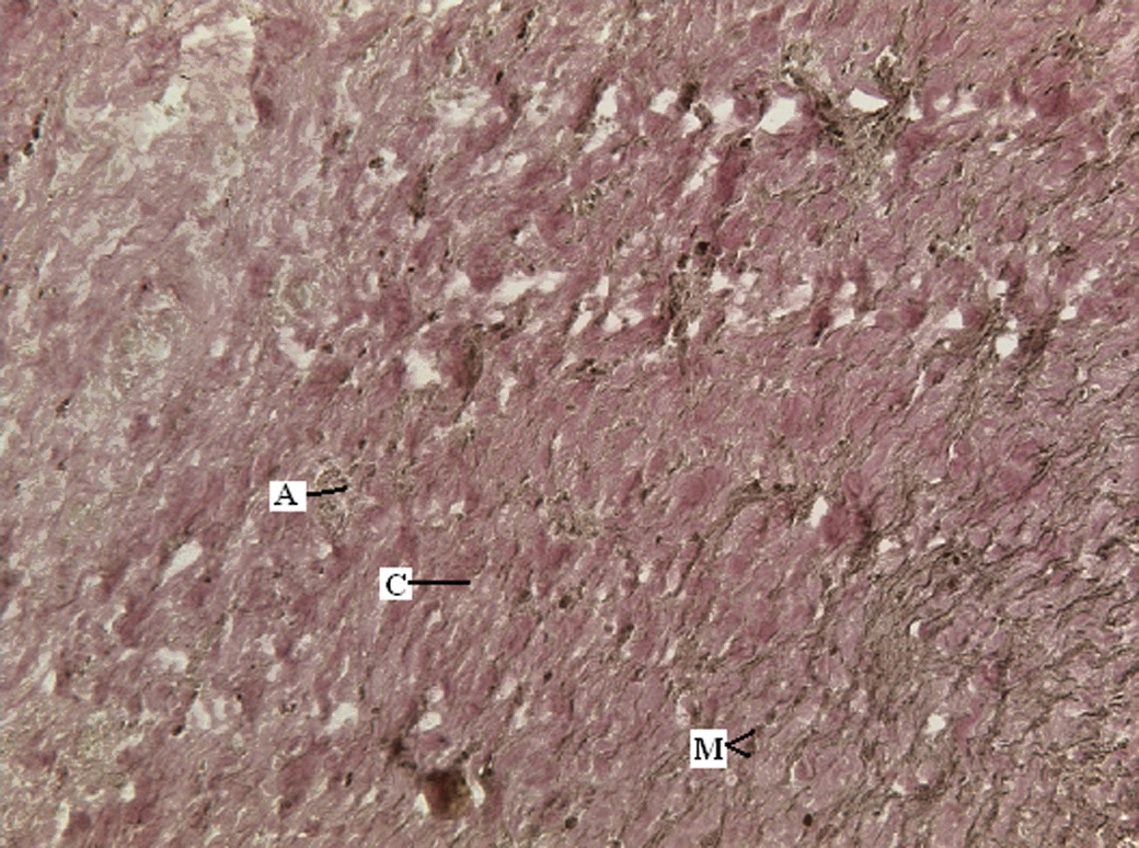
The wounds in the animals treated with the fruit skin extract were clean. The wounds of control animals that received plain petroleum jelly appeared to be hard and crusty with undermined margins and were generally unclean with a biofilm glaze on the surface. The granulation tissue of these animals showed wavy strands of sparse collagen deposition, inflammatory cells, and more macrophages (Fig. 3). The histological evaluation of the granulation tissue obtained from the experimental animals demonstrated thick bands of reddish collagen and scanty inflammatory cells and pigment-laden macrophages (Fig. 4). The histological findings of animals treated with standard drug were similar to experimental animals (Fig. 5).
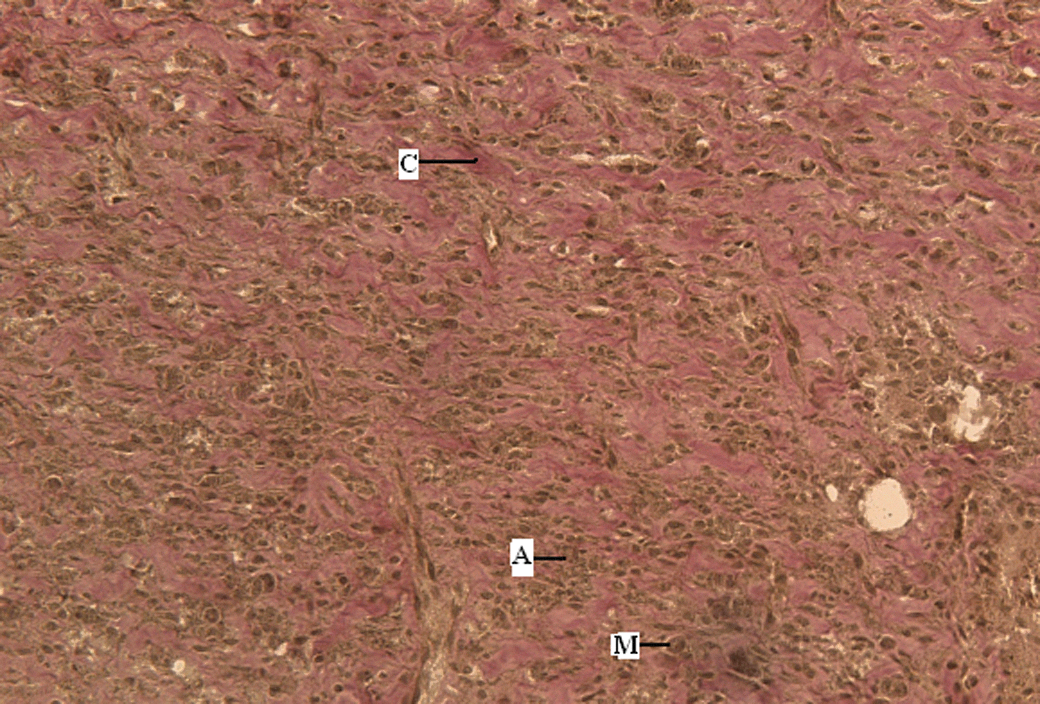
van Gieson's staining of granulation tissue of an animal treated with petroleum jelly shows wavy strands of sparse collagen deposition (C), inflammatory cells (A), and more macrophages (M). Color images available online at
van Gieson's staining of granulation tissue of an animal treated with Punica granatum shows thick bands of reddish collagen (C), scant inflammatory cells (A), and pigment-laden macrophages (M). Color images available online at
van Gieson's staining of granulation tissue of an animal treated with mupirocin shows bands of collagen (C), scant inflammatory cells (A), and macrophages (M). Color images available online at
Discussion
The results of our experiment showed that wounds treated with the P. granatum extract exhibited a higher rate of wound contraction and had higher hydroxyproline content compared to the untreated and mupirocin-treated wounds. An increase in hydroxyproline content indicates an increase in the rate of wound healing, as enhanced hydroxyproline is a reflection of increased cellular proliferation and therefore increased collagen synthesis. Collagen is a major protein in the extracellular matrix that contributes to wound strength and provides a structural framework for the regenerating tissue. 13 In addition, an increase in dry tissue weight indicated the presence of higher protein content, which is predominantly due to enhanced collagen synthesis in the formation of scar tissue. 14
The wound healing activity of P. granatum is potentiated by the phytochemical components present in the extract. The preliminary phytochemical analysis of the extract revealed the presence of saponins, triterpenes, tannins, alkaloids, flavonoids, and cardiac glycosides. Flavonoids are known to reduce lipid peroxidation not only by preventing or slowing the onset of cell necrosis, but also by improving vascularity. 15 Saponin stimulates the wound healing process through changes in the extracellular matrix metabolism, accompanied by modification of transforming growth factor-β receptor expression in fibroblasts. 16 Alkaloids are known to enhance wound healing activity due to their strong antioxidant activity and strong radical-scavenging power. 17 It has been reported that the peel in particular possesses relatively higher antioxidant activity than seed and pulp and therefore might be a rich source of natural antioxidants. 7,8
Similar types of wound healing activity were reported by Manjunatha et al., 18 who showed that the aqueous and methanol leaf extracts of Vernonia arborea have a high wound healing potency due to their phytochemical constituents. In our study, the wound healing activity of fruit skin of P. granatum might have been due to the effect of phytoconstituents like alkaloids, tannins, and flavonoids present in it. The specific component responsible for the increased wound healing activity was not investigated, hence, further phytochemical studies are needed to isolate and identify the specific active compound(s) responsible for the pharmacological activities.
Footnotes
Acknowledgments
The authors sincerely thank Dr. A.V.C. Rao, Terry Ramsingh, and Sabana Meyers for their technical assistance.
Author Disclosure Statement
No competing financial interests exist.