
Abstract
Studies suggest that the traditional applications of Kigelia pinnata leaves have beneficial effects against oxidative stress–mediated diseases and cancers. The pulverized dried leaves of K. pinnata were extracted with hexane, ethyl acetate, and methanol sequentially, and the crude extracts were fractionated by silica gel column chromatography with solvent gradient of increasing polarity. 3-hydro-4,8-phytene, trans-phytol, (9Z,12Z)-methyl octadeca-9,12-dienoate, and two oil fractions were obtained. The chemical compositions of chromatographic fractions were determined using gas chromatography–mass spectroscopy. The structure elucidations of the isolated compounds were based on FTIR, MS, and NMR spectral data analyses. These along with the crude extracts were examined for their antioxidant activities using ferric reducing antioxidant power (FRAP), 2,2-diphenyl-1-picrylhydrazyl (DPPH) radical scavenging, and 2,2-azinobis(3-ethyl-benzothiazoline-6-sulfonic acid) (ABTS) assays. Total phenolic contents were also determined. The crude extracts and purified compounds were evaluated on the rhabdomyosarcoma human cancer cell for their cytotoxicity using 3-(4,5-dimethylthiazol-2-yl)-2,5-diphenyltetrazolium bromide (MTT) cell viability assays. The methanol extract was richer in phenolics and was most potent as antioxidant and cytotoxic agent among all the substances tested. Among the fractions and pure compounds, the two oil fractions showed more cytotoxicity potency, with IC50s of 143.4±0.5 and 147.9±1.3 ng/mL, which is more significant than the reference standard, cyclophosphamide (165.6±1.0 ng/mL). 3-hydro-4,8-phytene showed lower antioxidant and cytotoxicity potential (IC50=1818±5.2 μg/mL and 171.7±0.8 ng/mL, respectively). Trans-phytol did not show a high cytotoxic power (IC50=769.8±4.3 ng/mL). The comparatively high cytotoxicity index of (9Z, 12Z)-methyl octadeca-9,12-dienoate (IC50=153.3±0.1 ng/mL) indicated that it may be one of the principal cytotoxic agent in the ethyl acetate extract. These results suggest that the leaves of K. pinnata possess tumor cytotoxic potential and could be part of a drug combination for future cancer chemotherapy.
Introduction
C
K. pinnata DC., Bignoniaceae [syn Kigelia africana (Lam.) Benth], an endangered tropical tree (Fig. 1) used extensively in African folkloric medicine, is a multipurpose medicinal plant outstanding for its anticancer properties among other things. 5,6 The extracts of the stem bark and fruit have been screened for their cytotoxic activities, and both showed promising results against melanoma and renal carcinoma, 7 whereas the root bark showed the activity against KB cells. 8 The leaf and fruit extracts have been tested for its antioxidant potentials. 6,9 Other reported bioactivities of the plant include antimalarial 10,11 anti-inflammatory, 12 antidiarrhoeal, 13 antileprotic, 14 and treatment of gynecological disorders. 15 Review of the literature revealed some composition of the leaves, flowers, and roots. 6,16 –18 The presence of naphthoquinones in the stem bark 8,19,20 and the presence of coumarins, 21 iridoids, 22 and flavonoids in the fruit 23 have been confirmed. Although extracts of K. pinnata plants have previously been evaluated for antioxidant and antiproliferative properties, this is the first report of the isolation, antioxidant, and human anticancer cell evaluation of constituents from K. pinnata leaves. The rhabdomyosarcoma (RD) cancer cell line is used in this study. RD is a highly metastatic tumor that develops from connective tissues in the body, such as muscles, fat, bones, membranes that line the joints, or blood vessels. 24

Kigelia pinnata in its natural habitat. Color images available online at
Materials and Methods
Chemicals
3-(4,5-dimethylthiazol-2-yl)-2,5-diphenyltetrazolium bromide (MTT), α-tocopherol (Sigma-Aldrich), 2,2-azinobis (3-ethyl-benzothiazoline-6-sulfonic acid) (ABTS; Sigma-Aldrich), 2,2-diphenyl-1-picrylhydrazyl (DPPH; Sigma-Aldrich), gallic acid (Sigma-Aldrich), Folin–Ciocalteu reagent (Sigma-Aldrich), Na2CO3 (NAAFCO), aluminium chloride, potassium acetate (J.T. Baker), phosphate buffer, K3Fe(CN)6, trichloroacetic acid (TCA; Qualikem), ferric chloride (Riedel-de Haen), HCl, and potassium persulfate were obtained in analytical grade, whereas solvents, silica gel 60 F254 for thin layer chromatography (TLC), silica gel 60 with mesh 70–230 μm for gravity column chromatography, and vanillin spray reagent were obtained from the Department of Chemistry, University of Ilorin.
Instruments
Absorbance measurements (ferric reducing antioxidant power [FRAP], phenolics, DPPH, and ABTS assays) were recorded on an UV/Vis Spectrumlab 23A Spectrophotometer. A gas chromatography–mass spectroscopy (GC-MS) system (GCMS-QP 2010 PLUS; Shimadzu) interfaced with a finigan MAT ion trap detector was used with the RTX5MS column packed with 100% dimethylpolysiloxane. The column temperature was initially held at 60°C for 5 min with injection volume of 1 μL and then programmed to rise at the rate of 5°C/min to 250°C. The injector temperature was set at 200°C, whereas the detector (mass spectrophotometer) temperature was maintained at 250°C. Helium was used as the carrier gas at a linear velocity of 46.3 cm/sec and pressure of 100.2 kPa. Ionization mode was electron impact at a voltage of 70 eV. The identification of the chemical components was carried out using the peak enrichment technique of reference compounds, and as final confirmation of the peak identification by GC-MS, their spectral were compared with those of NIST library mass spectra.
Samples and sample preparation
The leaves of K. pinnata were collected from a fruiting tree in Ado-Ekiti metropolis, Nigeria, during the summer and taxonomically authenticated at the herbarium of the Department of Botany, University of Lagos, Lagos, Nigeria. A voucher specimen number LUT/3525 was obtained. The leaves were dried at room temperature and blended into powder.
Extraction and isolation
The powdered plant material (240 g) was exhaustively extracted with n-hexane at room temperature for 6 days to afford 3.06 g of brownish green syrup (tagged KPLH) after concentration. The residual plant material was further extracted with ethyl acetate and thereafter with methanol to afford 22.1 and 7.58 g concentrated crude extract (tagged KPLE and KPLM), respectively. The KPLH, KPLE, and KPLM were fractionated separately in a silica gel open column using n-hexane and ethyl acetate as well as ethyl acetate and methanol in an increasing order of polarity. KPLH afforded 46 fractions of 10 mL each. Fractions with similar TLC profile were combined and concentrated using the rotary evaporator. The first 17 fractions of the KPLH were predominantly hydrocarbons and were analyzed as previously reported. 25 Combined fractions 25 and 26 indicated the presence of fatty acids (FAs) and β-tocopherol (Table 1), among others, whereas fractions 37–46 yielded clean yellow oil, which was also analyzed by GC-MS (Table 2). KPLE fractions 5–8 yielded a golden yellow oil trans-phytol (C20H40) (Fig. 1), fraction 9 afforded a light yellow oil (9Z,12Z)-methyl octadeca-9,12-dienoate (or methyl linoleate, C19H34O2; Fig.1), while combined fractions 27–31 of KPLE eluted with hexane/dichloromethane/ethanol in a ratio of 2:1:1 on a silica gel PTLC plate afforded 3-hydro-4,8-phytene (C21H40; Fig. 1). The details of the isolation processes and spectroscopic data of UV/Vis, IR, 1 HNMR, 13 C-NMR, GC-MS/MS of the isolated compounds have been reported previously. 25 KPLH, KPLE, KPLM, isolated compounds, and the selected combined fractions that were obtained in significant amounts and characterized were subjected to antioxidant assays, which include FRAP, DPPH, ABTS, and cytotoxicity assays. Fractions and purified compounds that were assayed were coded: KPLH 25–26 (KPLHA; Table 1), 3-hydro-4,8-phytene (KPLHB; Fig. 2), KPLH 37–46 (KPLHC; Table 2), trans-phytol (KPLEA; Fig. 2), and (9Z,12Z)-methyl octadeca-9,12-dienoate (KPLEB; Fig. 2). The samples were prepared in appropriate concentrations and sterile filtered using a Sterifix® microfilter 0.2 μm and stored (4°C) until used.

3
A/H, area per height; R T, retention time; KI, Kovats indices.
Determination of total phenolic index
Total phenolics in the samples were determined with Folin–Ciocalteu reagent using the method of Ebrahimzadeh et al. 26 About 2.5 mL of 10% Folin–Ciocalteu reagent was added to 0.5 mL of each sample (duplicates) already prepared in methanol to be 1 mg/mL. Two milliliters of Na2CO3 (20 mg/mL) was added to the mixture. The resulting mixture was shaken and incubated at 50°C for 30 min. The absorbance of the samples was measured at 765 nm using the UV/visible spectrophotometer. The concentrations of the extracts were extrapolated from a calibration curve of gallic acid using the formula y=0.646x. Results were expressed as milligrams of gallic acid equivalent/gram of powder dissolved in methanol.
Determination of antioxidant activity
The antioxidant capacities of the extracts, fractions, and isolates were analyzed using the FRAP, the free radical scavenging capacity (DPPH), and the ABTS radical cation scavenging capacity (ABTS) assays.
FRAP assay
The reducing powers of the extracts were evaluated according to standard methods. 27,28 The mixture containing 2.5 mL of 0.2 M phosphate buffer (pH 6.6) and 2.5 mL of K3Fe(CN)6 (10 mg/mL) was added to 1.0 mL of the extract dissolved in distilled water. The resulting mixture was incubated at 50°C for 20 min, followed by the addition of 2.5 mL of TCA (100 mg/mL). The mixture was centrifuged at 3000 g for 10 min to collect the upper layer of the solution (2.5 mL), which was mixed with distilled water (2.5 mL) and 0.5 mL of FeCl3 (1 mg/mL). The absorbance was measured at 700 nm against the reference blank. Higher absorbance of the reaction mixture indicates higher reductive potential, and the IC50 values were calculated. The IC50 value (in μg/mL) is the effective concentration at which the absorbance was 0.5 for the reducing power.
Assay of DPPH radical scavenging activity
The DPPH spectrophotometric assay was carried out according to the standard procedure 29,30 with minor modifications. The DPPH free radical was prepared at a 0.1 mM concentration (25 mg/L) in methanol, following the prescribed procedure. The radical was protected from light after preparation. Stock solutions of the analytes (1 mg/mL) were diluted to final concentrations of 500, 250, 200, 100, and 50 μg/mL in methanol. One milliliter of 0.1 mM DPPH methanol solution was added to solutions of the extracts or standards (α-tocopherol and gallic acid separately). The absorbance at 518 nm was monitored in the presence of different concentrations of extracts. Blank experiment was also carried out to determine the absorbance of DPPH before interacting with the extract. The absorbance was recorded to check the stability of the radical throughout the time of analysis. The antioxidant activity, AA, was calculated using the following equation: 31 AA=100×[(Abscontrol− Abssample)]/(Abscontrol).
Assay of ABTS radical scavenging activity
ABTS radical cation decolorization test, a spectrophotometric method, 32 widely used for the assessment of the antioxidant activity of various substances, was used for the determination of the hydrophilic content of KPLE and KPLM. ABTS radical was generated by the oxidation of ABTS with potassium persulfate. The procedure followed the method of Arnao et al. 33 with some modifications. The stock solutions included 7.4 mM ABTS+ solution and 2.6 mM potassium persulfate solution. The working solution was prepared by mixing the two stock solutions in equal quantities and allowing them to react for 12 h at room temperature in the dark. The solution was then diluted by mixing 1 mL ABTS+ solution with 60 mL methanol. The samples (1.0 mL) were allowed to react with 1.0 mL of the ABTS+ solution for 2 h in a dark condition. Then, the absorbance was taken at 734 nm using the spectrophotometer. The ABTS antioxidant capacity (AOC) was calculated and compared with α-tocopherol using the following equation: AOC=100×[(Abscontrol − Abssample)]/(Abscontrol), where Abscontrol and Abssample are the absorbances of the control and the sample tested, respectively.
Cell culture
Rhabdomyosarcoma RD human tissue cancer cell line was used for the determination of the cytotoxic activity. The RD cell line is used as a general cytotoxicity assay. Because it is a general cytotoxicity assay, it is not possible, using the assay, to determine the mechanism of action of the extracts and compounds. DNA damaging agents, antimitotics, and so on are all measured as general cytotoxic agents in the assay. Although it is not possible to determine the mechanism involved with this assay, it is possible to detect a wide range of compounds with different mechanisms of action. The RD cancer cell line used in the study was obtained from the Department of Virology, University of Ibadan. Cells were grown in minimum essential medium supplemented with 10% fetal bovine serum and penicillin/streptomycin-L-glutamine and cultured in a humidified atmosphere of 5% CO2 and 95% air at 37°C in the Forma Scientific incubator. Cells were seeded in 96-well culture plates (Corning®) at the density of 2×10⁵ cells/mL in 100 μL/well of RD medium. The cell lines were maintained for 1 day to allow cell stabilization. 34
Cytotoxicity evaluation–cell viability assay
The cytotoxicity evaluation was performed using the MTT assay. 35,36 After a 24 h period of incubation at 37°C under a humidified 5% CO2 atmosphere, the cell monolayers were confluent and the medium was removed from the wells. Different concentrations of the samples were prepared by dissolving the extract in DMSO and then diluting it with PBS medium. One hundred microliters of cells/well was exposed to sample concentrations in PBS (1.0, 10.0, and 100 μg/mL) 100 μL each. As a cell control, only 200 μL of medium was added to the cells. The plates were sealed with parafilm to avoid contamination and moisture loss. Cells were incubated for 24 h with test samples. Each concentration was tested in triplicate. The MTT assay was used to determine the cell viability. After 24 h of incubation, the medium was removed from all wells and 50 μL of the MTT (Sigma®; 1 mg/mL) solution prepared in the cell culture medium was added to each well and the plates were incubated for 3 h. After the incubation, the MTT solution was removed without disturbing the cells and 100 μL of DMSO was added to each well to dissolve the formazan crystals. The plates were gently shaken to dissolve the crystals completely, and the absorbances were read on a multiwell spectrophotometer (Emax Precision Microplate Reader) at 490 nm. The IC50 was defined as the cytotoxic concentration of each sample that reduced the absorbance of treated cells by 50%. Using optical density, the percentage cytotoxicity of RD cells was calculated as [(A − B)/A]×100, where A and B are the OD490 of untreated and treated cells, respectively. Using the inhibition response curve for the cell line, the IC50 was determined on GraphPad Prism 5 software through a nonlinear regression analysis. Cyclophosphamide, an anticancer drug, was used as a positive reference.
Data analysis
The 50% cytotoxic concentration (IC50) was calculated from dose–response inhibition curves after nonlinear regression analysis. The results represent the mean±standard error of the mean values of three different experiments.
Results and Discussion
GC-MS analyses
The two oil fractions, KPLHA and KPLHC, are predominantly FAs. KPLHA revealed a total of 15 compounds with the major compounds being elaidoic acid, C18H34O2 (22.18%), and cetylic acid, C16H32O2 (21.99%). The total free FA was obtained to be 76.74%. Alcohol accounted for 17.61%, which include trans-phytol (1.42%) and β-tocopherol (2.34%), among others. KPLHC contains five compounds with emery oleic acid (40.62%), an important free FA ester obtained as the main compound. FAs and their derivatives play important roles as antioxidant and antitumor agents. 37,38 Modulation of macrophage function by FAs has been demonstrated by several authors. 39,40
Total phenolic index, AOC by FRAP, DPPH, and ABTS assays
The antioxidant capacities of the K. pinnata extracts, fractions, and isolates were analyzed using the free radical scavenging capacity (DPPH), the FRAP, and the ABTS radical cation scavenging capacity (ABTS), and the phenolics content of the ethyl acetate and methanol extracts determined. The total phenolic content was also found to be significantly higher in KPLM than in KPLE (50.7±0.01 vs. 49.29±0.009; Fig. 3). The antioxidant activity increased in proportion to the phenol content. Recent reports have indicated that a high positive correlation exists between total phenols and antioxidant activity in many plant species. 41 The FRAP assay measured the ability of phenolics to reduce Fe3+ to Fe2+. The results of the FRAP method were similar to those of the DPPH method for KPLE and KPLM. The FRAP assay is based on the reducing ability of antioxidant compounds present in the sample. KPLM exhibited superior FRAP than KPLE and the standard, gallic acid as depicted in Figure 4.
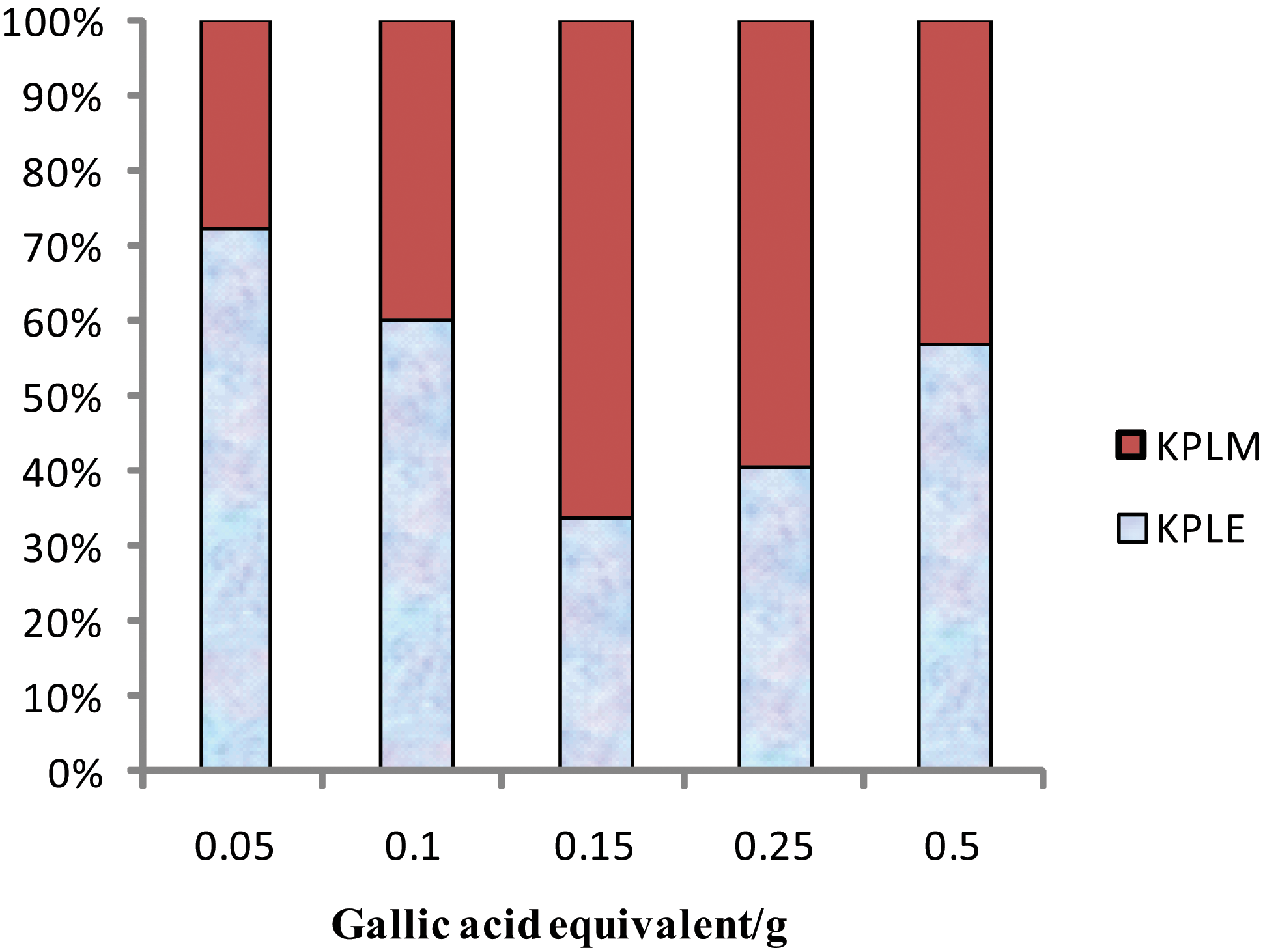
Total phenolic content of KPLE and KPLM. Data expressed as mg gallic acid equivalents/g extract, mean±%error (n=3). Color images available online at
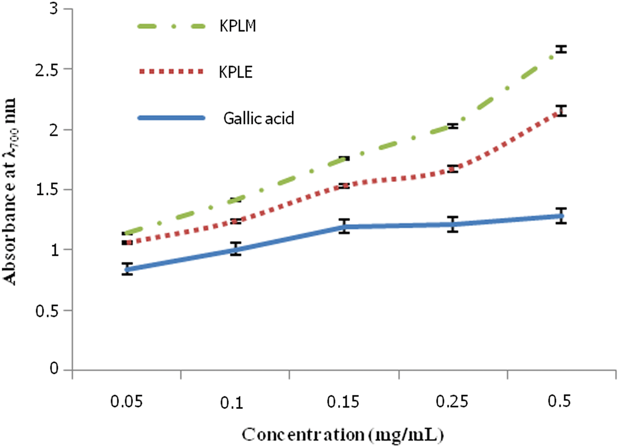
Ferric reducing antioxidant potential. Results represent the mean±%error (displayed at 5% value) of triplicate values. Color images available online at
The antioxidant activities of the extract, fractions, and isolates used in this study are shown in Figure 5, and the IC50 values are shown in Table 3. For the DPPH antioxidant potential, KPLM showed a slight increase as concentration increases. The antioxidant potential was significantly higher than all other tested samples and the standard compounds (gallic acid and tocopherol). KPLH, KPLE, and KPLHA showed slight increases in DPPH antioxidant potential as concentration increased. However, KPLE had a higher response factor than KPLH at higher concentration, whereas KPLHB and KPLHC had lower antioxidant power in the DPPH assay (Fig. 5). It is clear from the results that the antioxidant effect of K. pinnata is not due to the presence of (9Z,12Z)-methyl octadeca-9,12-dienoate (KPLHB).
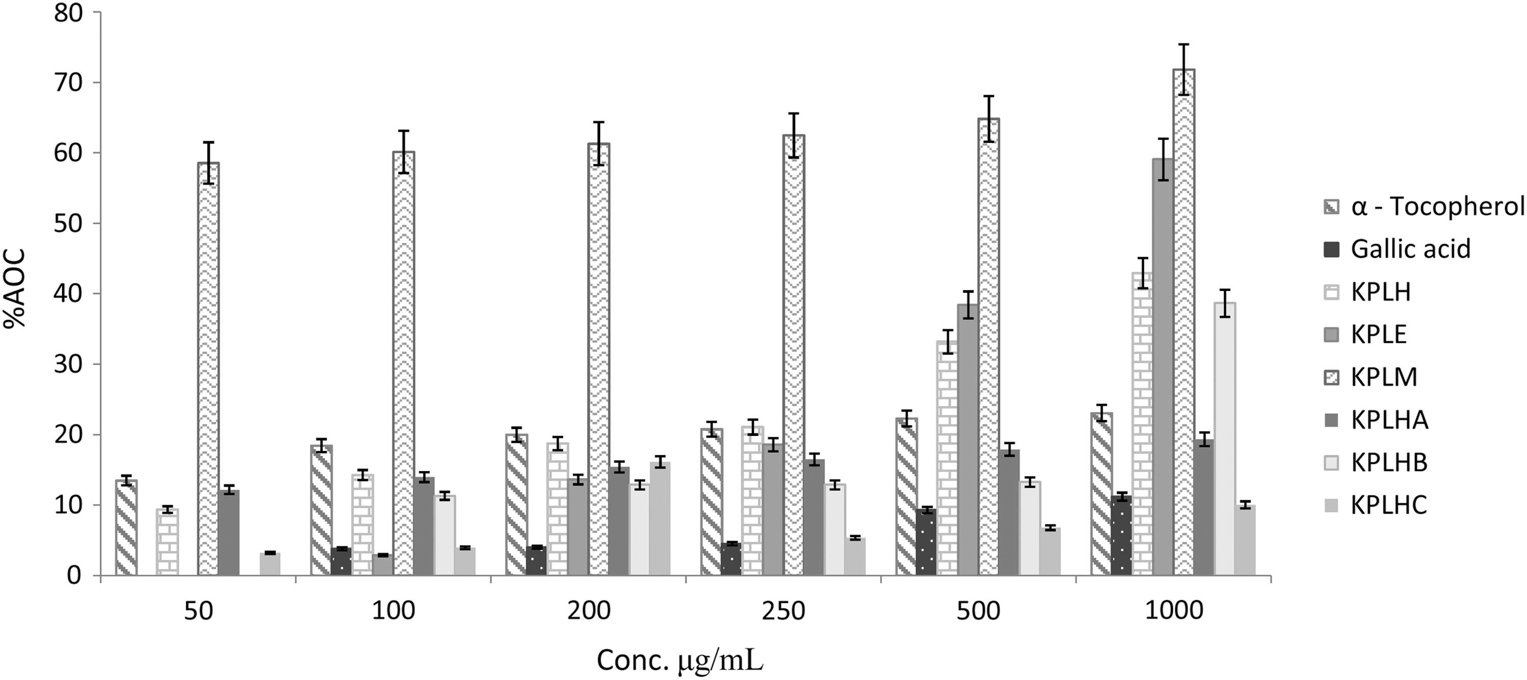
2,2-diphenyl-1-picrylhydrazyl (DPPH) antioxidant capacity of extracts and isolates. Error bar displayed at 5% value. Results represent the mean±percentage error (displayed at 5% value) of triplicate values.
Results represent mean±standard error of mean of triplicate determinations.
ABTS, 2,2-azinobis (3-ethyl-benzothiazoline-6-sulfonic acid); DPPH, 2,2-diphenyl-1-picrylhydrazyl; IC50, half-maximal inhibition concentration; NT, not tested; RD, rhabdomyosarcoma.
The ABTS test measures the relative antioxidant ability of extracts and isolates to scavenge the radical cation ABTS+ produced by the oxidation of 2,2′-azinobis-3-ethylbenzothiazoline-6-sulfonate. 42 In the ABTS assay, the methanolic extract KPLM had a lower IC50 (37.09±2.9) than the ethyl acetate extract, KPLE (57.94±1.7), which indicates that methanol extracted more hydrophilic components than ethyl acetate (Table 3). Also, the IC50 of KPLE (741±9.3) is quite higher than that of KPLM (532±1.9) in the DPPH assay.
KPLHA and KPLHC (with IC50=14.62±1.0 and 3.4±1.9 μg/mL, respectively) were oil fractions that apparently showed higher activity than KPLHB, a pure compound (Table 3). This straightly suggests that the activity of KPLHA and KPLHC is synergistic. It is submitted therefore that there is a possible antagonistic antioxidant interference in the hexane extract, KPLH (with IC50=15.08±5.1 μg/mL), whose DPPH IC50 value is higher than the values obtained for fractions KPLHA and KPLHC. This implies that fractionating the extract may yield fractions that have higher antioxidant value. It is impressive that KPLH has a lower IC50 value than KPLE and KPLM. There is no doubt that these extracts and fractions may neutralize free radicals by acting as rapid donors of a hydrogen atom to radicals.
Cytotoxicity index
The MTT assay is a laboratory test that measures changes in color for measuring the activity of enzyme that reduces MTT to formazan, giving a purple color. Yellow MTT (a tetrazole) is reduced to purple formazan in living cells. 43 KPLM has a higher cytotoxic effect (IC50, 151.3±0.9 ng/mL) than KPLE (460.6±2.2 ng/mL), KPLHB (171.7±0.8 ng/mL), KPLEA (769.8±4.3 ng/mL), KPLEB (153.3±0.1 ng/mL), and the reference drug, cyclophosphamide (165.6±1.0) (Table 3). However, fractions KPLHA and KPLHC showed the lowest IC50 values, which might be due to the strong synergistic effect of the compounds presented in Tables 1 and 2. KPLHA contains predominantly FAs compared strongly with cyclophosphamide (Fig. 6). KPLHC that contained emery oleic acid ester (40.62%), an important free FA ester, has been reported for its antiproliferative effects. 37 KPLHB, (9Z, 12Z)-methyl octadeca-9,12-dienoate, is a poor lipophilic antioxidant but exhibits a cytotoxicity potential (IC50=171.7±0.8), which is higher than that of the crude extract KPLE (460.6±2.2 ng/mL) and comparable with that of the reference, cyclophosphamide (165.6±1.0 ng/mL). This therefore indicates that the compound could be the major cytotoxic agent in the ethyl acetate extract. KPLM showed a persistent strong potential in polyphenolic, FRAP, DPPH, ABTS, and cytotoxicity assay. This might be due to the strong polar nature of the extracting solvent, methanol. KPLEB may be a principal cytotoxic agent in KPLE. Apparently, the cytotoxic activity of KPLEB was dampened by the presence of other compounds acting antagonistically to it. The free FA, 9,12-octadecadienoic acid (nonesterified) has been reported to possess average cytotoxic activity against five human cancer cell lines with ED50 (effective dose i.e. the concentration that exhibits 50% of its maximum activity) values ranging from 2.66 to 11.25 μM. 44 The high cytotoxic activity recorded in KPLEB (a methyl ester of 9,12-octadecadienoic acid) may be due to the decreased polarity or differences in the morphology of the cell types. Similarly, FA from the aerial part of Atractylodes macrocephala has been reported to exhibit high cytotoxic activity. 45
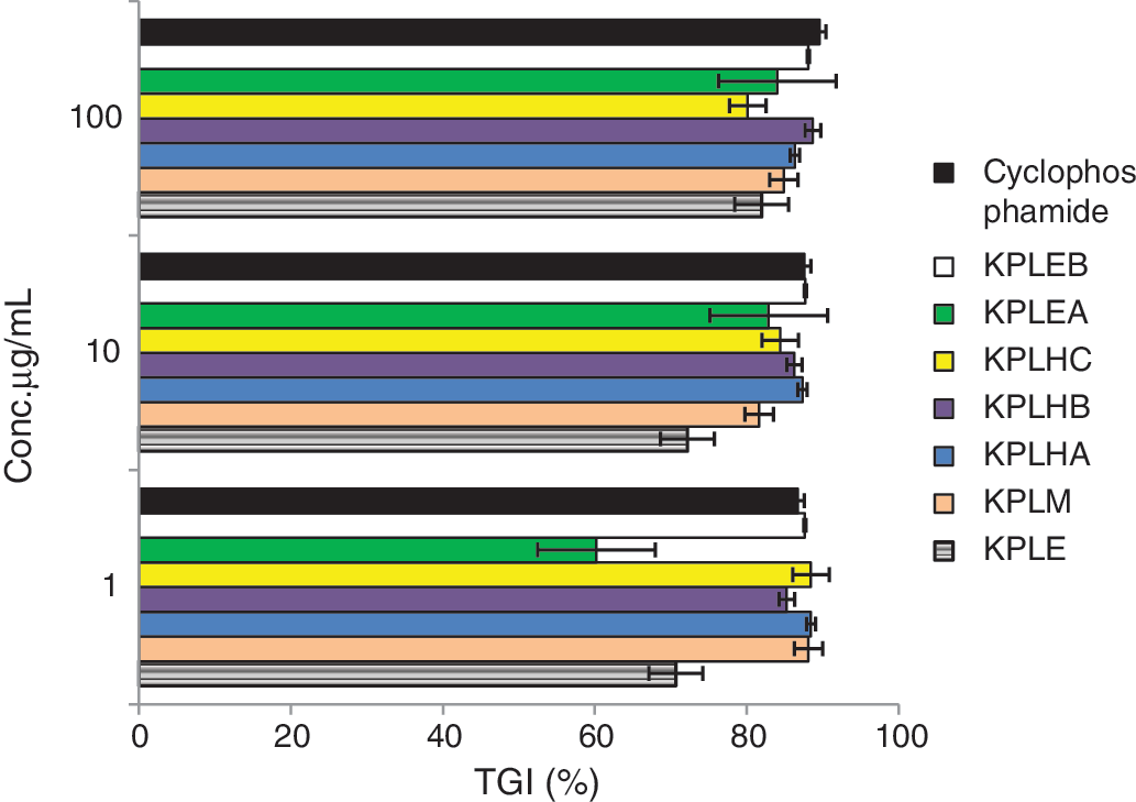
Antiproliferative activity of extracts, fractions, and major compounds of K. pinnata against cancer cell line. Results represent the mean±standard error of mean of triplicate determinations. Color images available online at
Various anticancer agents have been isolated from other parts of the K. pinnata. Verminoside and verbascoside isolated from the fruits showed a promising and selective anticancer potential when tested using the MTT assay. 46 Aqueous and organic extracts of the stem barks and fruits of K. pinnata have been shown to possess growth inhibitory effects against four melanoma cell lines and a renal cell carcinoma line (Caki-2) using two different (MTT and SRB) assays. Lapachol, a possible constituent of these extracts, together with known therapeutic antineoplastic agents, was also tested in the same way. Significant dose-dependent inhibitory activity was shown by the dichloromethane extract of the stem bark and lapachol. 7 The antitumor activity of Bignoniaceae family may be due primarily to its FAs and naphthaquinoids, such as lapachol. Lapachol present in K. pinnata root and lapacho tree 17,47 was considered for clinical administration as anticancer agent. 48 In vitro cytotoxic activity found in root bark extract of K. pinnata is attributed to γ-sitosterol, which was comparable to that of the standard used, lapachol. 45
In conclusion, this work describes the secondary metabolite profile of the leaf extracts of K. pinnata. The differences in the values produced by FRAP, DPPH, ABTS, and cytotoxicity assays, which possibly reflect the influence of different factors on the effectiveness of antioxidants and cytotoxicity, mean that antioxidant and cytotoxicity capacity may not be evaluated using only one assay protocol. The FRAP method measures total reductive power, the DPPH and ABTS measure free radical scavenging capacity, and the cytotoxicity assay evaluates the cell inhibition potential. The methanol extract of K. pinnata, KPLM, was the most potent cytotoxic substance tested. It is clear from the cytotoxic test that the cytotoxic effect of K. pinnata cannot be attributed to one single compound but rather synergy of many compounds. Further studies are needed to determine the entire composition of the methanolic extract, KPLM, and evaluate whether the identified compounds could hold promise as possible anticancer agents.
Footnotes
Author Disclosure Statement
The authors disclose that there are no conflicts of interest.