




Abstract
Acute monocytic leukemia (AML M5 or AMoL) is one of the several types of leukemia that are still awaiting cures. The use of chemotherapy for cancer management can be harmful to normal cells in the vicinity of the target leukemia cells. This study assessed the potency of the extracts from lesser galangal, turmeric, and ginger against AML M5 to use the suitable fractions in neutraceuticals. Aqueous and organic solvent extracts from the leaves and rhizomes of lesser galangal and turmeric, and from the rhizomes only of ginger were examined for their antiproliferative activities against THP-1 AMoL cells in vitro. Lesser galangal leaf extracts in organic solvents of methanol, chloroform, and dichloromethane maintained distinctive antiproliferative activities over a 48-h period. The turmeric leaf and rhizome extracts and ginger rhizome extracts in methanol also showed distinctive anticancer activities. The lesser galangal leaf methanol extract was subsequently separated into 13, and then 18 fractions using reversed-phase high-performance liquid chromatography. Fractions 9 and 16, respectively, showed the greatest antiproliferative activities. These results indicate that the use of plant extracts might be a safer approach to finding a lasting cure for AMoL. Further investigations will be required to establish the discriminatory tolerance of normal cells to these extracts, and to identify the compounds in these extracts that possess the antiproliferative activities.
Introduction
A
Spontaneous remission of AML M5 with bacteremia has been reported. 10 Such remissions are usually temporary, with relapse occurring within months of initial remission. The exact mechanism of remission is unknown. Common deliberate remission strategies usually include chemotherapy induction and hematopoietic stem cell (i.e., bone marrow) transplantation (HSCT) depending on the age of a patient, comorbidities, remission duration possibilities, and availability of a suitable HSCT donor. 5,11 These too do not offer a permanent cure, and the success rate is low especially among older adult patients. Current standard chemotherapy approaches have reached the limit of their efficacy in patients with AML. 12 In the past two decades, chemotherapeutic regimens have shown only little improvement, and therefore, the overall survival for patients has mostly been temporary. 5 There is a huge unmet need for less toxic, efficacious therapies for cancer patients than are currently available.
Clearly, there is a significant need to improve current cancer therapies and to search for novel therapies. For example, the World Health Organization (WHO) has taken an interest in the indigenous system of medicine; particularly, plant remedies. Plant-derived products have been shown to be valuable sources for the development of anticancer drugs. 13 However, the ease of isolation of the active compounds in plant extracts depends on the complexity and stability of the structures of such compounds. The active compounds in a plant extract may only work in combination, and in a delicate balance, to be effective. The extracts may therefore serve as effective nutraceuticals until the active components can be fully explored for effective treatment. Hence, it may be more appropriate to establish the potency of a plant in its extract form against cancer, which may also reduce the adverse effects associated with some drugs and other current methods of treatment of cancer patients. In the present study, we determined the anticancer activities of extracts from three medicinal plants, lesser galangal (Alpinia officinarum Hance Jam1), turmeric (Curcuma longa L.), and ginger (Zingiber officinale Rosc.), which are currently used as medicinal plants in the traditional treatment of various disease conditions in many parts of the world. Consequently, the goal of this study was to establish the potency of the extracts from lesser galangal (A. officinarum Hance Jam1), turmeric (C. longa), and ginger (Z. officinale) against AML M5, with a view to exploit the suitable fractions for use in neutraceuticals or as raw materials for the pharmaceutical industries.
Materials And Methods
Leaves and rhizomes of lesser galangal (A. officinarum Hance Jam1) and the rhizomes of ginger (Z. officinale) were obtained from two farms in Linstead, St. Catherine, Jamaica. The leaves and rhizomes of turmeric (C. longa) were obtained from a farm in Benin City, Nigeria. Plant leaves and rhizomes were rinsed in water, dried separately at 40°C, and ground to powder. About 150 g of powdered leaves was extracted using a standard eluotropic gradient based on solvent polarity beginning with deionized water (1000 mL) followed by 50% methanol, 100% methanol, chloroform, dichloromethane, hexane, and acetone. 14 The slurry was sonicated and extracted for at least 2 h. The extract was sieved through a cheesecloth and the filtrate was frozen (−80°C), and then freeze dried. The extract was redissolved in the initial solvent and stored at −80°C until used. The leaf residue was then extracted in the next sequential solvent. The entire extraction procedure was repeated for plant rhizomes.
Cell culture
Cell cultures of the AMoL cell line THP-1 were grown in the RPMI-1640 medium containing 10% fetal bovine serum, 50 IU/mL penicillin and antimicrobial-antimycotic solution, and maintained at 37°C in a humidified atmosphere containing 5% CO2. Cells were subcultured every 3 days and used for anticancer study experiments when culture cell density equaled 8×10⁵ cells/mL. 15
Antiproliferative activity test
Cell cultures of density up to 8×10⁵ cells/mL were transferred to 24-well plates in aliquots in quadruplicates. An equal volume of fresh culture media was added to each well, along with 2 mg/mL or 0.1 mg/mL solution of the plant extract. The culture was incubated at 37°C in 5% CO2 atmosphere for 24 h and tested for viability. 16,17 Cultures to which 2 mg/mL lesser galangal leaves extracts were administered were incubated up to 48 h. Cell cultures that were administered 1% (v/v) extraction solvents only in the place of plant extracts were used as respective positive controls. Replicates of cell cultures without any extract or solvent only administration were used as negative controls.
Cell viability
Cell viability was determined with the trypan blue staining test. 16 Living and dead cells were counted using a hemocytometer—each was normalized as a fraction/percentage of the total cell count. Cells were also subjected to the 3-(4,5-dimethylthiazol-2-yl)-5-(3-carboxymethyoxyphenyl)-2-(4-sulfophenyl)-2H-tetrazolium inner salt (MTS) assay for confirmation of viability.
Trypan blue staining test
Aliquots of 0.4% trypan blue solution and cell culture were introduced into a tube in equal volumes and mixed gently. A 10-μL aliquot of the mixture was loaded into the hemocytometer. Dead cells were counted by reason of the blue stain they had picked up. Living cells, which did not pick up the stain, were counted separately at the four quadrants of the hemocytometer. The percentage of dead cells was determined by the formula: average of the dead cell counts (from the four quadrants) multiplied by 104 multiplied by dilution factor (i.e., 2) divided by average of the total (i.e., dead plus living) cell counts (from the four quadrants) multiplied by 100%. 16
MTS assay
The MTS assay was conducted according to the Promega “CellTiter 96 Aqueous Nonradioactive Cell Proliferation Assay” technical bulletin. 17 An aliquot of 100 μL of phenazine methosulfate (PMS) was added to 20 μL of the MTS solution immediately before adding the cell culture and gently mixed. An aliquot of 20 μL of the combined MTS/PMS solution was added to 100 μL of cell culture. The culture plate was incubated for 4 h at 37°C in a humidified 5% CO2 atmosphere. Absorbance was read at 490 nm using the Bio-Rad Benchmark microplate reader.
Lesser galangal HPLC fractionation
The methanol extract from lesser galangal leaves was diluted 1 in 4 with methanol, filtered through a 0.45-μm membrane filter (Millipore, Bedford, MA, USA), and then chromatographed on an Agilent 1100 Series liquid chromatograph (Agilent Technologies, Palo Alto, CA, USA) equipped with the diode array detector (DAD) and timed fractionation modules. Gradient elution of extract in acetonitrile: water mixture (15:85–95:5 percentage ratio, v/v) was done with a C18 reversed-phase ZORBAX column (Agilent Technologies) of 4.6 mm×250 mm, 5-μm particle size dimensions, at a flow rate of 1 mL/min for 30 min. Sample effluents were collected in thirteen 2-min fractions. The activity in fractions was assessed using cell assays as previously described. Active fractions, based on cell activity assays, were further separated into eighteen 20-sec fractions, with activity assessed as previously described.
Statistical analysis
Data are given as means±standard error of the mean (n=4). Significant testing was performed using the Student's t-test for comparative analysis between two sets of data.
Results
Antiproliferative activities of plant extracts
Antiproliferative activities of extracts from lesser galangal (A. officinarum) and turmeric (C. longa) leaves and rhizomes and from ginger (Z. officinale) rhizomes were determined against the THP-1 AMoL cells. All organic fractions of lesser galangal (A. officinarum) leaf extracts showed an activity against the cultured cancer cells. The extracts in the organic solvents showed significantly effective antiproliferative activities with respect to their positive controls, within 24 h (Fig. 1A). The water soluble fraction showed no distinct antiproliferative activity. The antiproliferative activities were maintained by extracts up to 48 h without any significant solvent interference with the cancer cells (Fig. 1B).
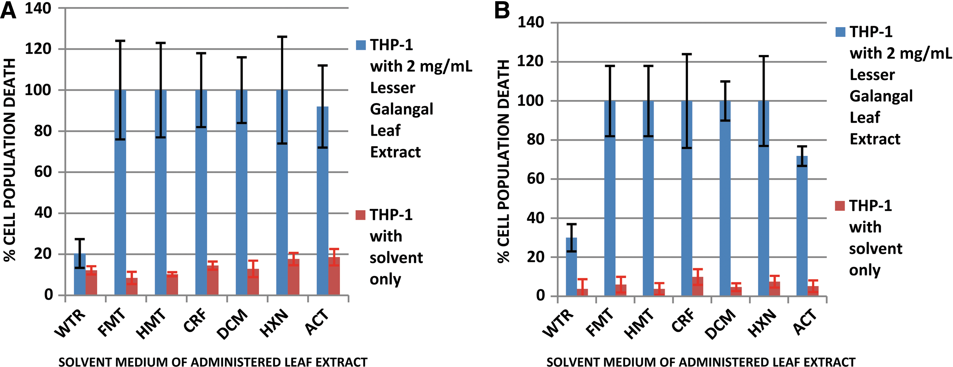
Percentage population death of THP-1 cells administered 2 mg/mL lesser galangal leaf extract and cultured for
When cell cultures were administered diluted quantity of lesser galangal leaf extracts, to a concentration of 0.1mg/mL, some differences in activity were observed. Only the extracts in methanol, chloroform, and dichloromethane showed a high antiproliferative activity. The 100% methanol extract showed a high activity of 78%±10% cell death, whereas chloroform and dichloromethane had antiproliferative activities of 100%±24% and 100%±28% cell death, respectively. However, the 100% methanol was observed to have the least interference on the cell culture as an extract solvent medium among the three. It showed a 2.3%±1.0% cell population death compared to 7.6%±3.0% and 4.8%±1.0% showed by chloroform and dichloromethane, respectively (Fig. 2).
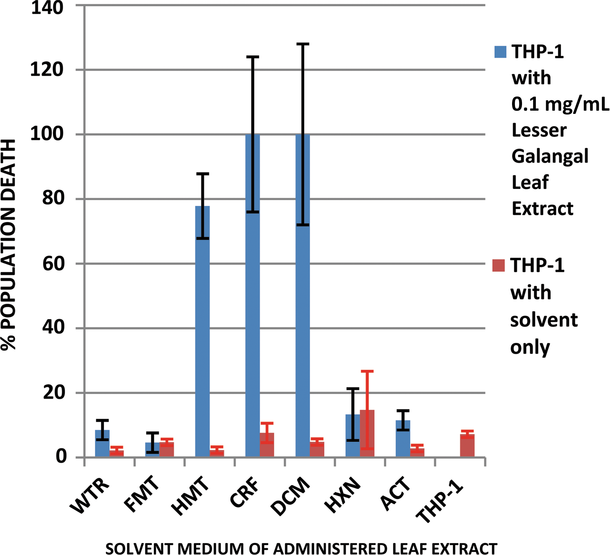
Percentage population death of THP-1 cells administered 0.1 mg/mL lesser galangal leaf extract and cultured for 24 h. THP-1, THP-1 cell culture only. Color images available online at
The lesser galangal rhizome extract was tested for the anticancer activity in a 24-h THP-1 culture. The chloroform, dichloromethane, and acetone extracts showed significant anticancer activities of 100%±35%, 100%±22%, and 92%±22% THP-1 population death. However, acetone had the least extract solvent medium interference on the cell culture (Fig. 3).
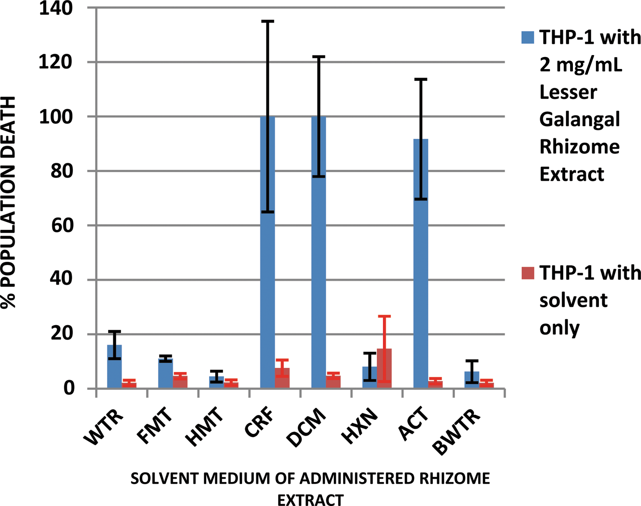
Percentage population death of THP-1 cells administered 2 mg/mL lesser galangal rhizome extract and cultured for 24 h. BWTR, boiled water. Color images available online at
The turmeric (C. longa) leaf and rhizome extracts were administered (2 mg/mL) separately to THP-1 cell culture. Among the leaf extracts obtained from different solvents, the 50% methanol extract showed the most significant antiproliferative activity of 100%±15% cell death with a minimal solvent interference of 1.3%±1.0% cell death (Fig. 4). The 50% methanol and 100% methanol rhizome extracts administered to THP-1 cell culture displayed the most significant antiproliferative activities of 76%±29% and 100%±27% cell death, without much interference from the methanol solvent-positive controls, which measured 4.2%±3.0% and 5.1%±6.0% cell death, respectively (Fig. 5).

Percentage population death of THP-1 cells administered 2 mg/mL turmeric leaf extract and cultured for 24 h. Color images available online at
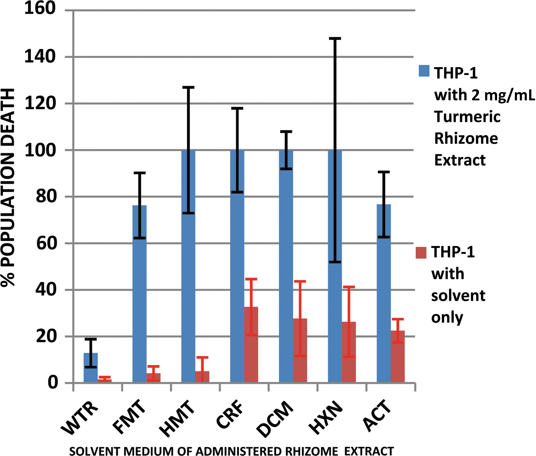
Percentage population death of THP-1 cells administered 2 mg/mL turmeric rhizome extract and cultured for 24 h. Color images available online at
The ginger (Z. officinale) rhizome had the highest antiproliferative activity in the 100% methanol extract (Fig. 6). It had a percentage THP-1 population death of 100%±27%, with the solvent causing a 2.8%±1.0% cell death.
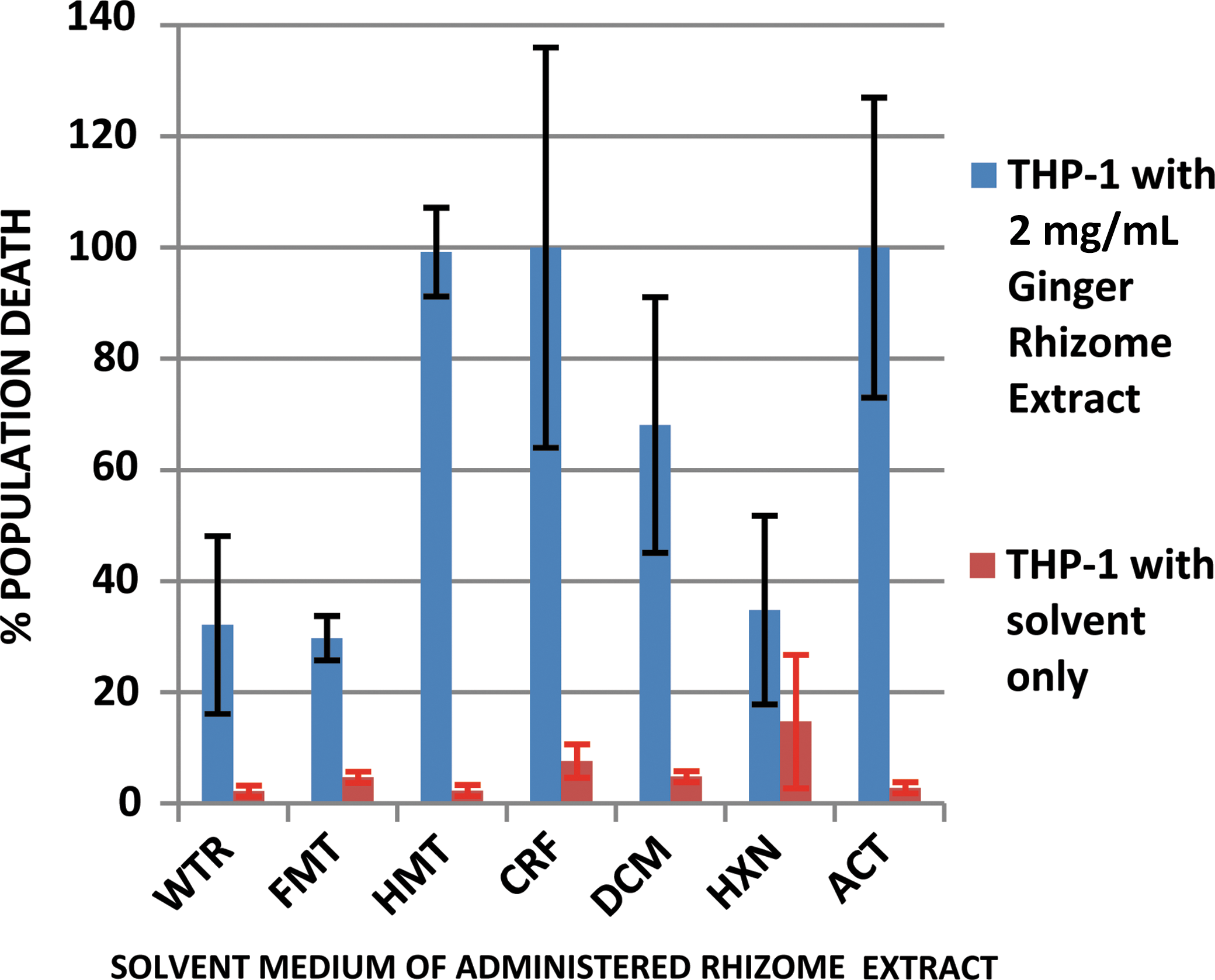
Percentage population death of THP-1 cells administered 2 mg/mL ginger rhizome extract and cultured for 24 h. Color images available online at
Antiproliferative profile of lesser galangal leaf methanol extract fractions
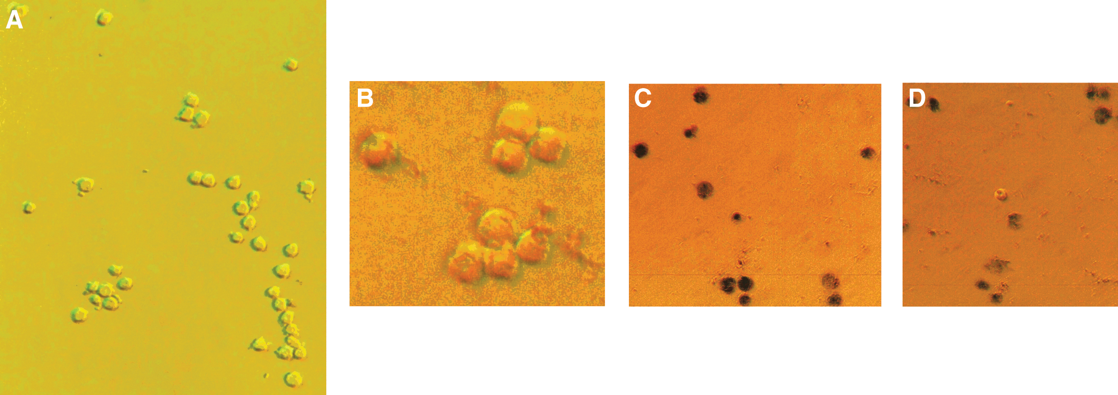
Separation of the 100% methanol extract of lesser galangal leaf was achieved using the reversed-phase high-performance liquid chromatography (RP-HPLC) method. The latter 6 of the 13 fractions, that is, fractions 8–13, showed high antiproliferative properties. Fraction 9 displayed the most impressive attack against cancer, with a THP-1 cell culture population death of 100%±5%. The solvent had a minimal impact of 2.3%±1.0% cell culture population death (see Figs. 7 and 8). Fig. 8A shows the cell culture of the AML THP-1 cells only without any plant extract. Fig. 8B shows the THP-1 cell culture to which HPLC fraction 4 of the lesser galangal leaf extract was added. The cells are seen to be alive. Fig. 8C shows the THP-1 cell culture to which HPLC fraction 9 of the extract was added. It reveals the cytotoxic effect of this extract fraction on the cells as the most potent relative to the other fractions. Fig. 8D shows the THP-1 cells to which the HPLC fraction 12 was added. This fraction is potent against the THP-1 cells, but not as much as fraction 9.

Population death of THP-1 cells administered reversed-phase high-performance liquid chromatography (RP-HPLC) gradient fractions of lesser galangal leaf extract in 100% methanol. THP-1/HMT, THP-1 cell culture administered with methanol solvent only. Color images available online at
The expanded fractionation profile of fractions, including the initial most effective antiproliferative fraction resulted in 18 new fractions. Several of them displayed high antiproliferative properties. They include fractions 01–04, 08, 12, 16–18. Fraction 16 showed the most impressive of all 18 fractions in attack against the THP-1 cancer cells. It achieved a population cell death of 99.2%±3.0% (Fig. 9). Fig. 10 is the HPLC fingerprint of the lesser galangal leaf 100% methanol extract. Fig. 10A shows the electrospray ionization (ESI) total ion current chromatogram of the extract fractionation. Fig. 10B shows the DAD chromatogram of the extract fractionation. Fig. 10C shows the ESI scan of the 22-min peak from Fig. 10A. Fig. 10D shows the UV signature of the 22-min peak.
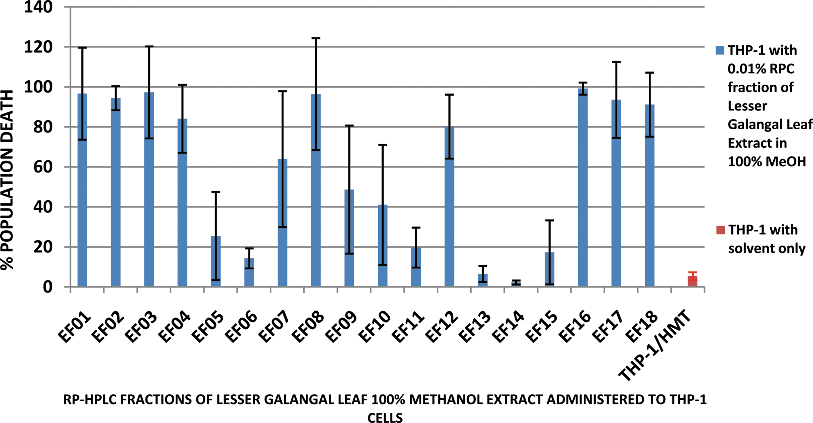
Percentage population death of THP-1 cells administered expanded RP-HPLC gradient fractions (EF) of lesser galangal leaf extract in 100% methanol. Color images available online at
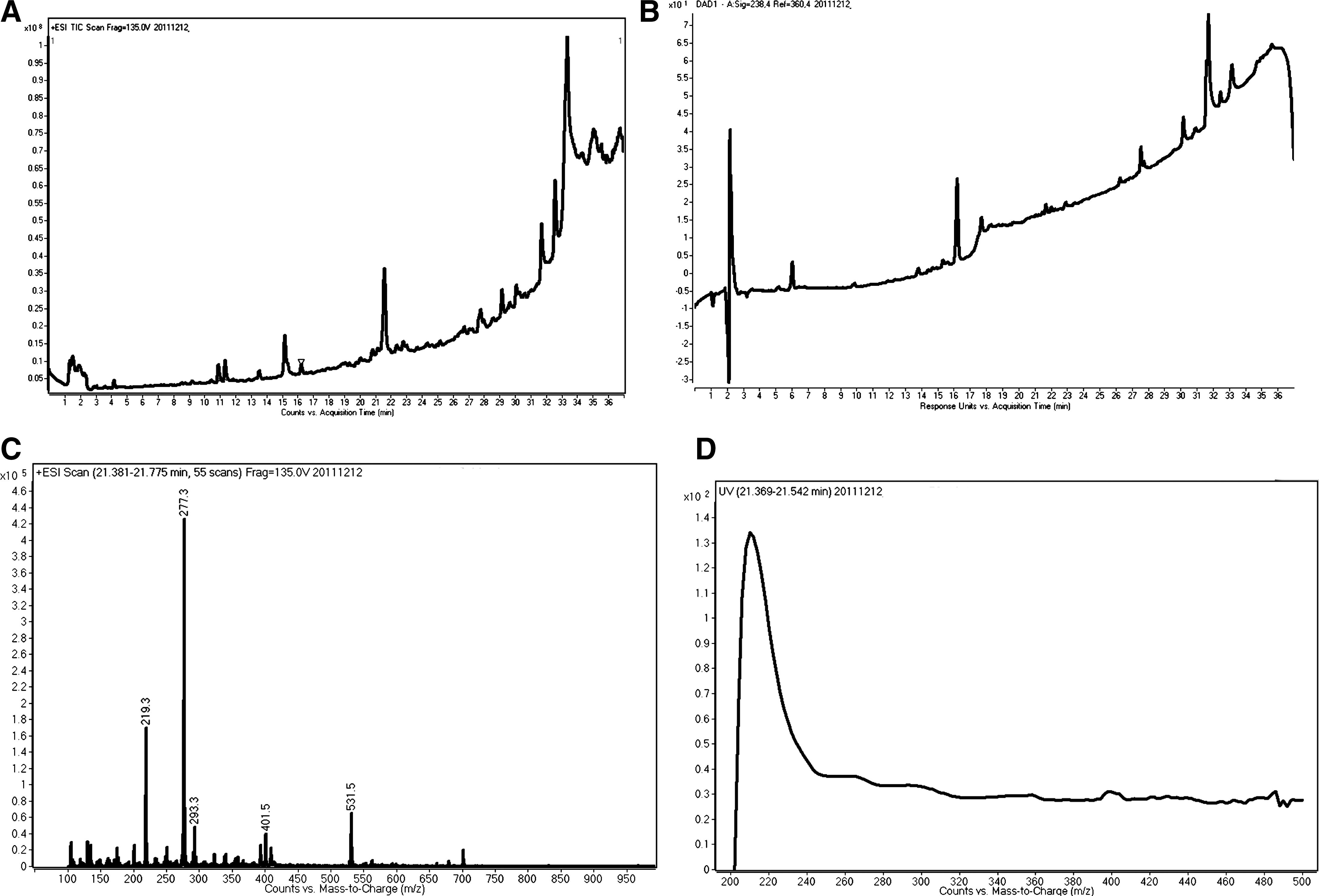
RP-HPLC fingerprint of the lesser galangal leaf 100% methanol extract.
Discussion
Current therapeutic approaches for remission of AML M5 include chemotherapy as a major strategy, and probably the only initial strategy for achieving a complete remission. 5 However, the use of chemotherapeutic agents like epipodophyllotoxins could cause the interruption of normal genetic sequences and illegitimate recombination of chromosomal fragments, and could lead to transforming mutations in some hematopoietic progenitors. 18 The need for novel noninvasive therapeutic strategies has been highlighted. 12 Although multiple strategies aimed at reducing the tumor burden in patients have been developed, relapse remains the most common cause of death among AMoL patients. 19 Natural agents that are selective, target specific, and will not lead to chromosomal and genetic aberrations are preferred. Desirable ideal agents should, in addition, use the strategy of apoptosis rather than cytotoxic necrotic activity to remove the leukemic cells. In pursuance of this target, extracts were obtained in this study from the leaves and rhizomes of lesser galangal (Apinia officinarum Hance Jam1) and turmeric (C. longa L.), and rhizomes only of ginger (Z. officinale Rosc.). The study was a preliminary probe for the antiproliferative activity against AMoL THP-1 cells. The three plants were studied together because they belong to the same family of zingiberaceae, which produces rhizomes, and are all used by herbalists for local medicinal remedies. Leaf and rhizome extracts from lesser galangal are known to be soluble in aqueous and organic solvents, which include methanol, ethanol, chloroform, acetone, etc. 20,21 However, only the aqueous and ethanol solvents are safe for use in nutraceuticals. Extracts from ginger and curcumin of turmeric have been reported to have poor aqueous solubility and, therefore, poor bioavailability, but strategies for improvement of such are being explored by several groups. 22 –25 Solubility of ginger extracts in water has been reported to be 0.69±0.03 mg/mL, but up to 3.19±0.38 mg/mL in simulated gastric fluid. 23 Curcumin extract aqueous solubility is said to range between 6.24±0.49 mg/mL and 19.62±0.84 mg/mL at different degrees of hot water. 26 Extracts from these plant organs were made by dissolution in deionized water, 50% methanol, 100% methanol, chloroform, dichloromethane, hexane, and acetone.
Lesser galangal leaf extracts from all organic solvents had a significant antiproliferative activity at 2 mg/mL extract in a culture medium, which was seen within 24 h of incubation, and remained so for the period of 48 h under study, without interference by the solvent-positive controls (Fig 1A, B). The result suggests that on direct exposure of the lesser galangal leaf extracts obtained from these solvents to the AMoL THP-1 cells, they would be effective in causing the death of the cancer cells, without any significant influence of the solvent media within a relatively short time frame. The active ingredient(s) in the extract responsible for controlling cancer cell proliferation is currently unknown. The organic solvent extract has been reported to contain some diarylheptanoids and flavonoids, which are believed to be effective as antiplatelet, antioxidant, antiproliferative, antiemetic, antihepatoxic, and anti-inflammatory agents. 27 –29 They are also known to contain fatty acid synthase inhibitors as anticancer agents, 30 and beta glycosides, which have been used in traditional medicine and as food preservatives. 31 In fact, Tabata et al. 20 reported the ability of diarylheptanoids derived from the A. officinarum to kill human neuroblastoma cells by apoptotic induction. Their report indicated that at concentrations of 10−8 M and 10−4 M, respectively, the two compounds isolated from the plant induced nuclear shrinkage and fragmentation, activated caspase-3 and caspase-9, and induced S-phase cell cycle arrest of the human neuroblastoma cells.
Dilution of the lesser galangal leaf extracts from 2 mg/mL to 0.1 mg/mL administered in culture and incubated for 24 h produced significant differences in THP-1 cell population death. Only the 100% methanol, chloroform, and dichloromethane extracts possessed significant antiproliferative activities, with no significant interference from the extract solvent medium (Fig. 2). The extract in 100% methanol produced an antiproliferative activity of 78%±10% AMoL THP-1 population death (P<.01). It showed a better statistical precision and the least extract medium interference in culture in comparison to the activities of the extracts in chloroform and dichloromethane 100%±24% and 100%±28% AMoL THP-1 population death, respectively. The limitation of significant effective activity to extracts in only three organic solvents (Fig. 2) suggests that they may either contain different, more potent, and effective active ingredients than those contained in the other extraction solvents, or if the same compound(s), may be of higher effective concentrations. The extract in 100% methanol was of interest for further probe since it had the least interference by its extraction solvent, and thus could be the least toxic in vitro against normal cells. It was therefore subjected to HPLC fractionation to enable further analyses of the potency of its content(s). It is worthy of note that the lesser galangal rhizome extract showed significant antiproliferative activity in less number of organic solvents (Fig. 3) at a 2 mg/mL concentration in culture, against AMoL THP-1 cells, than did the leaf extracts. Besides, the rhizome extract was made in boiled water and tested for the antiproliferative activity in light of the common traditional practice of rhizome tea production in boiled water. This preparation is believed to be effective as an analgesic and anti-inflammatory and to relieve dysmenorrhea, among other effects. However, the rhizome extract in boiled water showed no significant antiproliferative activity against the AMoL THP-1 cells within 24 h in culture.
The turmeric leaf and rhizome extracts were administered at 2 mg/mL in culture for the antiproliferative activity against the AMoL THP-1 cells. While the turmeric leaf extracts in 50% methanol, acetone, and chloroform were the most impressive from this plant tissue in demonstrating an effective antiproliferative activity, the rhizome presented the extracts both in 50% and 100% methanol as the most effective antiproliferative extracts, in light of their very low levels of extraction solvent interference (Figs. 4 and 5). Although the leaf extracts in 100% methanol and dichloromethane had a mean activity of 100% activity, they had very wide standard error margins and relatively higher levels of extraction solvent toxicity interference in causing cancer cell death. Nonetheless, the antiproliferative activity recorded by turmeric leaf and rhizome extracts in organic solvents is acknowledged. Turmeric has been reported to contain curcuminoids, which have been said to display antioxidant, immune enhancement, and detoxification characteristics in birds. 32 Curcumin has also been reported to induce apoptosis against human colon cancer, 33,34 human HL-60, 35 and murine WEHI 36 acute promyelocytic leukemia cells. The demonstration of the antiproliferative activity of turmeric extracts in this study against AMoL THP-1 cells points to the wide-ranging sphere of antiproliferative activity of turmeric.
Ginger rhizome extracts in 100% methanol, chloroform, and acetone showed a mean value each of 100% antiproliferative activity against AMoL THP-1 cells when administered in a 2 mg/mL concentration in culture. They also had very low values of extraction solvent interference (Fig. 6). Ginger is widely used as a spice, an antiemetic and carminative agent, and for its essential oils. It has also been reported to induce apoptosis in HL-60 leukemia 37 and nonsmall cell lung cancer 38 cells. It is also reported to be chemopreventive and anti-inflammatory. 39,40
The 100% methanol extract of lesser galangal leaf was fractionated using RP-HPLC into 13 fractions. Fractions 8–13 showed high antiproliferative properties at 0.1 mg/mL, but only fraction 9 displayed the highest level of antiproliferative activity with a THP-1 cell culture population death of 100%±5% and a minimal solvent interference of 2.3%±1.0% cell culture population death (Fig. 7). This suggests that fraction 9 contains the active agent against the AMoL THP-1 cells in enough concentrations to produce the total activity. An expanded fractionation profile showed that fractions 01–04, 08, 12, 16–18 displayed high antiproliferative properties, but fraction 16 had the highest activity against the AMoL THP-1 cells with a population cell death of 99.2%±3.0% (Fig. 9). The profile results mean that the active antiproliferative agent is in the greatest concentration in fraction 16 or else more than one compound might be responsible for the antiproliferative activity against the AMoL THP-1 cells. Identification of the active agent(s) in these fractions is underway.
In conclusion, our study establishes the antiproliferative activities of the organic extracts of the medicinal plants assessed, especially A. officinarum. Findings from this study indicate that the use of plant extract might be a promising approach to finding a lasting treatment for AMoL. We found one fraction of A. officinarum methanol extract to be the most potent, and expanded fractionation showed fraction 16 to be most effective against AML M5 cells. Although the anticancer activity of diarylheptanoids derived from Alpinia offinarum by apoptotic induction has been reported, 20 further work will be needed to establish the discriminatory tolerance of normal cells to these extracts in comparison to AML M5 cells. Besides, preparative work will be required to isolate and identify the active principles in the fractions found to possess antiproliferative properties that may be useful in the future development of drugs against AML M5.
Footnotes
Acknowledgments
We hereby thank Rayn International Holding Ltd. (Linstead, Jamaica) for the supply of the lesser galangal (A. officinarum Hance Jam1) used in this study, the Center for Coastal Studies and TAMU-CC for supplies, and Dr. Lillian Waldbeser for supplying the THP-1 cells.
Author Disclosure Statement
There are no conflicts of interest by the authors of this article.