
Abstract
A high dietary intake of polyphenols has been associated with a decreased risk of cardiovascular disease and cancer, attributed in part to their antioxidant activity and pro-apoptotic effects. Aquamin is a multi-mineral algal extract that enhances bone mineralization, relieves osteoarthritis, and aids digestion; however, Aquamin has not demonstrated antioxidant activity. In the present study, Aquamin was supplemented with 8% Enzogenol, a pine bark extract with a high phenolic content, and 2% Sunphenon, a green tea extract that also has a high phenolic content to produce a mixed product (A:E:S). The antioxidant activity of A:E:S was compared with that of its constituent compounds and also with catechin and epigallocatechin by measuring total phenol content, ferric-reducing antioxidant potential, and 2,2-diphenyl-2-picrylhydrazyl hydrate. The cytotoxic and apoptotic effects of the compounds were also measured in the U937, human monocytic blood cell line. A:E:S demonstrated an antioxidant activity that was equal to that of the compounds used in its preparation. Aquamin was not cytotoxic in the U937 cell line; however, A:E:S was cytotoxic and the primary mechanism of cell death was apoptosis. The biological effects of Aquamin were enhanced by supplementation with Enzogenol and Sunphenon to include antioxidant effects and the ability to induce apoptosis in U937 cells.
Introduction
P
The food supplement Aquamin is a natural seaweed–derived multi-mineral from the red algae, Lithothamnion species, which is rich in calcium, magnesium, and trace amounts of other minerals. It has been shown to relieve the symptoms of osteoarthritis, 7 enhance the mineralization of bone, 8,9 aid digestion, 10 and to have anti-inflammatory properties in vitro 11,12 and in vivo. 13 Enzogenol is a water-soluble, powdered extract from the bark of New Zealand Pinus radiata trees, and it contains the active components procyanidin dimers, trimers, oligomers, and polymers, formed from catechin and EC, taxifolin, and other flavonoids and phenolic acids. 6 Clinical and in vitro studies have shown that Enzogenol has antioxidant and anti-inflammatory benefits, may reduce markers of cardiovascular disease risk, improve brain functioning, and protect against tumor development in experimental mice. 14 –17 Sunphenon 90LB is a green tea extract produced from the leaves of the Camellia sinensis plant that has a high content of green tea catechins, EGCG, EGC, and ECG. Sunphenon was reported to prevent 1,2-dimethylhyrazine-induced carcinogenesis in rats. 18
The objective of the present study was to compare the antioxidant potential and pro-apoptotic effects of Aquamin supplemented with Enzogenol (8% w/w) and Sunphenon 90LB (2% w/w) (A:E:S) with those of Aquamin, Enzogenol, or Sunphenon alone, and also with catechin and ECG, which have previously demonstrated beneficial effects in-vitro. A:E:S is sold commercially as AquaPT (Marigot Ltd., Carrigaline, Co. Cork, Ireland).
Materials And Methods
Chemicals
All reagents were supplied by Sigma-Alrich Ireland Ltd (Wicklow, Ireland), unless otherwise stated. U937 cells were purchased for the European Collection of Animal Cell Culture (ECACC; Salisbury, United Kingdom). Cell culture plastics were supplied by Cruinn Diagnostics (Dublin, Ireland). Aquamin (Food and Drug Administration GRAS 000028; Marigot Ltd.) was prepared from the mineral-rich red marine algae, Lithothamnion species, harvested off the Atlantic coasts of Ireland and Iceland under approved licences. The calcified seaweed was separated from extraneous materials, sterilized, dried, and milled under ISO and HACCP certification. Enzogenol (ENZO Neutraceuticals Ltd., Paeroa, New Zealand) is an aqueous extract from the bark of New Zealand grown P. radiata trees containing ∼80% total proanthocyanidins and other water-soluble flavonoids, flavonoid conjugates, and phenolic acids. Sunphenon is a mixture of green tea polyphenols (Taiyo Kagaku Co., Mie, Japan) prepared by extracting Japanese green tea (C. sinensis var. sinensis) with hot water and then by partitioning with ethyl acetate as previously reported. 19
Antioxidant activity of the test compounds
ECG, catechin, Aquamin, Enzogenol, Sunphenon, and A:E:S were screened to measure their total phenol content (TPC) and their antioxidant potential by measuring ferric-reducing antioxidant potential (FRAP) and 2,2-diphenyl-2-picrylhydrazyl hydrate (DPPH) as previously detailed. 20 For the TPC, stock solutions of catechin, ECG, Enzogenol, and Sunphenon were prepared to a concentration of 1 mg/mL in distilled water. Aquamin was prepared to a concentration of 10 mg/mL in distilled water. The mix was prepared containing 1 mg/mL Enzogenol and the relative concentrations of Aquamin (11.25 mg/mL) and Sunphenon (0.25 mg/mL) to a ratio of 90:8:2 (w/w) A:E:S. TPC was determined by the Folin–Ciocalteau method as described by Singleton and Rossi. 21 A standard curve was prepared using gallic acid, and data were expressed as gallic acid equivalents.
Stock solutions prepared for the TPC were diluted 10-fold for the FRAP assay. The FRAP assay measures the formation of a blue-colored Fe2+-tripyridyltriazine (Fe2+-TPTZ) compound from the colorless oxidized Fe3+ form (Fe3+-TPTZ) by the presence of electron-donating antioxidants. Briefly, 2 mL of working FRAP reagent (acetate buffer:10 mM TPTZ:20 mM FeCl3·6H2O [10:1:1]) was prepared fresh for each experiment and was mixed with 100 μL test compounds; the absorbance at 593 nm was measured following a 30 min incubation. FRAP values were obtained by comparing the change in absorption for the test compounds with those obtained from increasing concentrations of Fe2+, and the data were expressed as FRAP equivalents.
Stock solutions prepared for the TPC were diluted 1:1 for the DPPH assay. DPPH is a free radical that is extensively used to test the ability of compounds to act as a free radical scavenger or hydrogen donor. A series of dilutions for each test compound was prepared, and 100 μL was added to 3.9mL 0.06 mM DPPH reagent prepared in methanol. Samples were incubated for 30min, the absorbance was measured at 515 nm, and an EC50, the concentration of compound required to decrease DPPH absorbance by 50%, was determined for each of the test compounds.
Maintenance of cells in culture
Human monocytic U937 cells were grown in suspension in RPMI-1640 medium supplemented with 10% (v/v) fetal bovine serum (FBS) and were maintained at 37°C/5% CO2 in a humidified incubator, in the absence of antibiotics. For experiments, cells were adjusted to a density of 1×10⁵ cells/mL in media containing reduced (2.5%) FBS. Stock solutions of catechin, ECG, Aquamin, Enzogenol, Sunphenon, and A:E:S were prepared in RPMI-1640 medium for addition to cells.
Cell viability
Cells were seeded in the wells of a 96-well plate and were exposed to increasing concentrations of the test compounds for 24 h. The MTT assay was conducted according to the instructions provided with the MTT I proliferation kit (Roche Diagnostics, West Sussex, United Kingdom) as previously described. 22 An IC50 value, which is the concentration of a compound that reduces cell viability to 50% of the untreated, control cells, was determined for each of the test compounds.
Cell death and apoptosis
The proportion of dead cells was quantified, after a 24 h incubation with the test compounds, using a fluorochrome-mediated viability assay previously described. 23 Briefly, cells were mixed 1:1 (v/v) with a solution of fluorescein diacetate (FDA) and ethidium bromide (EtBr), then incubated at 37°C for 5 min before being layered onto a microscope slide. Under these conditions, live cells fluoresce green, while dead cells fluoresce red. Samples were examined at 200×magnification on a Nikon fluorescence microscope using a blue (450–490 nm) filter. Cells (200) were scored for each slide, and data were expressed as the percentage of dead (red) cells.
The nuclear morphology of treated cells was assessed by fluorescence microscopy after staining with Hoechst 33342 as previously described. 23 Approximately, 4×10⁵ treated cells were harvested by centrifugation (200 g, 10 min) to form a pellet. Hoechst 33342 stain (200 μL, 5 μg/mL PBS) was added, and the samples were incubated at 37°C for 1 h. Stained samples were placed on a microscope slide and examined under UV light (330–380 nm) using a Nikon fluorescence microscope (400×magnification). A total of 300 cells in each sample were analyzed, and the percentage of condensed/fragmented (apoptotic) nuclei was calculated.
DNA fragmentation assay
Detection of small DNA fragments was carried out as previously described. 23 Briefly, 2×10⁶ cells were harvested, and the pellets were lysed; RNAse A (0.25 mg/mL) was added, and the samples were incubated at 50°C for 1 h. Proteinase K (5 mg/mL) was added, and the samples were incubated at 50°C for a further hour. Both the samples and a 100–1500 bp DNA standard (Promega) were loaded to the wells of a 1.5% agarose gel, and electrophoresis was carried out in TBE buffer [0.45 M tris(hydroxymethyl) aminomethane, 0.45M boric acid, and 2 mM EDTA, pH 8], at 3 V/cm. The gel was visualized using a UV transilluminator and photographed.
Caspase-3 activity
Caspase-3 activity was analyzed using a caspase-Glo® 3/7 assay kit supplied by Promega (Ireland). Briefly, U937 cells were seeded in the wells of a 96-well plate and exposed to test compounds for 6 h. The caspase-Glo® reagent was added to the cells at a volume of 1:1, and the cells were incubated for a further 3 h. The luminescence of the samples was measured, and the data were expressed as fold increase relative to an untreated, control sample.
Statistical analysis
Data represent the mean of at least three independent experiments±standard error. Statistical analysis was evaluated by one-way ANOVA followed by Dunnett's or Tukey's post-test (GraphPad Prism 4.0; GraphPad, Inc., San Diego, CA, USA); P<.05.
Results
Antioxidant potential of test samples
The TPC and antioxidant potential, as determined by the DPPH and FRAP assays, of both catechin and ECG were similar (Table 1). Enzogenol had a lower TPC and a significantly (P<.05) lower DPPH radical scavenging activity than catechin and ECG; however, the FRAP activity of Enzogenol was equal to that of catechin and ECG (Table 1). Sunphenon had a similar TPC to catechin and ECG, and the DPPH radical scavenging activity of Sunphenon did not significantly differ from that of catechin and ECG but Sunphenon demonstrated significantly higher (P<.05) FRAP activity than any of the other compounds investigated. Aquamin did not demonstrate antioxidant activity by any of the methods employed in the present study (Table 1). The A:E:S mix demonstrated an antioxidant activity higher than that of Enzogenol and lower than that of Sunphenon which reflected the fact that the composition of the mix comprised 100% of the Enzogenol concentration investigated and 25% of the Sunphenon concentration investigated.
The antioxidant potential of test compounds as determined by TPC, FRAP, and the DPPH radical scavenging assay. Data represent the mean of three independent experiments±SE.
Values in the same column with different superscript letters differ significantly (P<.05, ANOVA followed by Tukey's).
TPC, total phenol content; FRAP, ferric reducing antioxidant potential; DPPH, 2,2-diphenyl-2-picrylhydrazyl hydrate; ECG, epicatechin gallate; A:E:S, Aquamin supplemented with Enzogonol and Sunphenon; SE, standard error; ND, not detected; ANOVA, analysis of variance.
IC50 values for test compounds
Aquamin did not demonstrate any cytotoxic effects in the U937 cell line as determined by the MTT assay (Fig. 1); therefore, an IC50 value for Aquamin could not be determined. Sunphenon was the most cytotoxic of the tested compounds (Fig. 1) and caused a reduction in viable cells to 50% of the control, untreated cells, at a concentration of 16.0 μg/mL (Table 2). The IC50 value of ECG was almost half that of catechin, and Enzogenol was the least cytotoxic of the tested compounds with an IC50 value of 111.8 μg/mL. The IC50 value for the A:E:S mix was 39.6 μL/mL (Table 2); this concentration corresponded to 215.7 μL/mL Aquamin, 19.8 μg/mL Enzogenol, and 5.1 μg/mL Sunphenon.
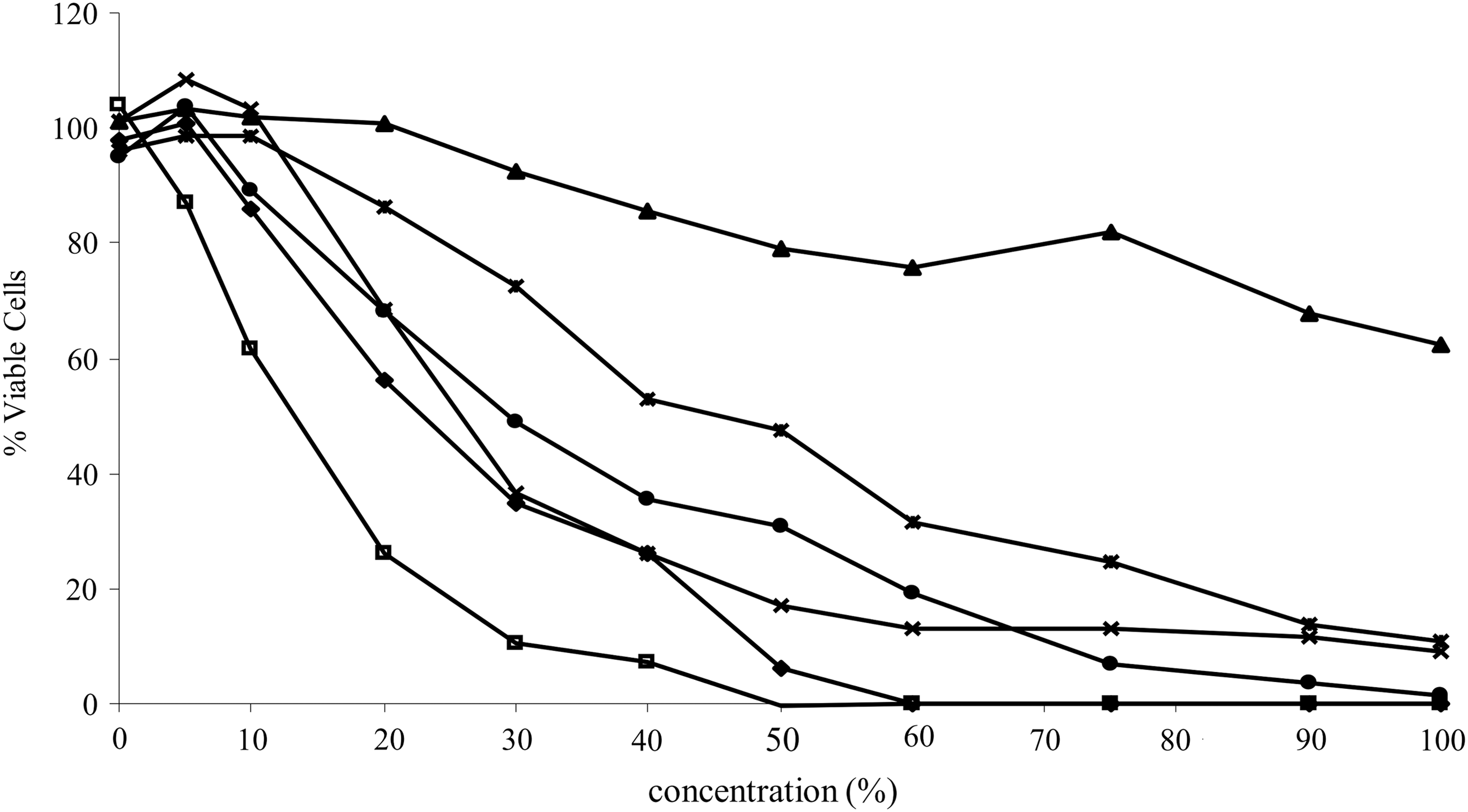
Cell viability was determined by the MTT assay after a 24 h incubation of the test compounds in U937 cells. Data represent the mean of three individual experiments. The concentration represents the percent of each compound added to the cells, where 100% represented 250 μg/mL catechin (◆); 250 μg/mL ECG (□); 2500 μg/mL Aquamin (▲); 500 μg/mL Enzogenol (*); 30 μg/mL Sunphenon (×); 60 μL/mL A:E:S (●). MTT, 3-[4,5-dimethylthiazol-2-yl]-2,5-diphenyl tetrazolium bromide; ECG, epicatechin gallate; A:E:S, Aquamin Supplemented with Enzogenol and Sunphenon.
The IC50 was determined by the MTT assay following a 24 h incubation of the test compounds in U937 cells. Data represent the mean of three individual experiments±SE.
Values in the same column with different superscript letters differ significantly (P<.05, ANOVA followed by Tukey's).
IC50, the concentration of the compound that reduced cell viability to 50% of the control cells; MTT, 3-[4,5-dimethylthiazol-2-yl]-2,5-diphenyl tetrazolium bromide.
Cell death
For the determination of cell death by the FDA-EtBr assay, catechin and ECG were added to U937 cells at a concentration of 100 μg/mL, and cells were incubated for 24 h. Concentrations of 200 μg/mL and 400 μg/mL Enzogenol were added to cells, and Sunphenon was added at concentrations of 20 and 40 μg/mL, as it was found to be ∼10-fold more cytotoxic than Enzogenol by the MTT assay (Fig. 1). The viability of U937 cells exposed to catechin did not significantly differ from that of control, untreated cells (Fig. 2). Cell death was significantly (P<.05) increased in the presence of 100 μg/mL ECG. Incubation with Enzogenol resulted in 23.7% cell death at 200 μg/mL, and this increased to 70.8% at the 400 μg/mL concentration. At 20 μg/mL, Sunphenon was not significantly cytotoxic; however, at the higher concentration (40 μg/mL), greater than 90% of cells were dead. The A:E:S mix was added to the cells at two concentrations, 0.125% and 0.5%, which corresponded to 1.4 mg/mL Aquamin:125 μg/mL Enzogenol:31.25 μg/mL Sunphenon and 4.5 mg/mL Aquamin:400 μg/mL Enzogenol:100 μg/mL Sunphenon, respectively. The lower concentration of the mix (0.125%) resulted in 32.6% cell death, and the higher concentration (0.5%) resulted in greater than 90% cell death.

Cell death as determined by the FDA-EtBr staining method and apoptosis as determined by the Hoechst staining method. U937 cells were incubated with the test compounds for 24 h. Cells (25 μL) were mixed 1:1 with FDA-EtBr that stains live cells green and dead cells red; 200 cells were scored at 200×magnification, and the percentage of dead cells was calculated. Cells were harvested by centrifugation and stained by Hoechst 33342, 300 cells were examined at 200×magnification, and the percentage of condensed and fragmented nuclei was calculated. Data represent the mean±SE of four independent experiments. *Significant difference from control dead cells (P<.05); †significant difference from control apoptotic cells (P<.05); ANOVA followed by Dunnett's post-test. FDA-EtBr, fluorescein diacetate-ethidium bromide; SE, standard error.
Apoptosis
Apoptotic cells were quantified as the percentage of condensed/fragmented nuclei identified after staining with Hoechst 33342. There was no significant increase in apoptotic nuclei in cells exposed to catechin (100 μg/mL) or the lower concentration of Sunphenon (20 μg/mL) after a 24 h incubation (Fig. 2). Incubation with EGC resulted in a significant increase (P<.05) in apoptotic nuclei to 42.8% (Fig. 2). At the lower concentration of Enzogenol (200 μg/mL), 47% of nuclei were identified as apoptotic but this decreased to 32.8% at the higher concentration (400 μg/mL). Sunphenon at 40 μg/mL resulted in 40% apoptotic nuclei after 24 h. The A:E:S mix induced a dose-dependent increase in apoptotic nuclei to 20% at the 0.125% concentration and 60% at the 0.5% concentration (Fig. 2). Apoptosis was also assessed by the DNA fragmentation assay (Fig. 3), and the addition of Enzogenol (400 μg/mL), Sunphenon (40 μg/mL), and the mix (0.5%) to U937 cells for 24 h caused the cleavage of DNA to fragments of 200 base pairs, which is the hallmark of apoptosis.

DNA Fragmentation assay. U937 cells were incubated with the test compounds for 24 h. DNA was isolated and loaded to the wells of an agarose gel, and electrophoresis was carried out. In apoptotic cells, the DNA is cleaved to fragments of 200 base pairs, which forms a ladder-like pattern. 1, mw marker; 2, control; 3, 100 μg/mL catechin; 4, 4.1 mg/mL Aquamin; 5, 400 μg/mL Enzogenol; 6, 40 μg/mL Sunphenon; 7, 0.25% A:E:S.
Caspase-3 activity
The activity of the apoptotic enzyme, caspase-3, was determined after a 6 h incubation in U937 cells and was expressed as the fold increase relative to control, untreated cells (Fig. 4). Catechin, at a concentration of 100 μg/mL, did not increase caspase-3 activity. Incubation of U937 cells with 100 μg/mL EGC resulted in a fourfold increase in caspase-3 activity at the 6 h timepoint. Enzogenol caused a dose-dependent increase in caspase-3 activity to 1.8-fold at the 200 μg/mL concentration and more than 5-fold at the 400 μg/mL concentration. Sunphenon induced a 7-fold increase at the 20 μg/mL concentration and a 5.5-fold increase in caspase-3 activity at the 40 μg/mL concentration at the 6 h timepoint. The A:E:S mix induced the greatest increase in caspase-3 activity, a ninefold increase at 0.125%, and a fourfold increase at the 0.5% concentration.

Caspase-3 activity. U937 cells were incubated with the test compounds for 6 h. Caspase-3 activity was determined using the caspase-glo 3/7 kit. Data represent 3 independent experiments±SE. *Significant difference (P<.05) from control, untreated cells; ANOVA followed by Dunnett's post–test.
Discussion
The antioxidant potential of the test compounds was determined by measuring their FRAP and DPPH radical scavenging activity and was compared with their TPC. Both of the monophenolic compounds, catechin and ECG had similar antioxidant activity (Table 1). Aquamin did not demonstrate antioxidant potential by any of the methods employed in the present study (Table 1). There was a strong correlation (R 2 =0.9422) between the TPC of all samples and their radical scavenging activity as determined by the DPPH assay. The correlation between TPC and the reducing power of the samples (FRAP) was poor (R 2 =0.1686), and both Sunphenon and Enzogenol displayed a higher reducing power (FRAP) relative to their TPC. FRAP values do not always correlate with other measures of antioxidant activity, as FRAP specifically measures only single-electron transfer (SET) while DPPH and TPC measure both SET and hydrogen atom transfer. 24 Roy et al. 25 also found a strong correlation between the TPC of green tea catechins and their DPPH radical scavenging activity, but the correlation with oxygen radical absorbance capacity (ORAC) activity was lower. Radical scavenging as determined by the DPPH assay is dependent on steric accessibility, and small molecules that have better access to the radical site will demonstrate better radical scavenging activity. 24 The high content of proanthocyanidins, which contains polymers, in Enzogenol may have resulted in its lower radical scavenging activity (DPPH) although the radical reducing (FRAP) activity of Enzogenol was equal to that of catechin and ECG. The A:E:S mix had an antioxidant activity that reflected the sum of Aquamin, Enzogenol, and Sunphenon used in its preparation, and, therefore, the high content of Aquamin contained within the mix did not hamper the antioxidant activity of Enzogenol and Sunphenon, which were present in the mix at much lower concentrations.
Polyphenols are consumed regularly as a part of a balanced diet and are not considered toxic even at high concentrations. 26 Amin et al. 27 found that polyphenols (EGCG and luteolin) were not cytotoxic to normal cell lines; however, several polyphenol compounds have demonstrated cytotoxic and antiproliferative effects in a number of cancer cell lines in vitro and possess anticarcinogenic and chemopreventative effects in several cancers in vitro and in vivo. 28 –31 In the present study, we investigated the cytotoxicity of the test compounds in the U937 human monocytic blood cell line using the MTT assay that quantifies metabolically active cells. Aquamin was not found to be cytotoxic at the concentrations investigated (Fig. 1). Sunphenon had the lowest IC50 value followed by ECG, catechin, and lastly, Enzogenol, which had the highest IC50 value and was, therefore, the least cytotoxic (Table 2). In order to induce a similar level of cytotoxicity, the concentration of Enzogenol required was more than fivefold lower (19.8 μg/mL), and the concentration of Sunphenon was threefold lower (5.1 μg/mL) within the mixed A:E:S product than the concentrations required to achieve an IC50 for each of the compounds in isolation (111.8 μg/mL Enzogenol and 16.0 μg/mL Sunphenon); therefore, the effective concentration of the compounds was lowered (Table 2).
Apoptosis is an important mechanism in chemotherapy and chemoprevention. 32 We found that higher concentrations of the compounds were required to induce apoptosis than the concentrations used to induce cytotoxic/antiproliferative effects, as determined by the MTT assay. The percentage of apoptotic cells was greater than total cell death at 200 μg/mL Enzogenol. The FDA-EtBr assay is a dye exclusion assay, and it measures cell membrane integrity; it is possible that the integrity of the cell membrane was still intact even though the nucleus was demonstrating apoptotic morphology. At the higher concentration of Enzogenol, apoptosis accounted for ∼46% of total cell death. The A:E:S mix would appear to have a greater portion of cells dying by apoptosis than the cells exposed to Sunphenon or the higher concentration of Enzogenol. Apoptosis was confirmed by the DNA fragmentation assay (Fig. 3) and by measuring the caspase-3 activity of U937 cells incubated with the test compounds. Apoptosis may be initiated through different pathways that generally converge at the activation of the effector caspase, caspase-3. 33 Caspase-3 activation occurs upstream of the externalization of phosphatidylserine and the cleavage of poly(ADP-ribose) polymerase, which results in the fragmentation of DNA to nucleosome-sized pieces. The time at which caspase-3 activity peaks is dependent on a number of factors, including the cell line, culture conditions, the concentration of the compound, and the duration of exposure. 34 Catechin did not induce apoptosis (Figs. 2 and 3) and also did not increase caspase-3 activity at 6 h (Fig. 4) or at 24 h (data not shown). EGC that induced apoptosis (Figs. 2 and 3) caused a fourfold increase in caspase-3 activity relative to the untreated control cells. Incubation of U937 cells with Enzogenol resulted in a dose-dependent increase in caspase-3 activity at 6 h. Caspase-3 activity was higher at the 20 μg/mL concentration of Sunphenon than at the 40 μg/mL concentration; it is possible that caspase-3 activity may have peaked at an earlier timepoint for the higher concentration of Sunphenon. Caspase-3 activity was used as a qualitative rather than a quantitative measure of apoptosis and confirmed that the mechanism of cell death occurred at least, in part, by apoptosis.
In conclusion, several health benefits have been attributed to Aquamin, but they did not demonstrate any antioxidant effects under the conditions of the present experiment. Aquamin supplemented with both Enzogenol and Sunphenon demonstrated antioxidant potential and also induced apoptosis in U937 cells. The mixed product (A:E:S) demonstrated enhanced cytotoxicity in U937 cells relative to Enzogenol and Sunphenon alone, as determined by the MTT assay, and there was also some evidence that, at certain concentrations, the A:E:S mix promoted apoptosis as the primary mode of cell death. Further investigations are required to determine the bioavailability and additional bioactivities of AquaPT and to support its role as a potential chemopreventative agent.
Footnotes
Acknowledgment
This work was funded by Marigot Ltd. All authors read and contributed toward the final manuscript.
Author Disclosure Statement
The authors declare that there are no conflicts of interest.