
Abstract
Previous studies have described the gastroprotective effects of essential oils that are derived from Citrus aurantium (OEC) and its main compound d-limonene (LIM) in a model of chemically induced ulcers in rats. However, these studies do not address the compound's healing effects on the gastric mucosa. Thus, the aim of this work was to evaluate the healing activity of OEC and LIM in acetic acid-induced gastric ulcers in rats, a model that reproduces human chronic ulcers. The obtained results demonstrated that lower effective doses of OEC (250 mg/kg) and LIM (245 mg/kg) induced gastric mucosal healing with a cure rate of 44% and 56%, respectively, compared with the control group (P<.05). During the 14 days of OEC or LIM treatment, none of the groups demonstrated toxicity in terms of body and organ weight or serum biochemical parameters. Both OEC and LIM treatment promoted an increase in epithelial healing, as confirmed by immunohistochemistry, which was greater in the animals that were treated with the positive control. In addition, both treatments increased cellular proliferation as measured by proliferating cell nuclear antigen and cyclooxygenase 2 expression in the gastric mucosa, vascular endothelial growth factor-mediated blood vessel formation in the margin of the ulcer, and production of gastric mucus, which fortifies the gastric protective barrier. We concluded that OEC and LIM, two common flavoring agents, promote gastric mucosal healing without any apparent toxic effect, resulting in better gastric epithelial organization in the treated rats.
Introduction
P
According to Akimoto et al., 6 there are no drugs that cause peptic ulcer remission without recurrence, thus highlighting the need for new molecules that have gastric protective actions without side effects. Indeed, novel therapeutics that improve gastric mucosal healing quality and promote protective factors can prevent disease recurrence.
The species Citrus aurantium L. (Rutaceae), which is popularly known in Brazil as orange-bitter or orange-sour, is commonly used for making jams and jellies and medicinal purposes. 7 Peels of C. aurantium fruit is used as tea form for treating gastrointestinal tract disorders and for its diuretic action and against tachycardia and rheumatism. 7,8 Previous studies have demonstrated gastroprotective effects of the essential oil from C. aurantium (OEC) and its main constituent, d-limonene (LIM), against chemically induced gastric ulcers. 8 The protective effects of OEC and LIM are directly related to increasing gastric mucus and PGE2 levels. However, it is not known whether these compounds also have effects on gastric mucosal healing because pretreatment is required before administration of the harmful agent to observe gastroprotection, which is a procedure that is not adopted in therapeutics. Thus, the aim of this work was to evaluate the ability of OEC and LIM to heal acetic acid-induced chronic gastric ulcers in rats, an animal model that is thought to be the most similar to human chronic ulcers. 9
Materials And Methods
Essential oil
Fruits were collected in July 2009 from Instituto de Biociências, Univ. Estadual Paulista (UNESP), Botucatu, São Paulo, Brazil. A herbarium voucher (Botu no. 23123) was identified and deposited at the Irina D. Gemtchujnicov Herbarium. Essential oil was water vapor extracted from fresh C. aurantium L. peels with a Clevenger-type device (Marconi, Brazil). The peels were mixed with distilled water inside a 5 L volumetric balloon and boiled. The extracted OEC was stored in an amber bottle at 5°C until use.
Reagents and isolated substances
Cimetidine, indomethacin, carbenoxolone, and d-limonene were purchased from Sigma (Sigma Chemical Co., St. Louis, MO, USA).
Animals
This study used 170–250 g male Wistar rats from the UNESP Central Animal House. The rats were fed a certified Nuvilab® (Nuvital) diet with free access to tap water and were housed in a 12-h dark–12-h light cycle at 24°C±3°C. All of the experiments were performed in the morning according to the recommendations of the Canadian Council on Animal Care. 10 The employed protocol was approved by the UNESP Institutional Animal Care and Use Committee (no. 231-CEEA).
Ulcer healing effects
Ulcer induction was based on the method described by Okabe and Amagase. 9 The rats were deprived of food for 24 h and kept in cages with raised wide mesh floors to prevent coprophagy. After fasting, the rats were anesthetized (ketamine 50 mg/kg, xylazine 10 mg/kg by intramuscular route) and a laparotomy was performed through a midline epigastric incision. After exposing the stomach, 0.05 mL of a 30% (v/v) acetic acid solution was injected into the submucosal layer greater curvature of the stomach. The stomach was bathed in saline to avoid adherence to the external surface of the ulcerated region. The stomach was then re-internalized and the cut was sutured. Animal treatments were administered by the oral route and started 24 h after the implant of gastric lesion during 14 consecutive days (once a day). Cimetidine (drug control), OEC, and LIM were administered at doses of 100 mg/kg, 250 mg/kg, and 245 mg/kg (n=6–7), respectively, because previous studies showed that these were the most effective doses. 8 The LIM dose was calculated based on its proportion in OEC (97%).
The animals received treatments by gavage 24 h after surgery, once a day for 14 consecutive days. During this period of treatment, body weight was recorded daily to evaluate if treatment affects animals. At the end of the treatment regimen, the rats were fasted for 24 h before death in a CO2 chamber and the stomachs were removed and opened at the greater curvature. The lesions were localized and scanned between two glass plates with HP scanjet 3800 before counting with the AVSoft program. The results were expressed as the total ulcerated area (mm2). The lesions were sectioned and fixed in alcohol, acetic acid, and formaldehyde (ALFAC solution) for 24 h at 4°C. After fixation, the samples were embedded in paraplast, cut into 10-μm-thick sections, and placed onto histological slides.
Histological analyses
The slides were stained with hematoxylin and eosin (H&E). 11 After locating the lesion, the regenerative mucosal thickness was measured to evaluate cicatrisation. Histology was analyzed using a Leica microscope and Leica Q-Win software 3.1 (Leica-England) at the image analysis laboratory of the Department of Morphology, UNESP-Botucatu.
Immunohistochemistry
Representative sections were deparaffinized, rehydrated, and immunostained by the ABC method. Nonspecific reactions were blocked with H2O2 and goat serum before incubation with a specific antiserum. The sections were rinsed in phosphate-buffered saline (PBS, 0.01 M, pH 7.4), followed by incubation in a secondary antiserum and an additional PBS wash. The ABC complex was prepared, and the staining was performed in a 3,3′-diaminobenzidine-tetrahydrochloride (DAB) solution containing 0.01% H2O2 in PBS. After immunostaining, the sections were lightly counterstained with Mayer's hematoxylin, and the immunoreactive cells were observed using a Leica microscope and Leica Qwin software. The slides were processed either without a primary antibody or without the primary and secondary antibodies as controls. The staining was performed for proliferating cell nuclear antigen (PCNA), vascular endothelial growth factor (VEGF), and cyclooxygenase 2 (COX-2).
Morphometry
Morphometric analysis was performed using an image analyzer that was connected to a microscope. The regeneration area and normal mucosal measurements were measured linearly until the muscular layer of the mucosa was reached. We followed a modified method of Ishihara and Ito. 12
Evaluation of subacute toxicity
As an additional parameter of biological activity, we assessed possible subacute toxicities of OEC and LIM in the treated animals. We investigated mortality and body weight changes and performed a macroscopic analysis of the vital organs (i.e., heart, lungs, liver, spleen, and kidneys). In addition, serum was collected to analyze glucose, gamma glutamyl transferase (γ-GT), urea, creatinine, aspartate aminotransferase, and alanine aminotransferase levels. An SBA-200 biochemical analyzer (CELM, Brazil) was used for sample analysis.
Statistical analysis
Results are expressed as the mean±SEM, and statistical significance was determined by a one-way analysis of variance followed by the Dunnett's or Tukey's test. P values<.05 were defined as significant.
Results
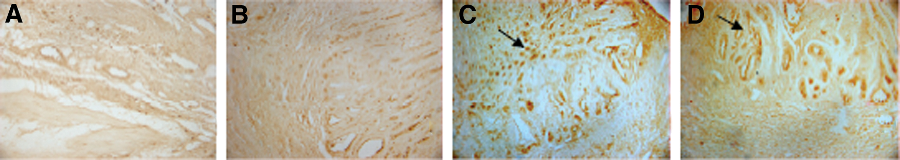
The results presented in Table 1 demonstrate gastric mucosal healing induced by OEC and LIM (44% and 56%, respectively). We observed that treatment with either OEC or LIM significantly reduced the lesion area and increased the epithelial height compared with vehicle-treated rats. The H&E-stained photomicrographs demonstrate that the vehicle-treated group (Fig. 1A) had gastric gland dilation with relative secretion into the lumen. In the cimetidine-treated animals (Fig. 1B), we observed gastric gland dilation and an increase in secretion into the lumen. Fig. 1C (250 mg/kg OEC group) and D (245 mg/kg LIM group) demonstrates increased secretion within the gastric glands and better structural organization. The OEC (Fig. 2C)- and LIM (Fig. 2D)-treated rats had increased PCNA-positive cells compared with the vehicle (Fig. 2A) or cimetidine (Fig. 2B)-treated groups. These results demonstrate that there was a significant increase in cell proliferation in the healing regions of the gastric mucosa in the OEC- and LIM-treated rats. As shown in Fig. 3, there was a significant increase in blood vessel density in the submucosal region of the OEC- and LIM-treated rats compared with those that were treated with vehicle. Interestingly, Fig. 4C and D shows that COX-2 was increased in the mucosa of the OEC- and LIM-treated rats. Electron microscopy suggests that the gastric glands of the OEC (Fig. 5C)- and LIM (Fig. 5D)-treated rats contained a large amount of mucus.
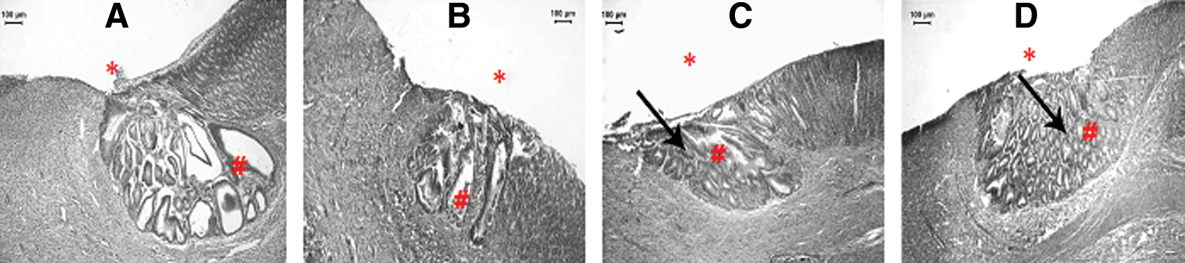
Histological sections of the stomach lesions induced by acetic acid in the rats:

Immunohistochemical analysis of proliferating cell nuclear antigen in the gastric mucosa of rats submitted to experimentally induced gastric ulcer:
Immunohistochemical analysis of vascular endothelial growth factor (VEGF) in the gastric mucosa of rats submitted to experimentally induced gastric ulcer:
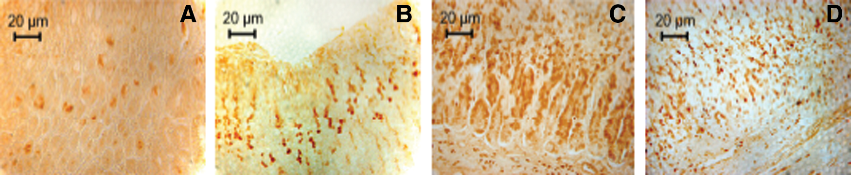
Immunohistochemical analysis of cyclooxygenase 2 (COX-2) in the gastric mucosa of rats submitted to experimentally induced gastric ulcer:
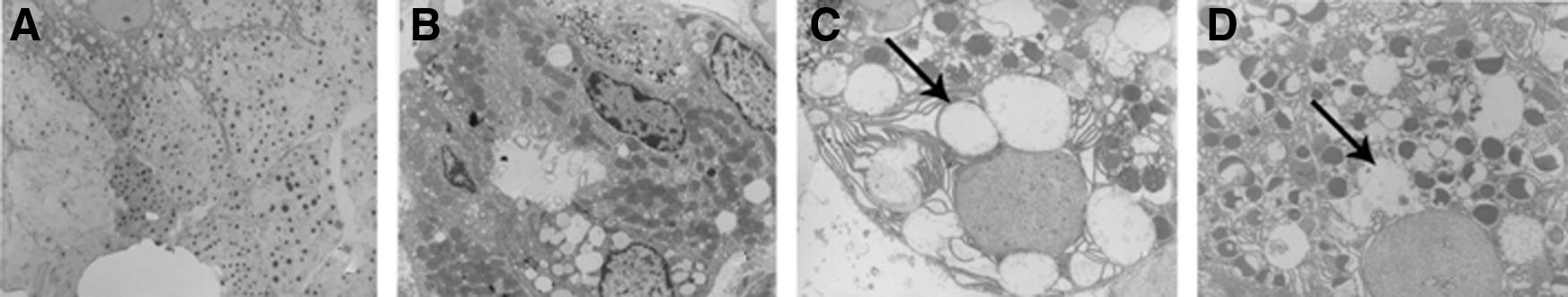
Electron microscopy of the gastric mucosa of rats submitted to experimentally induced gastric ulcer:
The results are expressed as mean±SEM.
Area of the lesion: ANOVA followed by the Dunnett's test, F(4,24)=5.71 (* P<.05, ** P<.01). The height of the epithelium: ANOVA, F(5,31)=28.19 (** P<.01).
OEC, C. aurantium essential oil; LIM, d-limonene; ANOVA, one-way analysis of variance.
At the end of the 14-day treatment period, blood was collected from all of the groups for biochemical assessment to determine whether there was any toxicity associated with OEC or LIM treatment. The results presented in Table 2 show that there were no significant changes in the serum biochemical parameters in the OEC- or LIM-treated rats compared with the vehicle-treated group. In addition, there were no significant differences in the vital organ weights (i.e., heart, lung, liver, kidneys, and pancreas). The rats were weighed daily during the treatment period, and the OEC- and LIM-treated rats displayed similar body weight compared with the vehicle-treated rats (vehicle: 279.2±11.26 g, OEC: 265.0±15.39 g, and LIM: 273.8±13.0 g).
The results are expressed as mean±SEM. Aspartate aminotransferase (AST), alanine aminotransferase (ALT), and gamma glutamyl transferase (γ-GT) are presented in U/L, and creatinine, urea, and glucose are presented in mg/dL. ANOVA, Dunnett's test, P>.05 in relation to the control groups treated with vehicle. n=5.
Discussion
The rat model of acetic acid-induced ulcers is the most similar model to gastric ulcers in humans. 9 Ulcer healing can be divided into three stages: 0–3 days, 3–10 days, and 10–20 days. Days 0–3 consist of ulcer development, including the development of necrotic tissue, ulcer implantation, inflammatory infiltration, and ulcer margin formation. The rapid healing phase occurs during days 3–10 and involves epithelial cell migration and ulcer contraction. Slow healing occurs between days 10–20 and includes angiogenesis, granulation tissue remodeling, and complete ulcer crater re-epithelialization. 13 Histologically, gastric ulcers consist of the ulcer margin, which is formed by the adjacent nonedge necrosis that defines the injury, and the ulcer base, which is composed of necrotic tissue. The ulcer healing process is complex and involves migration, proliferation, re-epithelialization, angiogenesis, and granulation tissue formation. 14 All of these processes are controlled by growth factors, transcription factors, and cytokines. 14 Granulation tissue is generated by the proliferation of connective tissue cells, such as macrophages, fibroblasts, and endothelial cells, and the formation of microveins through angiogenesis. 15 Granulation tissue growth and microvascular system formation are stimulated by FGF, VEGF, PDGF, and various cytokines. 16 The present results demonstrated that the OEC- and LIM-treated rats had a significant reduction in the ulcer lesion area, with healing rates of 44% and 56%, respectively, compared with the vehicle-treated rats (Table 1). We also observed that the OEC- and LIM-treated rats had a significant increase in the epithelial regeneration height compared with the vehicle-treated control group. The difference in the epithelial height demonstrated regenerative cell proliferation at the ulcer site, which contributes to the new epithelial formation and lesion healing. Although the cimetidine-treated rats had similar healing rates as the OEC- and LIM-treated rats, the epithelial regeneration height was not greater in the cimetidine-treated rats. To evaluate the quality of the OEC- and LIM-induced healing, histological sections were H&E stained and immunohistochemistry was performed for PCNA, VEGF, and COX-2, which are proteins and enzymes involved in the healing process. Figure 1 demonstrates that the OEC- and LIM-treated rats showed greater epithelial renewal in the ulcer margin compared with the vehicle- and cimetidine-treated rats, and the formation of the gastric glands was much more ordered. OEC and LIM treatment also promoted mucous gland elongation in the regeneration region, which improved the quality of the regenerated epithelial glandular structures. This result is very important in terms of avoiding relapses because according to Tarnawski, 14 vascular and glandular disruption, dilation of gastric glands, and increased connective tissue are abnormalities that cause gastric ulcer recurrence. PCNA staining demonstrated that both the OEC and LIM treatments promoted cell proliferation in the regenerating region (Fig. 2), which favors rats that received these treatments, exhibited improved epithelial height (Table 1). Cell proliferation is very important for gastric mucosal reconstitution, and studies have suggested that the balance between proliferation and apoptosis is critical for maintaining gastric mucosal integrity. 17 Decreased blood volume is a predisposing factor for ulcer onset 5 because this parameter may cause tissue hypoxia and anoxia. Thus, treatments that confer increased blood vessel density have an important role in gastric mucosal maintenance and regeneration. Immunohistochemistry for VEGF revealed increased angiogenesis in the lesion border in the OEC- and LIM-treated rats (Fig. 3) compared with the vehicle- or cimetidine-treated groups. Angiogenesis is essential for healing because the new vessels supply the new cells with nutrients and promote epidermal growth. 18 In the present study, we found that both OEC and LIM enhanced gastric mucosal restoration and healing. COX-2 expression, as measured by immunohistochemistry, was increased in the OEC- and LIM-treated rats (Fig. 4). According to Konturek et al., 19 COX-2 plays an important role in healing at the ulcer margin. Indeed, the PGE2 produced by COX-2 increases cell proliferation and promotes angiogenesis and mucosal integrity. Importantly, Moraes et al. 8 has shown that both OEC and LIM can modulate gastric mucosal PGE2 production. The increased COX-2 expression that was observed in the present study explains the increase in cell proliferation and angiogenesis in the gastric mucosal region, which increased scarring and improved healing quality in the OEC- and LIM-treated rats. This increase in COX-2 expression can explain the increase in protective mucus in the stomach of the OEC- and LIM-treated rats. Figure 5 demonstrates that a large amount of mucus was secreted from the stomach glands of the OEC- and LIM-treated rats.
The ability of OEC and LIM to increase protective mucus secretion is considered gastroprotective. 9 Increased mucus secretion is important for accelerating ulcer healing because it forms a protective barrier around the newly formed epithelial cells and protects them from harmful factors, such as acidic pH and proteolytic enzymes that are present in gastric juices. 20 Importantly, we did not observe any toxicity from the 14-day OEC and LIM treatments. In addition, there were no significant differences in the rat body weights throughout the treatment period. Moreover, there were no significant differences among the groups in terms of vital organ weight (i.e., heart, lungs, spleen, kidneys, and liver) or biochemical measurements (Table 2). These results are promising because ulcers require chronic treatment, and changes in these parameters could preclude the use of the OEC and LIM for peptic ulcer treatment. However, animal models of chronic exposure must be carried out before a potential toxicity or adverse effects can be ruled out.
The results from the present study suggest that both the OEC and LIM significantly improve the gastric mucosal healing. In addition, neither OEC nor LIM displayed any evidence of toxicity during the 14-day treatment period. Taken together, these results demonstrate that OEC and LIM are effective and safe treatments for experimentally induced gastric ulcers in rats, being promising compounds to be tested in other models, and perhaps, further in humans. Thus, we can hypothesize that the gastric mucosal healing promoted by OEC is caused by LIM.
Footnotes
Acknowledgments
This work was supported by the FAPESP (Fundação de Amparo a Pesquisa do Estado de São Paulo) and CNPq (Conselho Nacional de Desenvolvimento Científico eTecnológico).
Author Disclosure Statement
No competing financial interests exist.