
Abstract
Naringin, a well-known flavanone glycoside of grapefruit and citrus fruits, was found to be as an effective anti-inflammatory compound in our previous lipopolysaccharide-induced acute lung injury mouse model via blockading activity of nuclear factor κB. The current study sought to explore the anti-inflammatory effects of naringin on chronic pulmonary neutrophilic inflammation in cigarette smoke (CS)-induced rats. Seventy Sprague-Dawley rats were randomly divided into seven groups to study the effects of CS with or without various concentrations of naringin or saline for 8 weeks. The results revealed that naringin supplementation at 20, 40, and 80 mg/kg significantly increased body weight of CS-induced rats as compared to that in the CS group. Moreover, naringin of 20, 40, and 80 mg/kg prevented CS-induced infiltration of neutrophils and activation of myeloperoxidase and matrix metalloproteinase-9, in parallel with suppression of the release of cytokines, such as tumor necrosis factor-α and interleukin-8 (IL-8). IL-10 in bronchoalveolar lavage fluid was significantly suppressed after CS exposure, but dose dependently elevated by naringin. The results from hematoxylin and eosin staining revealed that naringin dose dependently reduced CS-induced infiltration of inflammatory cells, thickening of the bronchial wall, and expansion of average alveolar airspace. In conclusion, our data suggest that naringin is an effective anti-inflammatory compound for attenuating chronic pulmonary neutrophilic inflammation in CS-induced rats.
Introduction
C
The effects of CS exposure have been clearly demonstrated in large clinical studies, CS-induced rodent models, and in vitro cell models. 4 –8 It was demonstrated that oxidative stress caused by CS-exposure increases inflammatory cell influxes to the lung, which is followed by lipid peroxidation and increases in proinflammatory cytokine, such as tumor necrosis factor-α (TNF-α). 7 Moreover, interleukin-8 (IL-8) and myeloperoxidase (MPO), predominantly produced by neutrophils, were found to be elevated in human bronchial epithelial cells after CS-exposure, 9 whereas anti-inflammatory cytokine of IL-10 was reduced in patients with bronchial asthma, COPD, and in smokers. 10 Matrix metalloproteinase-9 (MMP-9), which is the most elastolytic of the MMPs and predominantly produced by macrophages, was significantly increased in the lungs of rats induced by CS and dose dependently attenuated by a selective phosphodiesterase 4 (PDE4) inhibitor, Zl-n-91. 11 Thus, the CS-exposure model in rodents might be useful for investigating anti-inflammatory effects of a novel drug that attenuate chronic pulmonary inflammation.
Naringin (4′,5,7-trihydroxy-flavonone-7-rhamnoglucoside), a well-known flavanone glycoside of grapefruit and citrus fruits, has been reported to exhibit antioxidative effects and inhibit lipid peroxidation in biological membranes. 12–13 Moreover, growing evidence has indicated that naringin exerts anti-inflammatory effects both in vitro and in vivo. 12,14 –16 It is well established that naringin significantly prevented lipopolysaccharide (LPS)-induced endotoxic shock in mice and production of NO in RAW 264.7 macrophage cells. 15 Furthermore, our previous study investigated the anti-inflammatory effects of naringin in an acute pulmonary inflammatory mouse model with LPS-induced acute lung injury, and demonstrated that naringin significantly attenuated LPS-induced infiltration of inflammatory cells and the release of proinflammatory cytokines via suppressing activation of the nuclear factor κB (NF-κB). 16 However, there is no information regarding the effects of naringin on chronic pulmonary neutrophilic inflammation induced by CS, which appears to not be affected by treatment with glucocorticoids as reported in previous studies. Naringin is an effective cAMP-specific PDE4 inhibitor, 17 and accumulating preclinical and clinical evidence suggests that selective PDE4 inhibitors are effective on inhibition of pulmonary inflammation and emphysema in COPD animal model and patients, respectively. 11 Thus, the main goal of the current study was to evaluate the anti-inflammatory activity of naringin on chronic pulmonary neutrophilic inflammation in CS-induced rats.
Materials and Methods
Animals
Seventy male and female Sprague-Dawley rats, weighting between 190–230 g, were obtained from the Guangdong Medical Laboratory Animal Center (Guangdong, China). All rats were housed in a temperature (22°C±2°C) and humidity-controlled room with free access to both fresh water and standard laboratory food. All experimental procedures were approved by the Animal Care and Use Committee of the School of Life Sciences, Sun Yat-sen University (No. 2011082102).
Reagents
The commercial nonfiltered cigarette contains 12 mg tar and 1.2 mg nicotine per cigarette (trade name: Ye Shu, purchased from the Guangdong Tobacco Industrial Co. Ltd., Guangdong, China). Naringin was extracted by our laboratory (extracted from Citrus grandis Tomentosa by water per ethanol extraction, and purified by 1–10 recrystallizations to obtain a purity >98.3% as determined by peak area normalization). Rat TNF-α, IL-8, IL-10, and MMP-9 ELISA Kits were purchased from R&D Corporation (R&D Systems, Inc., Minneapolis, MN, USA). MPO and bicinchoninic acid (BCA) assay kits were purchased from Jiancheng (Nanjing, China).
Dosage rationale of naringin
C. grandis Tomentosa is a medicinal and edible plant that contains naringin of 5% to 20% in the dry peel. According to Chinese Pharmacopoeia, the daily usage and dosage of dry peel of C. grandis Tomentosa is 3–6 g, which means the daily intake of naringin for rats is 13.5–108 mg/kg. So naringin dosages from 5 to 80 mg/kg were designed in the current study.
Apparatus
The smoking apparatus consists of three major parts, including a metal chamber with a glass door, a cigarette burning with inhalation apparatus, and a ventilation apparatus on the top of the chamber. A 100 cm (length)×60 cm (width)×60 cm (height) metal chamber separated into two layers with sufficient space for exposing 24 rats at a time. The cigarette burner contained 12 cigarette holders, a plastic wye injection casing, and a 300-mL glass injector, which could burn up 12 cigarettes in 3 min at a time and inject CS into the chamber by manual control of one-way valves. The ventilation apparatus would pump out all of the CS in the chamber within 15 min after exposure. During the experiment, temperature in the chamber was kept between 19°C–23°C with the help of a condensation interlayer, carbon monoxide was controlled between 310–380 ppm, carbon dioxide was controlled between 10415–11406 ppm, oxygen was kept between 19.2%–20.5%, and total suspended particles was kept between 70 and 110 mg/m3.
Animal administration and modeling procedures
Rats were randomly divided into the following seven groups and dealt with the following steps, respectively (n=10).
Group a: Con group. Animals were placed into the same type of apparatus as described in CS-induced group, and exposed to fresh air instead of CS for 8 weeks.
Group b: CS group. Animals were placed into the chamber and exposed to successive periods of CS for 1 h in the morning and 1 h in the afternoon, 7 days a week. The amount of cigarette exposure was increased as follows: 3 cigarettes on the 1st day, 7 cigarettes on the 2nd day, 12 cigarettes on the 3rd to 5th day (30 min at each time), and 12 cigarettes on the 6th day (1 h at each time) to the end of 8 weeks.
Group c: CS+Nar1 group, Group d: CS+Nar2 group, Group e: CS+Nar3 group, Group f: CS+Nar4 group, and Group g: CS+Nar5 group. Rats in these groups were exposed to CS as described in the CS group above. One hour before the first CS exposure on each day, animals were intragastricaly administered with naringin of 5, 10, 20, 40, or 80 mg/kg, respectively.
On each 7th day, bodyweights of rats in each group were weighed by electronic balance after 12 h fasting. Feed intake in each group was weighed by the weight loss method on the morning of every day.
Analysis of immunocytes and cytokines in bronchoalveolar lavage fluid
Fourteen hours after the last exposure to CS, animals were anesthetized with chloral hydrate (3.5%) and sacrificed by heart bloodletting using vacuum tubes from the left side of heart. Trachea and chest of rats were surgically exposed, and then a gavage needle (No. 16) was inserted into the trachea. The left lung lobes were ligated, while the right lung lobes were used for lavage. The bronchoalveolar lavage fluid (BALF) was performed as previously reported. 16 After centrifuging the BALF samples for 10 min (3000 g, 4°C), the sediment cells were resuspended in 0.5 mL PBS and counted by ABX MICROS 60. The supernatants were stored at −20°C until used for analysis of TNF-α, IL-8, and IL-10 according to the manufacturer's instructions.
Analysis of MPO and MMP-9 activities in lung tissue
Lung MPO activity was measured using the colorimetric method with a UV-VIS spectrophotometer (TU-1901, Purkinje General Instrument Co., Ltd., Beijing, China), using a commercial MPO assay kit. Total protein was extracted from the lung tissue by the RIPA lysate (Beyotime, Shanghai, China) and measured with the BCA assay kit according to standard steps. Then, the content of MMP-9 in the tissue lysate was analyzed by ELISA.
Lung histological examination by hematoxylin and eosin staining
The left lung lobes and the remaining trachea of rats were then removed and perfusion fixed via a tracheal cannula with 10% neutral formalin at a hydrostatic pressure of 20 cm H2O. After 10 min, the trachea was ligated and the lung was immersed in 10% neutral formalin for 7 days. After fixation, the left lung lobes were imbedded in paraffin wax and cut into sections of 3–4 μm, and then suitable sections were selected and stained with hematoxylin and eosin (H&E) for histological examination. Slides were scanned with a Mirax Scan (Carl Zeiss MicroImaging GmbH, Göttingen, Germany) at ×100 and ×400 magnifications, At least two nonconsecutive slides per block were analyzed for influx of inflammatory cells, average alveolar airspace, and thickness of bronchial wall by corresponding Mirax Viewer software as previously described. 18
Statistical analysis
Unless otherwise stated, data are presented as mean±S.D. Analysis of variance was used to compare experimental animals to control group, while comparisons between multiple groups were performed using the Tukey's Multiple Comparison test. For all experiments, P<.05 was taken as statistically significant.
Results
Effect of naringin on CS-induced body weight changes of rats
As shown in Figure 1, although the initial body weight was similar between all groups, the rats in the CS group weighed significantly less than those rats in the Con group after the 8-week exposure period. After the 8-week exposure, the body weight of rats in the Con group increased to 351.4±81.7 g, whereas the CS group rats enhanced to 271.7±65.4 g only, which means the loss ratio of body weight was 60.7% as compared with the Con group. The body weight of CS-induced animals treated with 5 and 10 mg/kg naringin showed no significant differences as compared to that in the CS group. However, body weight of CS-induced rats treated with naringin of 20, 40, and 80 mg/kg elicited a pronounced increase as compared to that in the CS group after 8 weeks of CS exposure.
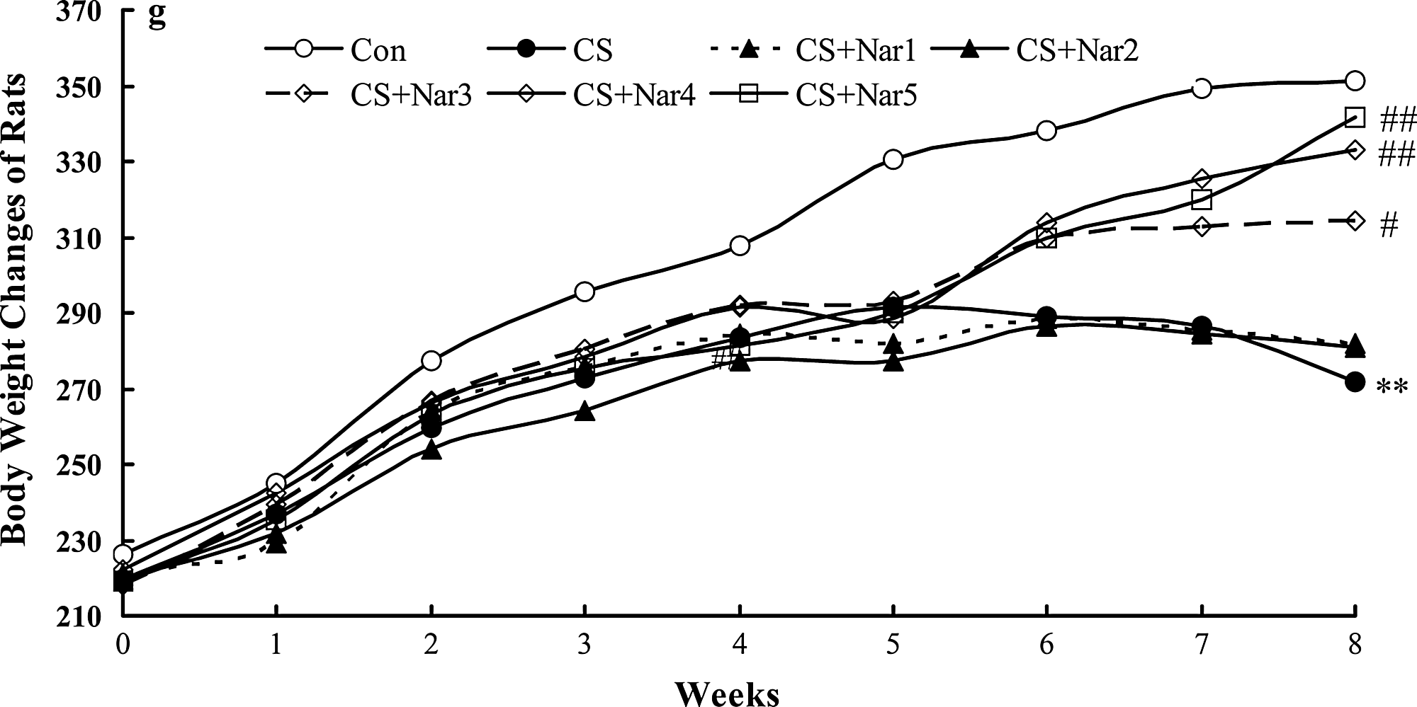
Effect of naringin on cigarette smoke (CS)–induced body weight changes of rats. Rats were repeatedly exposed to CS or fresh air (control [Con] group) for 8 weeks, the body weight of rats was weighed on each 7th day after 12-h fasting. Data are given as mean (n=8–10; value of S.D. is omitted). Significant differences were found: **P<.01 compared with the control group; # P<.05, ## P<.01 compared with the CS group.
Effect of naringin on CS-induced influx of leukocytes in BALF of rats
As shown in Table 1, the number of total leukocytes in BALF of CS group rats was significantly increased to 4.0-fold as compared to that in the Con group. Naringin dose dependently attenuated CS-induced influx of leukocytes in BALF. The total leukocytes in BALF of CS-induced rats treated with naringin of 20, 40, and 80 mg/kg elicited a pronounced decrease as compared to that in the CS group after 8 weeks of CS exposure. Moreover, the percentage of neutrophils in the BALF of CS group rats was significantly increased to 2.9-fold as compared to that in the Con group, while percentages of macrophages and lymphocytes partially, but not significantly, decreased suggesting that there is a remarkable chronic pulmonary neutrophilic inflammation after 8 weeks of CS exposure. Naringin of 20, 40, and 80 mg/kg significantly reduced the percentage of neutrophils in BALF of CS-induced rats. As compared with CS group rats, the number of neutrophils, macrophages, and lymphocytes in BALF of rats treated with naringin of 80 mg/kg was significantly decreased by 84.6%, 49.5%, and 61.8%, respectively.
Data are presented as mean±SD (n=8–10).
Significant differences were found: * P<.05, ** P<.01 compared with normal control group; # P<.05, ## P<.01 compared with cigarette smoke (CS) group.
Nar1–5, Naringin (5, 10, 20, 40, and 80 mg/kg).
Effect of naringin on CS-induced release of TNF-α, IL-8, and IL-10 in BALF of rats
Release of inflammatory mediators was additionally determined in BALF. As shown in Table 2, after 8 weeks of CS exposure, the contents of TNF-α and IL-8 in BALF were significantly elevated to 2.9- and 2.1-fold, respectively. Moreover, Naringin dose dependently reduced CS-induced release of TNF-α and IL-8. At the highest tested dose of naringin (80 mg/kg), maximum inhibition for TNF-α and IL-8 was 73.9% and 101.1%, respectively.
Data are presented as mean±SD (n=8–10).
Significant differences were found: ** P<.01 compared with normal control group; # P<.05, ## P<.01 compared with CS group.
C, contents; IR, inhibition rate; GR, growth rate; IL, interleukin
Otherwise, content of IL-10 in BALF was markedly decreased by 55.5% after 8 weeks of CS exposure, but dose dependently elevated after treated with naringin. In the group of rats treated with naringin of 80 mg/kg, the content of IL-10 was slightly higher than that in the Con group.
Effect of naringin on CS-induced release of MPO and MMP-9 in lung tissue of rats
MPO, as an important proinflammatory and pro-oxidant mediator, is mainly released from activated neutrophils. 9 As shown in Table 3, activity of MPO in the lungs of CS group rats was significantly increased to 3.4-fold as compared to that in the Con group. Naringin of 10, 20, 40, and 80 mg/kg significantly inhibited the activity of MPO in the lungs of CS-induced rats, with inhibition rates of 51.0% to 86.5%.
Data are presented as mean±SD (n=8–10).
Significant differences were found: ** P<.01 compared with normal control group; # P<.05, ## P<.01 compared with CS group.
MMP-9, matrix metalloproteinase-9; MPO, myeloperoxidase.
MMP-9, mainly secreted by lung macrophages, is considered an osteolytic enzyme and has been associated with loss of pulmonary elastin levels and adult onset emphysema in COPD. 11 As shown in Table 3, content of MMP-9 in the lung tissue of the CS group was significantly increased to 2.3-fold as compared to that in the Con group. Naringin of 10, 20, 40, and 80 mg/kg significantly inhibited the content of MMP-9 in lung tissue lysate of CS-induced rats, with inhibition rates of 26.9% to 64.6%.
Effect of naringin on CS-induced histological changes
To investigate the effects of naringin treatment on the infiltration of inflammatory cells, average alveolar airspace, and thickness of bronchial wall, lung sections were prepared and observed by H&E staining. As shown in Figure 2, the lung section of the Con group showed a normal bronchoalveolar structure with few infiltration of inflammatory cells, average alveolar airspace of appropriate 90 μm2, as well as bronchial wall thickness of nearly 29 μm. CS exposure markedly induced influx of inflammatory cells, thickening of bronchial wall, and expansion of average alveolar airspace. Moreover, naringin dose dependently reduced CS-induced infiltration of inflammatory cells, thickening of bronchial wall, and expansion of alveolar airspace, especially at doses of 20, 40, and 80 mg/kg, which exhibited significant differences as compared to that in the CS group. These results suggested that naringin could attenuate CS-induced damage of lung tissue by reducing the infiltration of inflammatory cells, expansion of alveolar airspace, as well as weakening the secretion and proliferation of airway epithelium.
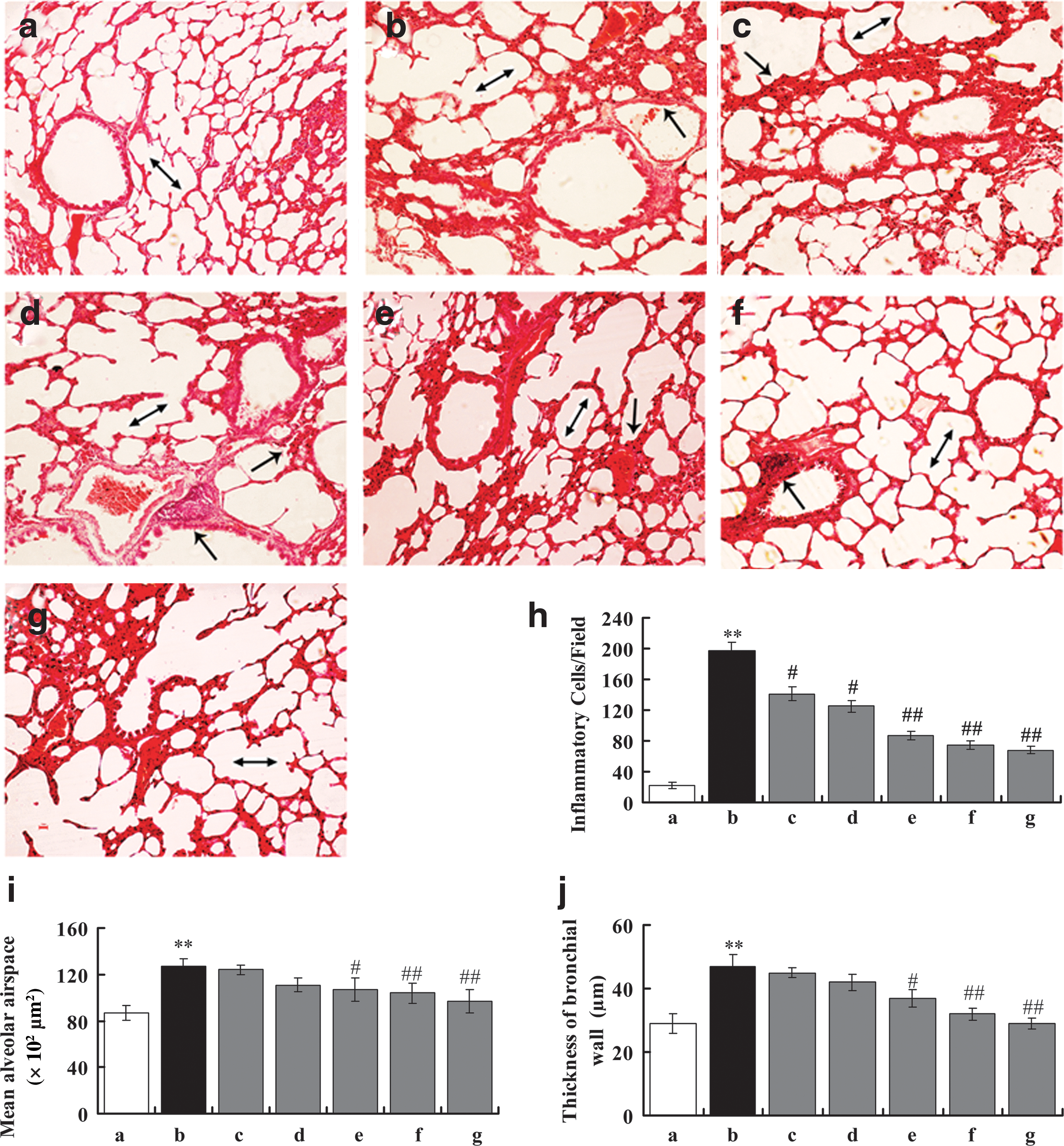
Effect of naringin on CS-induced histological changes. The left lung lobes of rats were stained by hematoxylin and eosin. Slides were scanned at×100 magnification.
Discussion
Various models of CS exposure in laboratory rats have been described over the years. 2 –6 It is demonstrated that CS exposure directly damages the airway epithelium and activates macrophages and lymphocytes, which then generate preinflammatory cytokines TNF-α and IL-8, and activate neutrophils, which leads to chronic pulmonary neutrophilic inflammation. 7 Moreover, the neutrophils help recruit macrophages, which secretes MMP-9 and a number of inflammatory cytokines contributing to the pathogenic nature of macrophages. 19 Our CS-induced rat model showed characteristic pathological changes consistent with chronic pulmonary neutrophilic inflammation. Moreover, our study demonstrated that naringin significantly reduced the infiltration of neutrophils and both activities of MPO and MMP-9 in the lung of CS-induced rats, which in parallel with the suppression in the release of cytokines, such as TNF-α and IL-8. These results suggest that naringin prevents the inflammation induced by CS, probably through regulation of these signal molecules.
The body weight of naringin-treated rats was greater than CS-induced rats after 8 weeks of CS exposure. This was probably because naringin prevented lung inflammation, as previous studies demonstrate that severe lung inflammation in mice caused by CS exposure is associated with weight loss. 20 Moreover, the feed intake in each group was weighed on the morning of each day. Our results showed that at the highest tested dose of naringin (80 mg/kg), the feed intake of rats was increased by 21.4% as compared to that in the CS group (data not shown).
As previous studies describe, oxidants and MMPs complement each other in the potential to destroy lung tissue. 21 –23 Moreover, it is reported that MMPs also play a role in regulating inflammation through the generation of cytokines, such as TNF-α. 24 We observed that the increase of MMP-9 activity in the CS group was associated with expansion of average alveolar airspace after exposure to CS for 8 weeks. Moreover, CS-induced rats treated with naringin of 20, 40, and 80 mg/kg displayed lower MMP-9 activity and smaller alveolar airspace, probably due to less cell inflammatory influx into the airspace, and the suppression of oxidative stress provoked by CS.
Clinical studies have shown that IL-10 was reduced in patients of bronchial asthma and COPD. 25 Moreover, IL-10 was reported to inhibit the secretion of TNF-α and IL-8 from macrophages and tip the balance in favor of antiproteases by increasing the expression of endogenous tissue inhibitors of MMPs. 26 In the present study, we demonstrated that CS exposure significantly inhibited the release of IL-10, which was associated with previous clinical studies. Moreover, naringin of 80 mg/kg markedly elevated the release of IL-10, as well as strongly suppressed the activity of NF-κB. 16 Previous reports have revealed that IL-10 was very effective at inhibiting the activity of NF-κB. 27 This means that IL-10 should suppress the CS-induced activity of NF-κB, which reduces release of proinflammatory cytokines and infiltration of inflammatory cells.
In conclusion, our study demonstrated that the infiltration of inflammatory cells, activation of MPO and MMP-9, and increases in TNF-α and IL-8 in the lungs of CS-induced rats were associated with decreased IL-10. In this study, naringin at doses 20, 40, and 80 mg/kg elevated the release of IL-10, prevented the infiltration of neutrophils, and decreased activities of MPO and MMP-9, which resulted in the suppression of TNF-α and IL-8 release. Our data suggest that naringin is an effective anti-inflammatory compound to attenuate chronic pulmonary neutrophilic inflammation in CS-induced rats.
Footnotes
Acknowledgment
This work received funding from the National Natural Science Foundation of China (No. 81173475).
Author Disclosure Statement
The authors declare no conflict of interests.