
Abstract
Ethnobotanical surveys indicated that in the traditional medicines worldwide, several Juniperus species are utilized as antihelmintic, diuretic, stimulant, antiseptic, carminative, stomachic, antirheumatic, antifungal, and for wound healing. In the present study, essential oils obtained from heartwood samples of Juniperus virginiana L., Juniperus occidentalis Hook. and Juniperus ashei J. Buchholz were evaluated for wound healing and anti-inflammatory activities by using in vivo experimental methods. The essential oils were obtained by the supercritical carbon dioxide extraction method. Linear incision and circular excision wound models were performed for the wound-healing activity assessment. The tissues were also evaluated for the hydroxyproline content as well as histopathologically. To evaluate the anti-inflammatory activity of the essential oils, the test used was an acetic acid-induced increase in capillary permeability. The essential oil of J. occidentalis showed the highest activity on the in vivo biological activity models. Additionaly, the oil of J. virginiana was found highly effective in the anti-inflammatory activity method. The experimental data demonstrated that essential oil of J. occidentalis displayed significant wound-healing and anti-inflammatory activities.
Introduction
T
Several species of junipers, including Eastern Red Cedar (Juniperus virginiana L.), Western Juniper (Juniperus occidentalis Hook.), and Ashe Juniper (Juniperus ashei J. Buchholz; all members of the Cupressaceae family) are very abundant conifers in the United States. In fact, all three are often considered pest species because of their encroachment onto rangeland and pastures and the subsequent loss of forage production. 10 –12 Eastern Red Cedar wood is known for its aromatic smell, toxicity, and repellency to several species of insects, including clothing moths, 13 flour beetles, 14 cockroaches, 15 and ants. 16,17
Cedarwood oil (CWO) has great potential as a safe natural bioactive agent, including wood preservation against both insects 18 –20 and microbes, 21 as an insecticide, 22 acaricide, 23 and mosquito 24 and cockroach repellent. 15 These species of junipers represent a vast underutilized resource with great potential and economic opportunities.
The present study was conducted to assess the wound-healing and anti-inflammatory potential of subextracts of essential oils obtained from Eastern Red Cedar, Western Juniper, and Ashe Juniper in animal models.
Materials and Methods
Juniper heartwood samples
Heartwood samples from trunk samples of Eastern Red Cedar (Woodford County, Illinois), Western Juniper (Harney County, Oregon), and Ashe Juniper (Edwards County, Texas) were from freshly cut trees (three trees per species). Sapwood was removed using a band saw, and then the trimmed heartwood was turned into sawdust using a compound miter saw. Sawdust samples were held in a sealed glass jar to prevent loss of volatile components until being extracted.
Supercritical carbon dioxide extractions
The supercritical carbon dioxide (SC-CO2) extractions were performed in an extractor previously described by King et al. 25 Approximately 100–150 g of sawdust were placed in a 500-mL stainless steel vessel (Thar Process, Inc.). The extraction conditions were 80°C and 55.5 MPa with a flow rate of ∼3 L/min (expanded CO2). The CWO was collected in a 20-mL glass vial. Water was removed from the collected CWO by adding ∼0.5 g anhydrous sodium sulfate to the CWO, mixing thoroughly, and centrifuging.
Chemical analyses
Solutions of CWO in hexane (∼200 ng/μL) were analyzed by gas chromatography to determine the composition of the oil. CWO extracts were analyzed by 0.5-min split-delay splitless injection onto a Hewlett-Packard (HP) 5890 Series II GC equipped with a flame ionization detector and an SP-2380 column (60 m×0.25 mm inner diameter; 0.20-μm film thickness; Supelco) using helium as the carrier gas at a linear flow velocity of 18 cm/sec. The temperature program was 60°C for 1 min, rising by 5°C/min to 250°C. The injector and detector temperatures were 235°C and 250°C, respectively. Injections were made using an HP model 7683 auto-injector and sample volumes were 1 μL. Chromatographic data were acquired using an HP Vectra VL2 computer and ChemStation software.
Biological activity tests
Animals
Male Sprague-Dawley rats (160–180 g) and Swiss albino mice (20–25 g) were purchased from the animal breeding laboratory of Saki Yenilli (Ankara, Turkey).
The animals were left for 3 days at room conditions for acclimatization. They were maintained on a standard pellet diet and water ad libitum throughout the experiment. A minimum of six animals were used in each group. The study was permitted by the Institutional Animal Ethics Committee and was performed according to the international rules considering the animal experiments and biodiversity rights.
Preparation of test samples for bioassay
Incision and excision wound models were used to evaluate the wound-healing activity. For the in vivo wound models, test samples were prepared in an ointment base (vehicle) consisting of glycol stearate, 1,2-propylene glycol, and liquid paraffin (3:6:1) in 1% (w/w) concentration. 0.5 g of each test ointment was applied topically on the wounded site immediately after wound was created by a surgical blade.
The animals of the vehicle group were treated with the ointment base only, whereas the animals of the reference drug group were treated with 0.5 g of Madecassol® (Bayer; 00001199). Madecassol contains 1% (w/w) extract of Centella asiatica.
For the assessment of anti-inflammatory activity, test samples were given orally to test animals after suspending in a mixture of distilled H2O and 0.5% (w/v) sodium carboxymethyl cellulose (CMC). The control group animals received the same experimental handling as those of the test groups except that the drug treatment was replaced with appropriate volumes of dosing vehicle. Indomethacin (10 mg/kg) in 0.5% (w/v) CMC was used as the reference drug.
Wound-healing activity
Linear incision wound model
Animals, seven rats in each group, were anesthetized with 0.15 mL Ketalar®. The hairs on the dorsal part of the rats were shaved and cleaned with 70% (v/v) alcohol. Two 5-cm-length linear-paravertebral incisions were made with a sterile blade through the shaved skin at the distance of 1.5 cm from the dorsal midline on each side. Three surgical sutures were placed each 1 cm apart.
The ointments prepared with test samples, the reference drug (Madecassol®), or the ointment base (glycol stearate, propylene glycol, liquid parafin [3:6:1]) were topically applied on the dorsal wounds in each group of animals once daily throughout 9 days. All the sutures were removed on the last day and the tensile strength of previously wounded and treated skin was measured by using a tensiometer (Zwick/Roell Z0.5). 26,27
Circular excision wound model
This model was used to monitor wound contraction and wound closure time. Each group of animals (seven animals in each) was anesthetized by 0.01 mL Ketalar®. The back hairs of the mice were depilated by shaving. The circular wound was created on the dorsal interscapular region of each animal by excising the skin with a 5-mm biopsy punch; wounds were left open. 28 Test samples, the reference drug (Madecassol®; Bayer), and the vehicle ointments were applied topically, once a day, till the wound was completely healed. The progressive changes in the wound area were monitored by a camera (Fuji, S20 Pro) every other day. Later on, the wound area was evaluated by using AutoCAD program. Wound contraction was calculated as percentage of the reduction in the wounded area. A specimen sample of tissue was isolated from the healed skin of each group of mice for the histopathological examination. 29
Histopathology
The skin samples were collected for the histopathological alterations. Specimens were fixed in 10% buffered formalin, processed and blocked with paraffin, sectioned into 5 μm, and stained with hematoxylin & eosin and Van Gieson stains. The slides were examined with a light microscope (Olympus CX41 attached Kameram® Digital Image Analyze System) and graded as mild (+), moderate (++), and severe (+++) for epidermal or dermal remodeling. Re-epithelization or ulcus in epidermis; fibroblast proliferation, mononuclear and/or polymorphonuclear cells, neovascularization and collagen depositions in dermis were analyzed to score the epidermal or dermal remodeling. Obtained results were combined and staged for wound-healing phases as inflammation, proliferation, and remodeling in all groups.
Hydroxyproline estimation
Tissues were dried in a hot air oven at 60°C–70°C till consistant weight was achieved. Afterward, samples were hydrolyzed with 6 N HCl for 4 h at 130°C. The hydrolyzed samples were adjusted to pH 7 and subjected to chloramin T oxidation. The colored adduct formed with Ehrlich reagent at 60°C was read at 557 nm. 30 Standard hydroxyproline was also run and values reported as μg/Mg dry weight of tissue. 31
Anti-inflammatory activity
Acetic acid-induced increase in capillary permeability
Effect of the test samples on the increased vascular permeability induced by acetic acid in mice was determined according to the Whittle method 32 with some modifications. 33 Each test sample was administered orally to a group of 10 mice in 0.2 mL/20 g body weight. Thirty minutes after the administration, the tail of each animal was injected with 0.1 mL of 4% (w/v) Evans blue in a saline solution (intravenously) and waited for 10 min. Then, 0.4 mL of 0.5% (v/v) AcOH was injected intraperitoneally. After 20-min incubation, the mice were killed by dislocation of the neck, and the viscera were exposed and irrigated with distilled water, which was then poured into 10-mL volumetric flasks through glass wool. Each flask was made up to 10 mL with distilled water, 0.1 mL of 0.1 N NaOH solution was added to the flask, and the absorption of the final solution was measured at 590 nm (Beckmann Dual Spectrometer; Beckman). A mixture of distilled water and 0.5% (w/v) CMC was given orally to control animals, and they were treated in the same manner as described above.
Statistical analysis of the data
The data on percentage anti-inflammatory and wound healing was statistically analyzed using one-way analysis of variance. The values of P≤.05 were considered statistically significant. Histopathologic data were considered to be nonparametric; therefore, no statistical tests were performed.
Results
In the present study, we aimed to evaluate the wound-healing and anti-inflammatory activities of the essential oils obtained by SC-CO2 extraction.
The results of biological activity tests are presented in Tables 1 –4. As presented in Table 1, topical application of the ointment of J. occidentalis essential oil showed the best effect in the linear incision wound model. The tensile strength value of this experimental group was determined as 41.3%, which was found to be significant (P<.01) when compared to the vehicle control group with the value of 12.3% on day 10.
Percentage of tensile strength values: Vehicle group was compared to Negative control group; the extracts and the reference material were compared to vehicle group. Statistical significance is indicated: ** P<.01; *** P<.001.
Percentage of contraction values: Vehicle group was compared to Negative control group; the extracts and the reference material were compared to vehicle group. Statistical significance is indicated: * P<.05; ** P<.01; *** P<.001.
P<.01; *** P<.001 significant from the control.
SEM, standard error of the mean.
P<.05; ** P<.01; *** P<.001 significant from the control.
In Table 2, the contraction values of vehicle, negative control, oils, and reference drug-treated groups are shown. The essential oil obtained from J. occidentalis was found to have wound-healing potential, with the contraction value of 39.75% and 52.44% for J. occidentalis on day 10 and 12. On the other hand, the reference ointment-treated group showed 71.31% and 100.00% contraction on day 10 and 12. The other experimental groups did not exert any significant effect on this model.
Hydroxyproline levels of treated tissues were evaluated for the assessment of collagen synthesis. As shown in Table 3, a significant increase in the hydroxyproline content was observed for the tissues treated with the essential oil of J. occidentalis. However, the experimental group animals treated with the essential oils of J. virginiana and J. ashei did not show any significant enhancement on the hydroxyproline content.
Phases in wound-healing processes (inflammation, proliferation, and remodeling) were observed within the experimental groups with different degrees (Figs. 1 and 2). The reference drug and ointment prepared from the essential oil of J. occidentalis treated groups demonstrated faster re-epithelization, respectively, compared to the other groups that showed delayed wound-healing processes. Remodeling was seen in all groups except the individual animals in some groups. The best remodeling was recorded in the reference group than in the J. occidentalis, J. virginiana, and J. ashei groups, respectively.

Histopathological view of Juniperus virginiana, Juniperus occidentalis, and Juniperus ashei on linear and circular excision wound models.
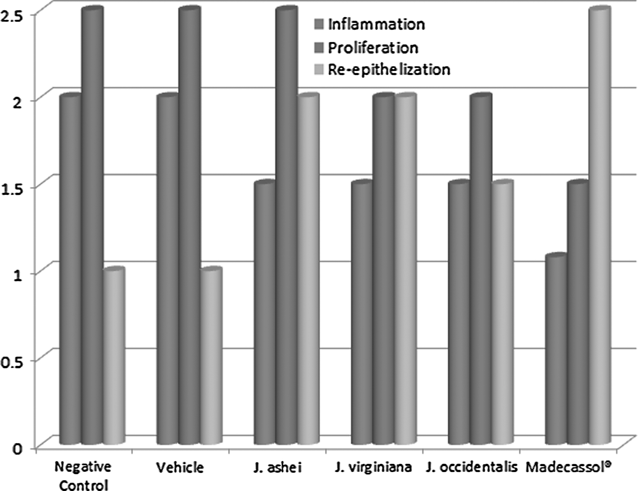
Wound-healing phases of the experimental group animals.
The Whittle method was performed for the evaluation of the anti-inflammatory activity. As presented in Table 4, at the dose of 200 mg/kg, the anti-inflammatory effect was observed for the essential oils of J. occidentalis and J. virginiana with the inhibitory values of 25.94% and 30.05%, respectively, while the essential oil of J. ashei showed no significant effect.
Gas chromatographic analyses are presented in Table 5 and Figures 3 –5. The results indicated that Western Juniper contained the highest levels of cedrol, while Ashe Juniper contained the highest levels of thujopsene.

Gas chromatogram of supercritical carbon dioxide (SC-CO2) extract of J. virginiana.

Gas chromatogram of SC-CO2 extract of J. occidentalis.
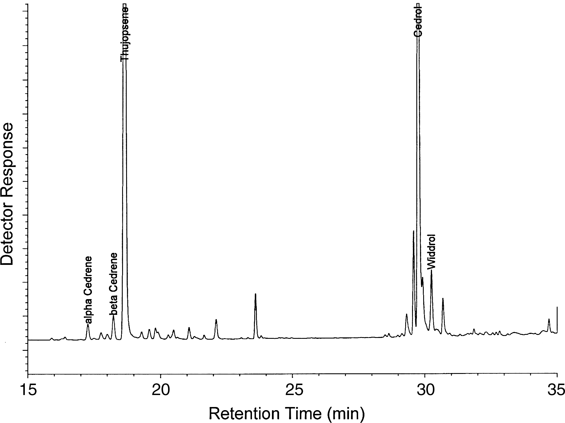
Gas chromatogram of SC-CO2 extract of J. ashei.
Discussion
CWO is generally obtained by steam distillation, however, because components of CWO can be degraded at the high temperature employed during steam distillation; solvent extraction is preferable to avoid such undesirable changes. 34 The degradation (i.e., dehydration) of the sesquiterpene alcohol cedrol to the sesquiterpene hydrocarbon α-cedrene is promoted by the acidic conditions present during extractions of wood at elevated temperatures with water (e.g., steam distillation). 35 Steam-derived CWO contains a very low ratio of cedrol to α-cedrene (i.e., 0.1, 0.6, and 1.1). 36 –39 When a solvent, such as hexane, SC-CO2, or liquid carbon dioxide (L-CO2) is used instead, the ratio of cedrol to α-cedrene is much higher (i.e., 10.1, 2.6, and 13.3). 35,40,41
Solvents, such as acetone, pentane, hexane, and methanol have all been used to obtain biologically active extracts from J. virginiana; however, these solvents are hazardous due to their toxicity and flammability. Carbon dioxide, on the other hand, is nontoxic, nonflammable, and leaves no solvent residue in the extract. Carbon dioxide has been demonstrated to effectively extract CWO and Eller and King found that SC-CO2 gave higher yields of CWO than previously reported for steam distillation. 41 In addition, the CWO obtained by SC-CO2 extraction more closely resembled the odor of the original wood than did CWO obtained by steam distillation.
Juniper essential oils are composed monoterpenes, sesquiterpenes, and other volatile compounds. 42 Both berries and oils are used as phytopharmaceuticals in Europe, mostly as a diuretic. 43 The oil of berries is also used as fragrances for soaps, perfumes, and cosmetics. 44 In the U.S., commercial products are produced from J. virginiana, J.occidentalis, and J. ashei. 45 These oils contain the sesquiterpenes α-cedrene, β-cedrene, cedrol, thujopsene, and widdrol as the major components in different ratios. 38 The bark of Juniperus species was found to have dimeric procyanidins and flavanoid monomers, which have important antioxidant properties. 46 Previous biological activity studies on junipers have revealed that some antibacterial activity was shown by J. occidentalis heartwood extracts against Staphylococcus aureus, Bacillus subtilis, and Mycobacterium smegmatis. 21
Juniper wood is also known for its resistance to both insect and wood rot decay. Zhu et al. found that CWO repelled termites. 47 Juniper has been shown to be resistant to both Formosan 48 and eastern subterranean 49,50 termite attack and has long been used for fence posts. 51,52 Particleboard-chip panels made from J. virginiana are moderately resistant to termite damage. 53 Eller et al. reported that both CO2-derived CWO as well as ethanol-derived CWO exhibited significant resistance to termite damage compared to untreated controls and the CO2-derived CWO conferred significant resistance toward the wood-rot fungus, Gloeophyllum trabeum. 2
Antitermitic compounds can be removed from J. virginiana heartwood by organic solvent (i.e., acetone, pentan, hexane, or methanol) extraction and these extracts are toxic to eastern subterranean termites. 18,19,40,49 An extract (acetone/hexane/water mix) of J. virginiana significantly reduced the termite attack when applied to southern pine by vacuum impregnation. 20
Previous studies revealed that the agents which anti-inflammmatory, antioxidant, and antimicrobial activities can contribute the wound-healing process. 54 –57 In the present study, it was demonstrated that J. occidentalis possesses both wound-healing and anti-inflammatory activities. Reported antiomicrobial and anitoxidant activities 21,58 could also contribute to the wound healing.
The gas chromatographic analyses were found to be the similar with the data reported by Adams 37 for steam-derived CWOs. Western Juniper was also found to have high levels of oxygenated sesquiterpenes cedrol and widdrol. Therefore, the wound-healing activity of the essential oil of J. occidentalis was attributed to the presence of oxygenated sesquiterpenes.
Results of the present study demonstrated that essential oil of J. occidentalis possesses wound-healing activity, which supports traditional utilization of Juniperus species in various folk medicines. In the literature survey, no reports relating to the wound-healing activity of the essential oil of J. occidentalis have been found. To the best of our knowledge, we herein report for the first time about the wound-healing effect of the essential oil of J. occidentalis.
Footnotes
Acknowledgments
The authors wish to thank Dr. Charles “Butch” Taylor (Texas A&M AgriLife Research, Sonora, TX, USA) and Dr. Jonathan Bates (USDA-ARS, Burns, OR, USA) for providing the Ashe Juniper and Western Juniper samples, respectively, used in these studies, as well as Jeffrey Teel (Functional Foods Research Unit, National Center for Agricultural Utilization Research, USDA-ARS, Peoria, IL, USA) for his technical assistance. Mention of trade names or commercial products in this article is solely for the purpose of providing scientific information and does not imply recommendation or endorsement by the U.S. Department of Agriculture. USDA is an equal opportunity provider and employer.
Author Disclosure Statement
The authors declare that there are no conflicts of interest.