




Abstract
Oxidative stress in diabetic tissues is a consequence of free radical accumulation with concurrently impaired natural antioxidants status and results in oxidative tissue damage. The present study investigated the protective effects of mangiferin against pancreatic β-cell damage and on the antioxidant defense systems in streptozotocin (STZ)-induced diabetic rats. Diabetes was experimentally induced by a single intraperitoneal injection of STZ. Oxidative stress biomarkers such as tissue malondialdehyde, hydroperoxides, reduced glutathione (GSH) content, and nonenzymatic antioxidants were measured. Biochemical observations were further substantiated with histological examination and ultrastructural studies in the pancreas of diabetic, glibenclamide and mangiferin-treated diabetic rats (dosage of 40 mg/kg body weight daily for 30 days). Oral administration of mangiferin and glibenclamide to diabetic rats significantly decreased the level of blood glucose and increased levels of insulin. Additionally, mangiferin treatment significantly modulated the pancreatic nonenzymatic antioxidants status (vitamin C, vitamin E, ceruloplasmin, and reduced GSH content) and other oxidative stress biomarkers. The histoarchitecture of diabetic rats showed degenerated pancreas with lower β-cell counts, but mangiferin treatment effectively regenerated insulin secreting islet cells. The electron microscopic study revealed damaged nuclear envelope and mitochondria and fewer secretory granules in pancreas of diabetic rats; however, mangiferin treatment nearly normalized pancreatic architecture. The present findings suggest that mangiferin treatment exerts a therapeutic protective nature in diabetes by decreasing oxidative stress and protecting against pancreatic β-cell damage, which may be attributable to its antioxidative properties.
Introduction
D
Major consequences of high blood glucose are often due to oxidative stress, which is one of the mechanisms causing chronic diabetic complications. 3 Free radicals are highly reactive oxygen species and causes oxidative injury to the organisms by damaging macromolecules. In normal physiological conditions, there is an acute equilibrium in the generation of oxygen free radicals and antioxidant defense systems, which leads to deactivation of the free radicals and protects the organisms against free radical toxicity. 4 Molecular and cellular tissue damages are mainly due to oxidative stress in a wide range of human diseases/disorders. 5,6
The treatment/management of diabetes is generally via diet control, exercise, and use of insulin and/or oral hypoglycemic agents. Yet, they usually have reduced efficacy over time, are ineffective against long-term diabetic complications, and also very costly. 7 Because of perceived effectiveness, fewer side effects in clinical trial experience, and relatively low cost, herbal drugs are a valuable source of natural medicines. 8
Although there are numerous known antidiabetic drugs available in the market, remedies from natural sources, especially from the traditional medicinal plants have demonstrated efficacy for managing diabetes. 9,10 There are various kinds of modern drugs available to treat several ailments including diabetes; however, nearly 80% of the people all over the world still rely on medicinal plants for their primary health care. 11 The World Health Organization (WHO) has emphasized the rational usage of traditional and natural indigenous medicines for treatment of diabetes mellitus. 12 Focusing on these we have chosen one of the well-known traditional medicinal plants (Salacia chinensis) and its primary bioactive compound (mangiferin) to investigate its efficacy against chemically induced diabetes in rats.
S. chinensis Linn is an important medicinal plant belonging to the family Hippocrateaceae and contains the active phytochemical mangiferin in the root. 13 Mangiferin is a C-glycosyl xanthone and traditionally used to treat many diseases in India, and it is consequently reported to have various pharmacological effects. 14 The effect of mangiferin may be mediated by the stimulation of antioxidant pathways, which decrease cellular oxidative stress in the immune system. 15 Mangiferin acts as an effective antioxidant molecule, and it has the potency to treat immunopathological disorders such as inflammatory diseases atherosclerosis, or septic shock. 16 In addition, mangiferin has numerous effects on macrophage function, comprising the inhibition of phagocytic activity and free radical activity. 16 Mangiferin effectively decreased blood glucose and increased insulin levels through increased utilization of glucose in the liver. 17
Currently, no systematic investigations exist in the scientific literature on the beneficial effect of mangiferin on pancreatic tissue oxidative stress in streptozotocin (STZ)-induced diabetes. The present study aimed to characterize the antioxidant and protective nature of mangiferin on hyperglycemia-mediated lipid peroxidation (LPO), in STZ-induced diabetic rats. These beneficial effects were compared with the well-known antidiabetic agent, glibenclamide.
Materials and Methods
Chemicals
STZ was obtained from Sigma Chemicals (St. Louis, MO, USA), stored at −4°C, and protected from light. All other chemicals were analytical grade.
Plant material and isolation of mangiferin
The S. chinensis roots were collected from Veenangaputtu, Karumpakkam, Thangal, and Kurumpuram, Puducherry, India. The plant material was identified by a plant taxonomist and has been deposited in the Center for Advanced Studies in Botany (voucher specimen no: 778; University of Madras) for future reference. Isolation of mangiferin was done by column chromatography as previously described. 13 The purity of mangiferin (99.4%) was confirmed through high performance liquid chromatography by authentic standard (Sigma Aldrich, St. Louis, MO, USA). The structure of isolated mangiferin was characterized by NMR studies.
Animals
Wistar albino male rats weighing around 160–180 g were procured from the Tamil Nadu Veterinary and Animals Sciences University, Chennai, India. Animals were acclimatized to standard animal house conditions for about 1 week and given free access to standard rat chow (Hindustan Lever Ltd., Bangalore, India) and water. The animal experiments were conducted in accordance with the ethical norms approved by the Ministry of Social Justices and Empowerment, Government of India and Institutional Animal Ethics Committee Guidelines (IAEC No. 02/004/06).
Experimental protocol
Diabetes was induced in rats with a single intraperitoneal injection of freshly prepared solution of STZ (55 mg/kg body weight) in 0.1 M cold citrate buffer, pH 4.5.
17
The rats were allowed to drink a 5% glucose solution overnight to overcome the drug-induced hypoglycemia. After allowing 1 week for the development and aggravation of diabetes, the experimental rats with moderate diabetes having persistent glycosuria and hyperglycemia (blood glucose range of above 250 mg/dL) were considered as diabetic rats and used for the experiment. The phytocompound and standard drug treatment were started on the 8th day after STZ injection and this was considered as the 1st day of treatment. A total of 24 animals (18 diabetic rats and 6 control rats) were separated into four groups of six animals in each group as follows: Group 1: Control rats Group 2: Diabetic control rats Group 3: Diabetic rats treated with mangiferin (40 mg/kg body weight daily) Group 4: Diabetic rats treated with glibenclamide (600 μg/kg body weight daily)
The animals were fasted overnight and then sacrificed by cervical decapitation after the end of the experimental period (30 days). Blood sample was collected in tubes containing EDTA for the estimation of glucose by O-toluidine method. 18 The assay of plasma insulin was carried out by RIA assay kit according to the standard protocol (for rats) supplied by Linco Research, Inc., (St. Charles, MO, USA) and estimated vitamin C, 19 vitamin E 20 , and ceruloplasmin. 21 The pancreatic tissue from control and experimental groups was excised, rinsed in ice-cold saline, and homogenized in Tris-HCl buffer, pH 7.4 with a Teflon Homogenizer at 4°C. Protein content present in the tissue homogenate was estimated by the method of Lowry et al. 22
Concentrations of TBARS (thiobarbituric acid reactive substances) and hydroperoxides were estimated by the method of Ohkawa et al. 23 and reduced glutathione (GSH) was estimated by the method of Ellman 24 in plasma and tissue homogenate.
Histological observation of pancreas
A slice of pancreatic tissue was fixed in 10% formalin solution for histopathological analysis at room temperature. After fixation, tissues were dehydrated in a graded series of ethanol, cleared in xylene, and embedded in paraffin wax; solid sections were cut at 4 μm thickness using a rotary microtome for further staining process. Sections were stained for pancreatic β-cells by standard staining protocol. The stained sections were examined under light microscope and photomicrographs were taken.
Transmission electron microscopic studies
A portion of pancreas (about 1 mm3) from each rat was fixed in 3% glutaraldehyde and then postfixed in osmium tetroxide and embedded in araldite (epoxy resin). One micron sections were cut and then stained with toluidine blue. Suitable areas for ultrastructural study were chosen after examining one micron sections under a light microscope. The sections (60–90 nm) were cut on an LKBUM4 ultra microtome using a diamond knife and sections were mounted on a copper grid and stained with uranyl acetate and Reynolds lead citrate. 25 The grids were examined under a Phillips EM201C transmission electron microscope.
Statistical analysis
Results were expressed as mean±SD for six rats in each group. All the grouped data were statistically evaluated with SPSS/10.0 software. Hypothesis testing methods included one-way analysis of variance followed by least significant difference test. Values were considered statistically significant at P<.05.
Results
Identification of mangiferin
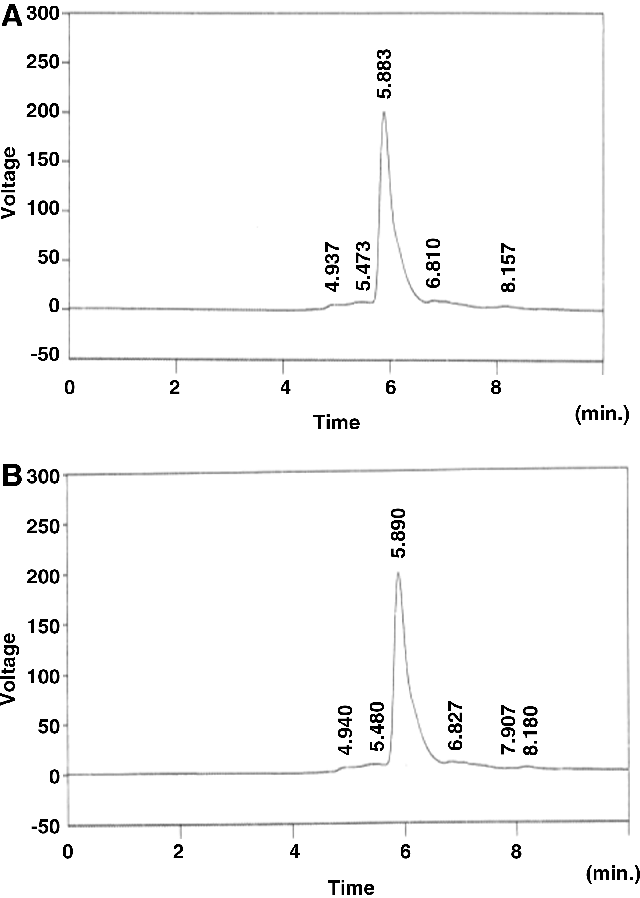
The high performance liquid chromatography analysis revealed that authentic mangiferin had a retention value of 5.883 (79.9%; Fig. 1A), while purified mangiferin had a retention value of 5.890 (80.04%; Fig. 1B). When compared with authentic mangiferin, the purified sample closely resembled the standard by 99.4%. The structure of mangiferin was also characterized by spectral studies.
1H NMR spectrum analysis
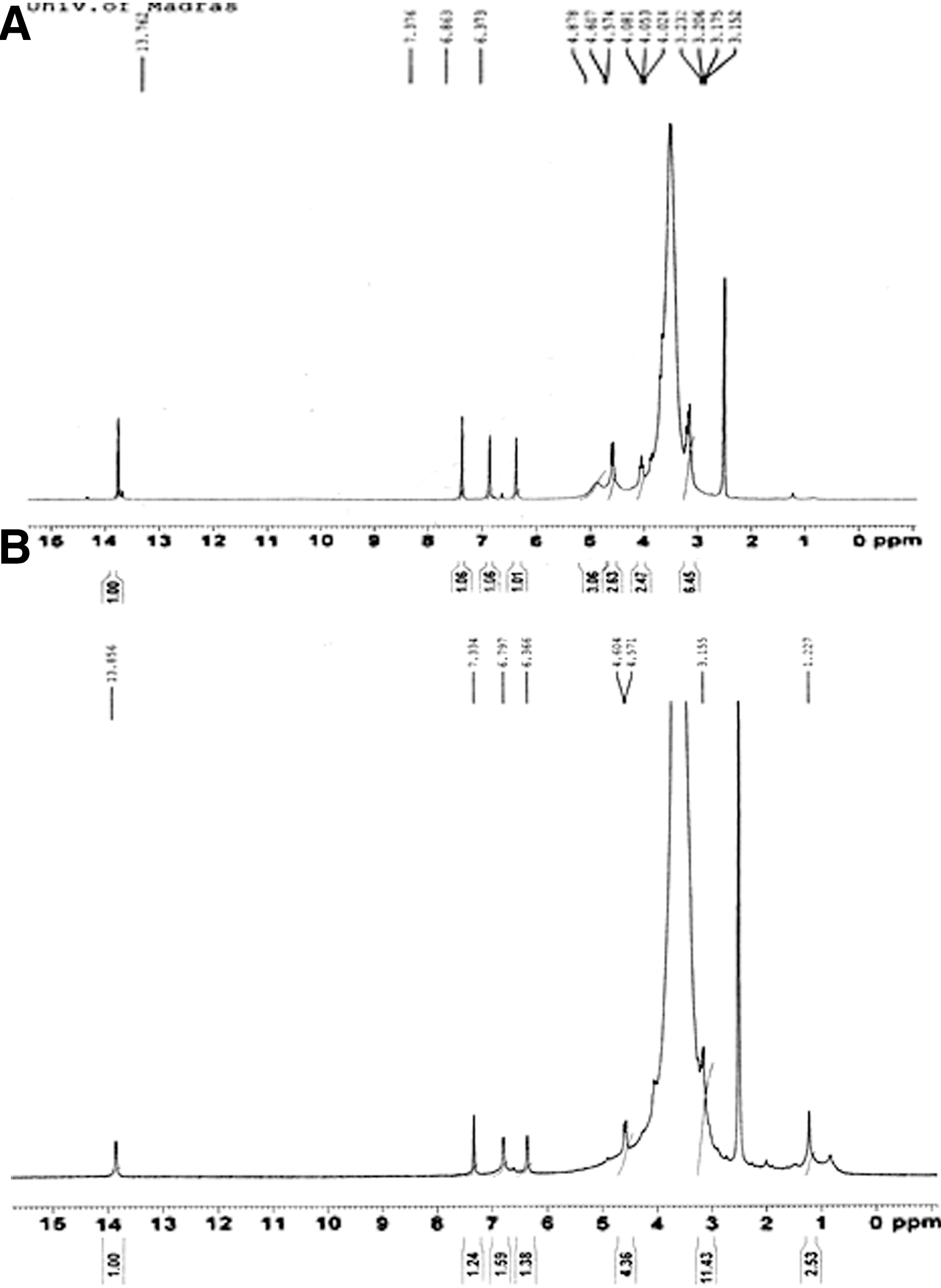
The 1H NMR spectrum of authentic mangiferin and isolated compound are shown in Figure 2. The multiplet appearing at 3.15–3.23 ppm are accounted for by the following three protons present in the mangiferin, (H-3′), (H-4′), and (H-5′). The multiplet at 3.8 ppm accounted for the two protons (H-6′). A double-doublet centered at 4.05 ppm are due to the one proton (H-2′). The doublet presented at 4.59 ppm was due to (H-1′) proton. The singlets at 6.37, 6.86, and 7.37 accounted for the protons (H-4), (H-5), and (H-8), respectively.
13C NMR spectrum analysis
The 13C NMR spectrum of authentic mangiferin and isolated compound are shown in Figure 3. The peak at 61.4 ppm was due to (C-6′) carbon. Similarly, 70.2, 70.5 (C-3′, 4′); 73.0 (C-5′); 81.4 (C-1′); 73.2 (C-4), 101.2 (C-1a); 102.5 (C-5); 107.5 (C-2); 107.9 (C-8); 111.6 (C-8a); 143.7(C-7); 150.7 (C-5a); 154.1 (C-6); 156.2 (C-4a); 161.7 (C-1); 163.8 (C-3); 179.0 (C-0).

Structure of compound
The isolated compound (Fig. 4) was confirmed as mangiferin, based on the 1H NMR and 13C NMR analysis.
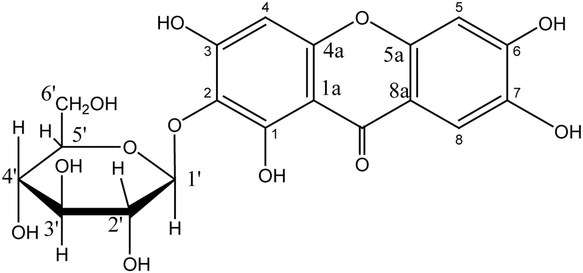
Structure of the isolated compound, mangiferin. Formula: C19H18O11; molecular weight: 422.35.
Biological activities of mangiferin
The levels of blood glucose and plasma insulin in control and experimental groups of rats are summarized in Figure 5. There were significant elevations in the level of blood glucose and concomitant decrease in the level of insulin in STZ-induced diabetic rats. The levels of blood glucose and insulin were reverted back to near normal levels in the diabetic rats treated with mangiferin and glibenclamide.

The levels of blood glucose
Figure 6 depicts the concentration of TBARS and hydroperoxides in plasma of control and experimental groups of rats. There was a significant increase in the levels of TBARS and hydroperoxides in plasma of diabetic rats, but oral administration of mangiferin and glibenclamide tended to restore them to similar concentrations as the normal rats.
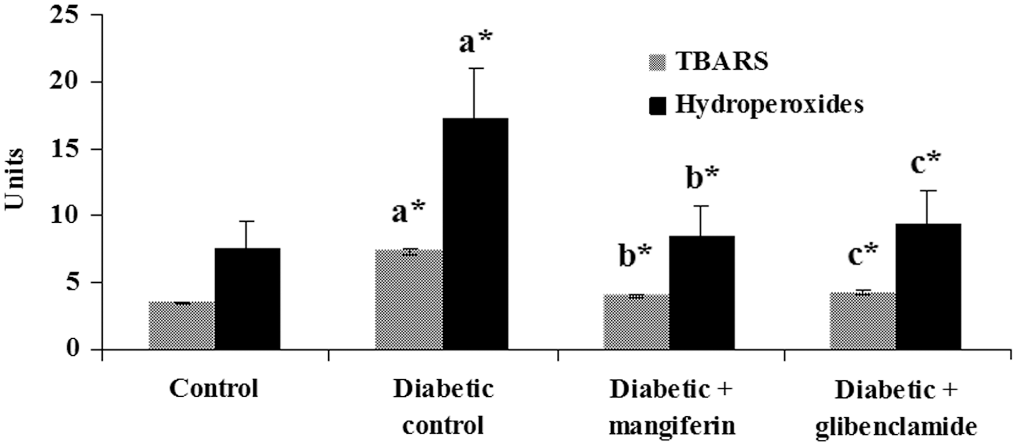
Levels of TBARS (thiobarbituric acid reactive substances) and hydroperoxides in plasma of control and experimental groups of rats. Data are expressed as mean±standard deviation for six animals in each group. One-way ANOVA followed by post hoc test LSD. Values are statistically significant at *P<.05: adiabetic control rats were compared with control rats; bmangiferin-treated diabetic rats were compared with diabetic control rats; cglibenclamide-treated diabetic rats were compared with diabetic control rats. Units: TBARS, nmoles of TBARS/mL of plasma; hydroperoxides, × 10−5 mM/dL of plasma.
Changes in the levels of vitamin C, vitamin E, and ceruloplasmin in the plasma of control and experimental groups of rats are shown in Figure 7. Decreased levels of vitamin C and concomitantly increased levels of vitamin E and ceruloplasmin were observed in STZ-induced diabetic rats when compared with control rats. Treatment with mangiferin and glibenclamide reversed these levels to near control levels when compared with diabetic rats.

Levels of nonenzymatic antioxidants in plasma of control and experimental groups of rats. Data are expressed as mean±standard deviation for six animals in each group. One-way ANOVA followed by post hoc test LSD. Values are statistically significant at *P<.05: adiabetic control rats were compared with control rats; bmangiferin-treated diabetic rats were compared with diabetic control rats; cglibenclamide-treated diabetic rats were compared with diabetic control rats.
Figure 8 shows the levels of TBARS, hydroperoxides, and reduced GSH in pancreas of control and experimental groups of rats. A marked increase in TBARS and hydroperoxides concentration and decrease in the level of reduced GSH were observed in pancreatic tissue of diabetic rats, which reverted back to near control levels after treatment with mangiferin and glibenclamide.
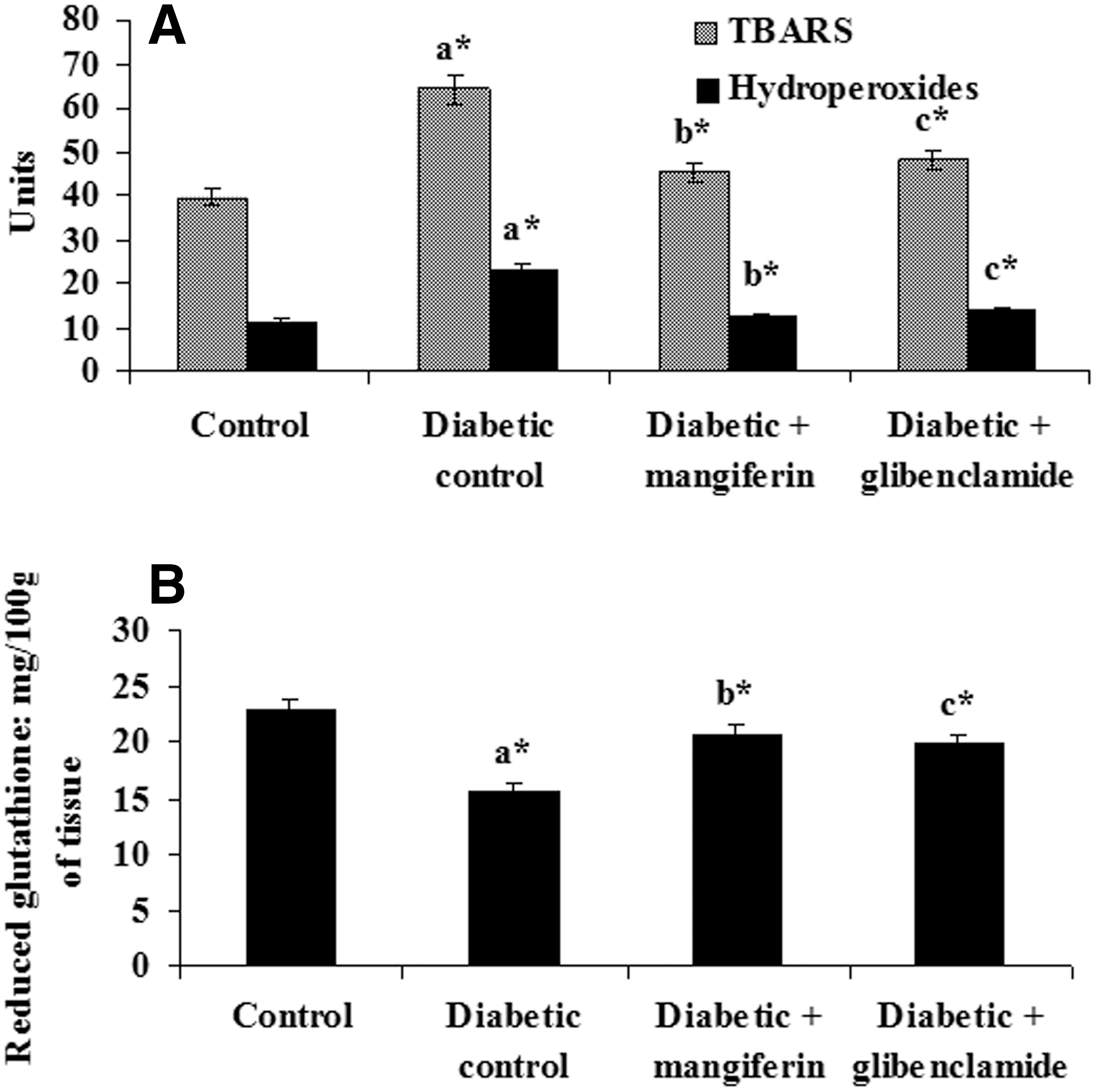
Levels of TBARS, hydroperoxides
Histopathological examination of the pancreas of control rats showed normal islets with clusters of purple stained β-cells (Fig. 9A). In the diabetic rats, pancreas showed destruction/absence of islets cells, when compared with control rats (Fig. 9B). In mangiferin-treated diabetic rats, the pancreas showed regeneration and hyperplastic islets cells (Fig. 9C). In glibenclamide-treated diabetic rats, the pancreas showed near normal architecture (Fig. 9D).
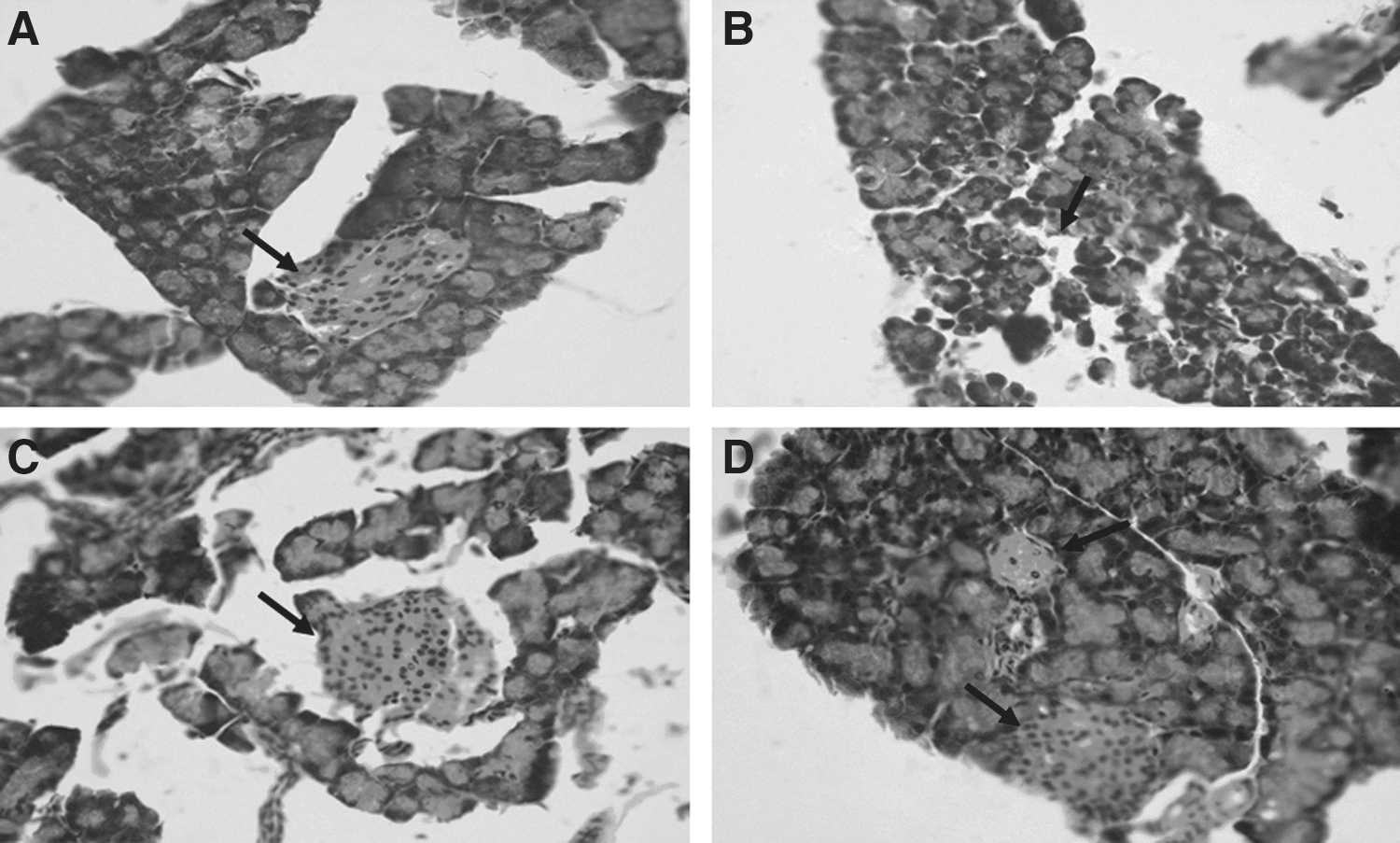
Histological observations in the pancreatic tissue of control and experimental groups of rats.
The electron microscopic analysis of pancreatic β-cells showed normal cellular arrangements in control rats (Fig. 10A). They showed normal nucleus, endoplasmic reticulum, and secretory granules. The pancreas of STZ-induced diabetic rats showed swelling of mitochondria with loss of cristae, damage of nuclear envelope, damaged mitochondria, and less number of secretory granules (Fig. 10B). The pancreas of mangiferin-treated diabetic rats showed near normal architecture of mitochondria, increased secretory granules, and normal nucleus with envelope (Fig. 10C). The glibenclamide-treated diabetic pancreas showed near normal architecture of normal nucleus, mitochondria, and increased secretory granules (Fig. 10D).

Transmission electron microscopic studies on pancreas of control and experimental groups of rats.
Discussion
STZ-induced hyperglycemia in experimental animals is considered to be a good preclinical model for the preliminary screening of natural products and other active agents against diabetes and it is also a nitric oxide donor in pancreatic β cells. Oxidative stress occurs as a result of excessive accumulations of oxygen free radicals during diabetes and its complication. Consequences of oxidative stress include the production of highly reactive oxygen radicals, which affect the cells, especially cell membrane through interaction with lipid bilayer and generate lipid peroxides that lead to pancreatic β-cell dysfunction and further major cellular organelle impairment. 26,27 The damage to β-cells is mainly due to nitric oxide and free radicals during diabetes, because they decrease the activities of free radical scavenging enzymes. 28
The treatment of diabetes with various natural extracts and their active compounds has been shown to effectively decrease hyperglycemia by decreasing free radical accumulation in experimental animals. 29 –31 Administration of mangiferin exerted a significant anti-diabetic effect, probably due to the stimulation of insulin secretion/action from remnant pancreatic β-cells, which enhanced glucose utilization/metabolism in peripheral tissues of experimentally induced diabetic rats. 32 Glibenclamide is a well-known second-generation standard anti-hyperglycemic agent, belonging to the class of sulfonylureas. The sulfonylurea agents are widely used for treating diabetes and its complications. It undergoes first-pass activity and the most frequently reported adverse effects are gastric disturbances like nausea, vomiting, anorexia, and increased appetite after oral treatment.
STZ is effectively used for the induction of diabetes mellitus in experimental animals. It is postulated to induce diabetes by the degeneration and necrosis of β cells of pancreatic islets, leading to the decline in insulin release. 33 In the present investigation, we observed that increased oxidative stress in diabetes due to STZ-induction decreased the release of insulin secretion from pancreatic β-cells (Figs. 6 –9).
Ascorbic acid is one of the water-soluble antioxidants that inhibits oxidative damage in the cell membrane caused by aqueous radicals and facilitates the maintenance of vitamin E level at optimal concentrations. 34 Alpha tocopherol (vitamin E) is the most important lipid soluble antioxidant and is involved in the sequence terminator of LPO and protecting cellular structures from attack of free radicals. 35 In the present study, significantly lower levels of vitamin C and increased level of vitamin E were noted in plasma of untreated diabetic rats compared with control rats, which are consistent with those of earlier researchers. 30,36 Oral administration of mangiferin significantly altered the levels of vitamins C and E in STZ-induced diabetic rats compared with diabetic untreated rats. These findings suggest that mangiferin effectively decreases oxidative stress through enhancing nonenzymatic antioxidant status due to scavenging of free radicals.
Ceruloplasmin is one of the most abundant proteins in plasma and consists of 95% of the total circulating copper in normal adults. In normal conditions, it acts as a potent free radical scavenger that oxidizes iron from the ferrous state to ferric state. Elevated levels of ceruloplasmin specifies the degree of oxidative stress owing to its ferroxidase activity and also generation of oxygen products comprising hydrogen peroxides. 37,38 The decreased level of ceruloplasmin were observed in diabetic rats administered with isolated mangiferin, which may be a tissue protective response to a decrease in circulating unbound Fe2+ and act as a known inhibitor for further free radical-induced oxidative damage and associated mechanisms.
GSH is an abundant tripeptide nonenzymatic biological antioxidant present in the liver, kidney, and other vital tissues. GSH plays a key role in the antioxidant defense system that protects the cellular system against the toxic effects of LPO. It functions as a direct scavenger of free radicals and also as a cosubstrate for peroxide detoxification through GSH peroxidases. 39 The depletion of tissue GSH is due to either decreased synthesis or increased deprivation of GSH by oxidative stress. 40 A noticeable depletion in the GSH content of pancreas was observed in diabetic rats. In the present study, it was observed that mangiferin was able to increase level of GSH in comparison to the diabetic control rats (Fig. 8B).
LPO is a specific feature of chronic diabetes and affects the cell membrane fluidity and alters the membrane-bound enzymes activities and their receptors, leading to membrane malfunction. 41 The secondary product of LPO is malondialdehyde (MDA), which is an indicator of tissue damage. 23,42 The observed elevation of MDA in diabetic untreated rats in the present investigation possibly resulted from increased intensity of LPO arising due to increased free radical production, as reported earlier 43 and also altered membrane-bound enzymes activities in STZ-induced diabetic rats. 18 Oral administration of mangiferin to diabetic rats brought about a significant decrease in the mean level of LPO. Mangiferin possibly scavenges free radicals, thereby stabilizing the endogenous antioxidant defense network and decreasing the level of LPO, as has been investigated for other natural antioxidants such as quercetin, 44 lycopene, 45 and curcumin. 46
Dietary natural supplements contribute to the prevention of diabetes and their complications by decreasing LPO and improving antioxidant status. Li et al. 47 reported that mangiferin significantly decreased LPO levels in kidney tissues and improved their antioxidant status. Further, mangiferin effectively protects renal tissues from cytotoxin through various antioxidant mechanisms. In the current investigation, we observed MDA formation and the LPO index was significantly increased in pancreatic tissue of STZ-treated animals. Treatment with mangiferin potently abrogated MDA levels, suggesting that mangiferin might act as a natural antioxidant principle to reduced oxidative damage.
The histological observation of pancreatic tissues provided additional support that mangiferin has a protective nature against oxidative damage. The STZ induction produced severe pancreatic injury such as a decrease of pancreatic islets diameter that was perhaps due to the decreased number of β cells. It is highly specific to β-cell toxicity and consequently causes diabetes; hence it is widely used to study β-cell damage in in vivo animal experiments. 48 STZ mainly eliminates the β-cell response in pancreatic islets to glucose. A temporary return of responsiveness to glucose then seems to be a permanent responsiveness, due to coincident damage of β-cells. 49
In the present investigation, shrinkage and vacuolization of pancreatic islets and decreases in the β-cell mass were observed in pancreatic tissue of diabetic rats through histological and ultrastructural studies. However, oral administration of mangiferin to diabetic rats appeared to preserve the remaining β-cell mass and to reduce vacuolization of pancreatic islets and swollen mitochondria and endoplasmic reticulum. Mangiferin possibly protected the pancreatic islets from free radical and hyperglycemic-mediated oxidative stress and preserved the integrity of pancreatic β-cells, thereby stimulating the remaining pancreatic β-cells to synthesize and secrete additional insulin to maintain glucose homeostasis. Such a sequence of events has been proposed by other investigators to strengthen the antioxidant defense system. 50 –52 These histological observations in diabetic rats treated with mangiferin in the present study appear to correlate with increased level of insulin, which would have brought about lowered levels of blood glucose in this group of rats.
These findings suggested a protective nature of mangiferin against structural and functional integrity of pancreatic β-cells. Further investigations will be needed to identify the specific molecular mechanism utilized by mangiferin to protect pancreatic tissue in STZ-induced toxicities in experimental rats.
Conclusions
Management of diabetes by mangiferin may be due to mangiferin-mediated insulin release and regeneration of β-cells. Histopathological observations depicted that STZ destroyed pancreatic β-cells, but that they were revived many folds with mangiferin treatment, this result demonstrates insulin-mimicking effect of mangiferin by regenerating/activate insulin-producing cells in pancreas.
Footnotes
Acknowledgment
The authors are grateful to Dr. C.S. Vijayalakshmi, MD, DCP (Consultant, Pathologist, Government Hospital, Royapettah, Chennai, India) for her help in interpretation of histopathological findings.
Author Disclosure Statement
No competing financial interests exist for any of the authors.