
Abstract
The Terminalia genus includes plants that are used in a variety of food, nutritional products, and traditional medicines. Aqueous bark extract of Terminalia paniculata (TPW) was screened for its antioxidant and analgesic potential. The major polyphenols were identified by high-performance liquid chromatography. In vitro antioxidant potential of TPW was investigated by 1,1-diphenyl-2-picryl hydrazyl (DPPH), 2,2′-azinobis-(3-ethylbenzothiazoline-6-sulfonic acid) (ABTS2−) radical assay, nitric oxide (NO) scavenging, superoxide scavenging (O2−), Fe2+ chelating (O-phenanthroline), and ferric reducing/antioxidant power (FRAP) assay. We evaluated the effects of TPW on cell viability, lipopolysaccharide (LPS)-stimulated reactive oxygen species (ROS), nitrite, and cytokines (interleukin [IL] 6 and tumor necrosis factor alpha [TNF-α]) in RAW 264.7 murine macrophages. Evaluation of analgesic activity of TPW was performed using acetic acid-induced writhing and hot plate test in mice. Phytochemical analysis showed the presence of four polyphenols, namely, gallic acid, ellagic acid, rutin, and quercetin. TPW showed maximum superoxide, ABTS2−, NO, DPPH inhibition, and Fe2+-chelating property at 400 μg/mL, respectively. FRAP value was 4.5±0.25 μg Fe(II)/g. TPW, per se, did not affect RAW 264.7 cell viability. In LPS-induced RAW 264.7 cells, TPW attenuated the elevation in ROS, nitrite, IL-6, and TNF-α levels. TPW (100–400 mg/kg, orally) significantly reduced the number of writhes in a dose-dependent manner compared with the control. Similarly, TPW (400 mg/kg, orally) evoked a significant increase in the maximum percentage effect in the hot plate test. The study suggests the efficacy of aqueous bark extract of T. paniculata as a potential antioxidant and analgesic agent.
Introduction
F
The plant kingdom serves as a rich source of bioactive compounds, many of which work as natural antioxidants. These include polyphenols such as phenolic acids, flavonoids, hydrolysable tannins, present in a variety of fruits, vegetables, herbs, cereals, sprouts, and seeds. 2 Several medicinal plants have been explored extensively for their natural antioxidant activity. 3 –5 In the last decade, dietary polyphenols have emerged as antioxidants pertaining to their free-radical scavenging property. Polyphenols have shown to exhibit an important role in the prevention of diseases, particularly inflammation and arthritis through cyclooxygenase (COX) and lipoxygenase inhibition. 6 –8
Various species of Terminalia trees are known for their anti-inflammatory, immunomodulatory, 9 –11 and wound-healing activities. 12 Terminalia paniculata Roth. (Combretaceae), one such species, is widely distributed in the semi-evergreen forests of the Western Ghats in India. The flower juice and bark of this tropical and deciduous tree is traditionally used for the treatment of parotitis, menstrual cycle abnormalities, and gastrointestinal disorders. 13 –15
Based on the ethnopharmacological evidence, the present study was carried out to determine the in vitro antioxidant and in vivo analgesic potential of the aqueous bark extract of T. paniculata (TPW) and to correlate these activities with the constituent polyphenols.
Materials and Methods
Chemicals
1,1-Diphenyl-2-picryl hydrazyl (DPPH); 4,6-tripyridyl-s-triazine, 2,2′-azinobis-(3-ethylbenzo-thiazoline-6-sulfonic acid) (ABTS), nitro blue tetrazolium (NBT), Folin-Ciocalteu phenol reagent, sodium carbonate, lipopolysaccharide (LPS), Griess reagent, ferric chloride (FeCl3), sodium nitrite, ascorbic acid, gallic acid, ellagic acid, rutin, and quercetin were from Sigma-Aldrich Co. (St. Louis, MO, USA). Dulbecco's modified Eagle's medium and fetal bovine serum were purchased from Invitrogen Corp. (Carlsbad, CA, USA). Sodium nitroprusside (SNP), α-napthyl-ethylenediamine (NEDD), and dimethyl sulfoxide (DMSO) were purchased from LobaChemie Pvt. Ltd. (Mumbai, Maharashtra, India). All the chemicals used, including the solvents, were of analytical grade, acquired from Merck Specialities Pvt. Ltd. (Mumbai, Maharashtra, India).
Aqueous extract preparation and standardization
The bark of the tree (specimen voucher: MCOPS/PHCOL/2009/2) was collected from Manipal, Karnataka, India, authenticated (Dr. G.K. Bhatt, Department of Botany, Poornaprajna College, Udupi, Karnataka, India) and TPW prepared according to a previously established method. In brief, T. paniculata bark (1000 g) was air dried at room temperature, powdered, and macerated in water as per Indian Pharmacopoeia for 7 days with intermittent shaking. The resulting solution (5000 mL) was concentrated under reduced pressure at 40°C and lyophilized (−40°C) to obtain a solid reddish-brown residue, which was stored in a refrigerator at 4–5°C, until use. The extract was standardized by high-performance liquid chromatography (HPLC) by comparing the retention time and UV spectra of the chromatographic peaks with those of the reference standards as reported earlier. 16 It was run with a two-pump linear gradient program for pump A (water containing 0.1% formic acid) and pump B (acetonitrile), initially started with a gradient of 10% B changing to 70% in 25.0 min and finally, to 10% in 35 min followed by washing for 40 min. Flow rate and injection volume were 1.0 mL/min and 20 μL, respectively.
In vitro antioxidant activity
DPPH scavenging effect
DPPH scavenging activity was measured as previously described. 17 Briefly, 0.1 mM solution of DPPH was prepared in methanol, and 0.5 mL of this solution was added to 1.5 mL of different concentrations of TPW (12.5–400 μg/mL) and ascorbic acid (12.5–400 μg/mL) prepared in methanol. After incubation at 37°C for 30 min, the absorbance at 517 nm was measured in triplicate, and the scavenging effect was calculated against control, that is, methanol.
Scavenging of ABTS2−
ABTS2− was produced by reacting 2 mM ABTS in distilled water with 2.45 mM potassium persulfate (K2S2O8), stored in the dark at room temperature for 8 h. To 0.2 mL of various concentrations of the TPW or ascorbic acid (in distilled water), 0.6 mL of ABTS solution was added and incubated for 20 min. Absorbance of these solutions were measured spectrophotometrically at 734 nm. 18
Nitric oxide radical inhibition assay
The reaction mixture containing SNP (10 mM, 4 mL), phosphate buffer saline (1 mL), and TPW or ascorbic acid (1 mL) was incubated at 25°C for 150 min. After incubation, 500 μL of the reaction mixture was added to 1 mL of sulfanilic acid reagent (0.33% in 20% glacial acetic acid [AA]), mixed, and allowed to stand for 5 min for completion of diazotization reaction. Next, 1 mL of NEDD was added, mixed, and allowed to stand for 30 min in diffused light. The absorbance was measured at 540 nm. 19
Scavenging of superoxide radical by alkaline DMSO method
1 mL of alkaline DMSO (1 mL DMSO containing 5 mM NaOH in 0.1 mL water) was added to the reaction mixture containing 0.1 mL of NBT (1 mg/mL solution in DMSO) and 0.3 mL of TPW or ascorbic acid in DMSO to give a final volume of 1.4 mL, and the absorbance was measured at 560 nm. 17
Reduction of ferric ions by ortho-phenanthroline color method
The reaction mixture contained 1 mL of ortho-phenanthroline (50 μg in 10 mL methanol), 2 mL of 200 μM FeCl3 (3.24 mg in 100 mL distilled water), and 2 mL of various concentrations of TPW (25–400 μg/mL) or ascorbic acid. The mixture was incubated, and absorbance was measured at 510 nm. 20
Ferric-reducing/antioxidant power assay
A previously described method was adopted with suitable modifications. 21 In brief, the stock solution of 10 mM 2,4,6-tripyridyl-s-triazine (TPTZ) in 40 mM HCl, 20 mM FeCl3.6H2O, and 0.3 M acetate buffer (pH 3.6) were prepared. The ferric-reducing/antioxidant power (FRAP) reagent contained 2.5 mL TPTZ solution, 2.5 mL FeCl3 solution, and 25 mL acetate buffer. It was freshly prepared and warmed to 37°C. Then, 900 mL FRAP reagent was mixed with 90 mL water and 30 mL TPW in distilled water and ascorbic acid solution. The reaction mixture was then incubated at 37°C for 30 min, and absorbance was recorded at 595 nm. An intense blue color complex was formed when ferric tripyridyl triazines (Fe3+ TPTZ) complex was reduced to the ferrous (Fe2+) form, and the absorption at 595 nm was recorded. The calibration curve was plotted with absorbance at 595 nm versus concentration of ferrous sulfate in the range 0–1 mM.
In vitro cell-based assays using RAW 264.7 macrophages
Cell viability assay using MTT reagent
Cell viability of RAW 264.7 cells pretreated with various concentrations of TPW (before presence or absence of LPS challenge) was assessed using 3-(4,5-dimethylthiazol-2-yl)-2,5-diphenyltetrazolium bromide (MTT) reagent according to a previously established method.
22
In brief, a monolayer of cells in the plate was exposed to various dilutions of the TPW (0–3000 μg/mL). Cells were later challenged with LPS (0.2 μg/mL) and kept for 24 h. MTT, a tetrazolium salt, is metabolically reduced by viable cells to yield a blue insoluble formazan product, which was measured at 540 nm using a micro plate reader (ELx800; BioTek Instruments, Inc., Winooski, VT, USA). Percentage cell viability was calculated as follows:
Reactive oxygen species assay
A previously described method
23
was adopted and slightly modified. Intracellular reactive oxygen species (ROS) levels were measured by detecting the fluorescence intensity of the oxidant-sensitive probe 2′,7′-dichlorodihydrofluorescein diacetate (H2DCFH-DA; Sigma-Aldrich). H2DCFH-DA is metabolized to nonfluorescent DCFA by intracellular esterases and to fluorescent DCF by free oxygen radicals. RAW 264.7 cells were seeded into a 96-well microplate at a density of 2.0×10⁴ cells/well. The next day, the medium in the wells was aspirated out and was replaced with same volume of fresh medium containing 50 μM DCFH-DA. An hour later, the contents of the wells were further aspirated out and were replaced with the same volume of medium containing LPS (0.2 μg/mL) for 24 h. Then, cells were treated with various doses of TPW (25–250 μg/mL), which had been confirmed to be dose dependent by cell viability assay using MTT reagent, and incubated for 24 h. Diphenylene iodonium (10 μM) was used as a standard. Next, the plate was read at an excitation wavelength of 488 nm and an emission wavelength of 525 nm using a fluorescence plate reader (FLx800; BioTek Instruments, Inc.). The experiments were performed with at least three independent replications, and the results from one representative experiment (n=6 wells) are presented. The percentage inhibition of ROS was calculated using the following formula:
Nitrite assay
RAW 264.7 cells were seeded into a 96-well microplate at a density of 2.0×10⁴ cells/well. The cells were treated with various doses of TPW (25–250 μg/mL), for which dose dependency had been established using MTT assay, and incubated for 24 h. Then, they were treated with 0.2 μg/mL of LPS, and incubated for 12 h. NO accumulated in culture medium was measured as an indicator of NO production based on the Griess reaction. 24 Each 100 μL of cell culture medium was mixed with 100 μL of Griess reagent (equal volumes of 1% sulfanilamide in 5% phosphoric acid and 0.1% naphtylethylenediamine), incubated at room temperature for 10 min, and then, the absorbance was read at 540 nm using a micro plate reader (ELx800; BioTek Instruments, Inc.). Fresh culture medium was used as the blank in all experiments. The amount of NO in the samples was measured with the sodium nitrite serial dilution standard curve.
Estimation of pro-inflammatory cytokines (TNF-α and IL-6) production
RAW 264.7 cells were plated in a 24-well culture dish at a density of 5×10⁵ cells/well and then incubated with TPW (0, 12.5, 25, 50, and 100 μg/mL) in the presence or absence of LPS (0.2 μg/mL) for 24 h. The cell culture supernatants with or without TPW treatment were used to measure the tumor necrosis factor alpha (TNF-α) and interleukin 6 (IL-6) levels using solid-phase sandwich enzyme-linked immunoadsorbent assay kits (Invitrogen Corporation, Carlsbad, CA, USA) according to the manufacturer's instructions. The concentrations of cytokines in the samples were calculated from a standard curve that was developed using a known concentration of recombinant TNF-α and IL-6.
In vivo analgesic activity
Animals
All experiments were performed using male Swiss albino mice (18–25 g) obtained from the Central Animal Research Facility, Manipal University. Animals were maintained in a room with controlled temperature 22±2°C for 12 h light/dark cycle with free access to food and water. Three hours before each experiment, the animals received only water, in order to avoid food interference with compound absorption. The Institutional Animal Ethics Committee (IAEC) approved the experimental protocol (No. IAEC/KMC/55/2009–2010). All protocols and experiments were conducted in strict compliance according to the ethical principles and guidelines provided by the Committee for the Purpose of Control and Supervision of Experiments on Animals.
AA-induced writhing in mice
A modified previously described protocol was used.
25
Briefly, the total number of writhings after an intraperitoneal administration of 1% (v/v) AA was recorded over a period of 20 min, starting 5 min after AA injection. Mice were pretreated orally with T. paniculata extract (200 and 400 mg/kg) or diclofenac (10 mg/kg), 60 min before administration of AA. The inhibition rate of writhes was calculated using the formula:
Hot plate test in mice
Mice were tested according to the method previously described.
26
Animals were placed on a hot plate (Inco Instruments, Ambala, Haryana, India) set at 55±1°C. Reaction time was recorded when the animals licked their fore and hind paws and jumped at several intervals of 30, 60, 90, and 120 min (cut off time 15 sec) after oral administration of TPW (200 and 400 mg/kg) or pentazocine (4 mg/kg). Data are expressed as time course of the maximum percentage effect (%MPE):
Statistical analysis
The statistical analyses were performed by one-way ANOVA, followed by Dunnett's post hoc test. The results were expressed as the mean±S.E.M. Differences are considered significant when P<.05. IC50 values were calculated by nonliner regression analysis by fitting the concentration–response relation for each drug (TPW and AscA) using Prism 5.03 Demo Version (GraphPad Software, Inc., La Jolla, CA, USA).
Results
Standardization of TPW polyphenols
TPW, used in the current study, was standardized using HPLC. The retention time and UV spectra of the chromatographic peaks obtained were compared with those of the reference standards. This confirmed the presence of polyphenols, namely, gallic acid, rutin, ellagic acid, and quercetin (Fig. 1), which were in accordance with results reported earlier. 16
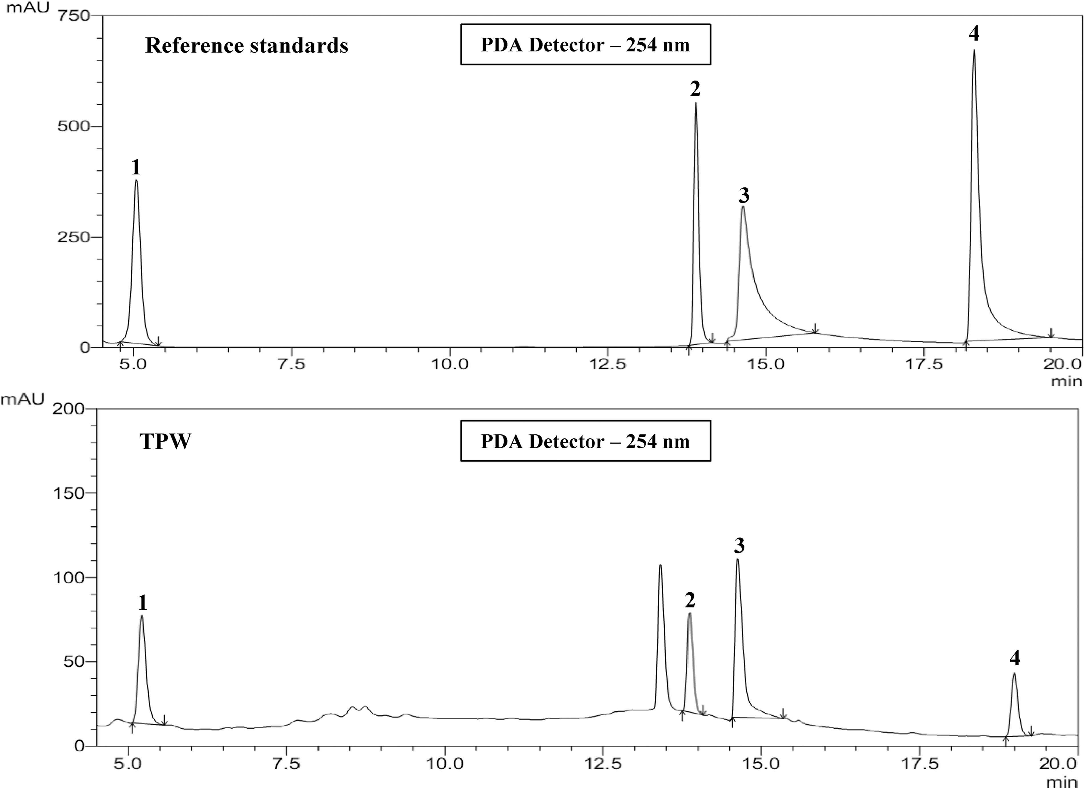
Analytical high-performance liquid chromatography chromatogram of an aqueous bark extract of Terminalia paniculata (TPW) at 254 nm. 1, gallic acid; 2, rutin; 3, ellagic acid; 4, quercetin. The chromatographic peaks of the analytes were confirmed by comparing their retention time (Rt) and UV spectra with those of the reference standards (≥97% purity).
In vitro antioxidant activity of TPW
In the present study, aqueous extract of T. paniculata exhibited strong scavenging effects in all antioxidant assays performed. An overall comparison showed TPW to exhibit a stronger scavenging effect on DPPH radicals (IC50=44.15±0.42 μg/mL), whereas it was lowest at reducing ferric ions (O-phenanthroline) (177.47±1.63 μg/mL). Furthermore, the antioxidant effect of TPW was evaluated by suppression of the superoxide anion radicals generated in a photochemical system. TPW exhibited a strong SOD-like effect suppressing O2 − release in a dose-dependent manner (IC50=98.16±0.70 μg/mL) as shown in Table 1. The ability of free radical scavenging activity of TPW in various antioxidant assays are presented next in decreasing order of reactivity: DPPH > ABTS > nitric oxide > superoxide > O-phenanthroline. The results showed that the TPW had modest antioxidative potential, as compared with ascorbic acid.
Data represented as mean±S.E.M of three replicates.
DPPH, 1,1-diphenyl-2-picryl hydrazyl; ABTS, 2,2′-azinobis-(3-ethylbenzothiazoline-6-sulfonic acid; TPW, aqueous bark extract of Terminalia paniculate; AscA, ascorbic acid (12.5–400 μg/mL).
In vitro activity in RAW 26.7 macrophages
Effect of TPW on cell viability
None of the concentrations of TPW (0–3000 μg/mL) impaired cell viability per se. In the presence of LPS, a drastic reduction in percentage cell viability (54.14±8.25) was observed. This was reversed in a dose-dependent manner by pretreatment of RAW 264.7 cells with TPW (0–300 μg/mL). A further increase in TPW doses for approximately 3000 μg/mL did not lead to any drastic changes in cell viability. Hence, TPW was considered safe at the doses tested (Fig. 2).
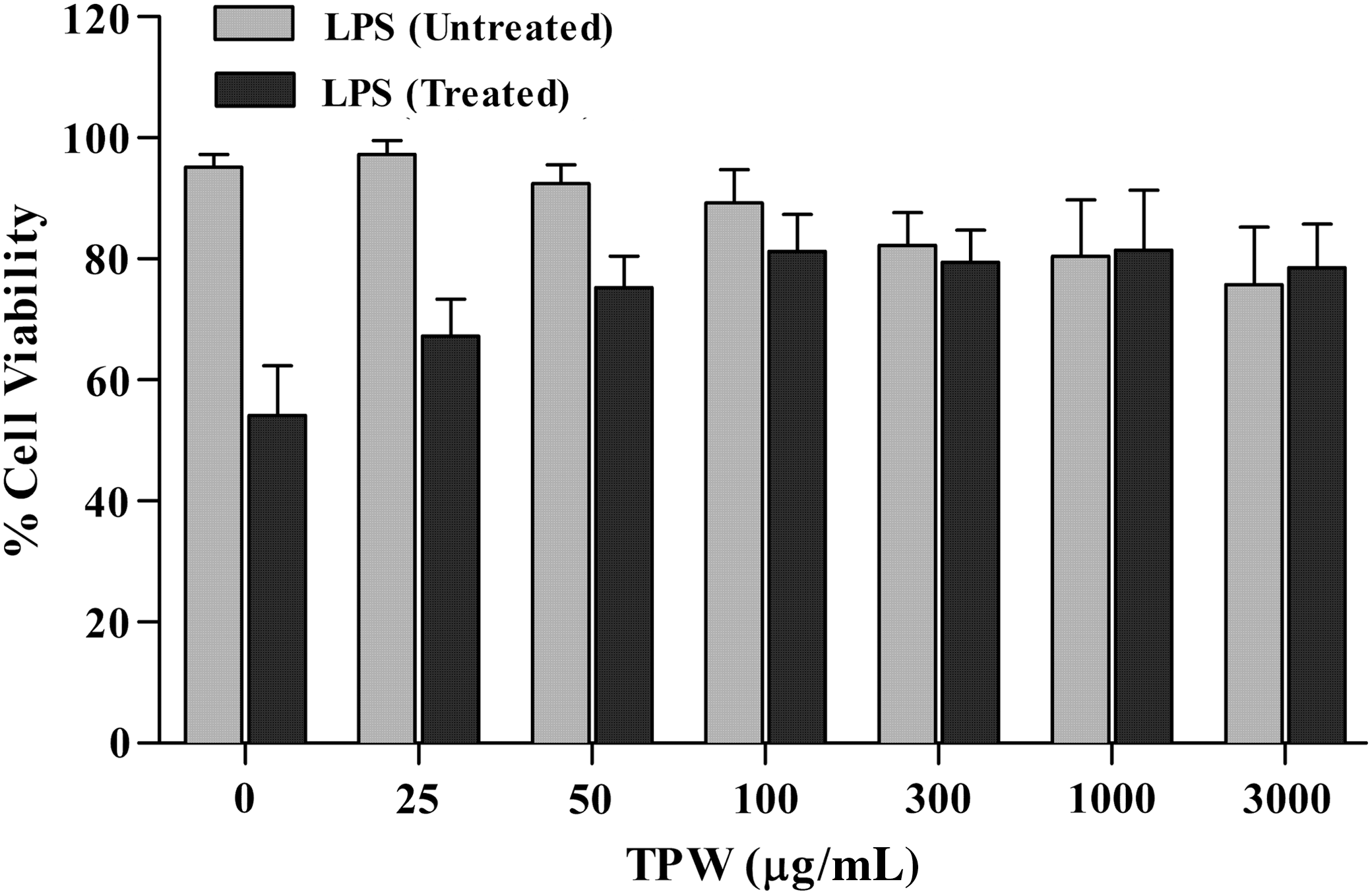
Effect of TPW on cell viability of RAW 264.7 cells in the presence/absence of lipopolysaccharide (LPS). RAW 264.7 cells pretreated with various concentrations of TPW were challenged with or without 0.2 μg/mL of LPS and observed after 24 h for cell viability using MTT reagent.
Effect of TPW on LPS-stimulated ROS generation
LPS increased intracellular ROS levels after 24 h. TPW significantly prevented LPS-induced increase of intracellular ROS levels after 40 h of culture. Diphenylene iodonium (NADPH-oxidase inhibitor) also attenuated the LPS-induced ROS elevation in RAW 264.7 cells. However, the response exhibited by TPW was biphasic, as it was not found to change linearly with an increasing concentration (Fig. 3).

Effect of TPW on reactive oxygen species (ROS) inhibition in the LPS-stimulated RAW 264.7 cells. RAW 264.7 cells were treated with various concentrations of TPW for 40 h, and then stimulated with LPS (0.2 μg/mL) for 24 h. Intracellular ROS levels were measured after staining with a fluorescent dye, DCFH2-DA. All the values were expressed as mean±S.E.M. (n=3) analyzed by one-way ANOVA followed by post hoc Dunnett's test. # P<.05 when compared with normal control, *P<.05 when compared with LPS control.
Effect of TPW on LPS-stimulated NO generation
A nitrite assay was performed to further investigate the protective effects of TPW against the cytotoxicity of LPS. TPW showed the decreasing NO generation in the dose-dependent manner from 100–250 μg/mL, indicating the lower production of NO (Fig. 4).
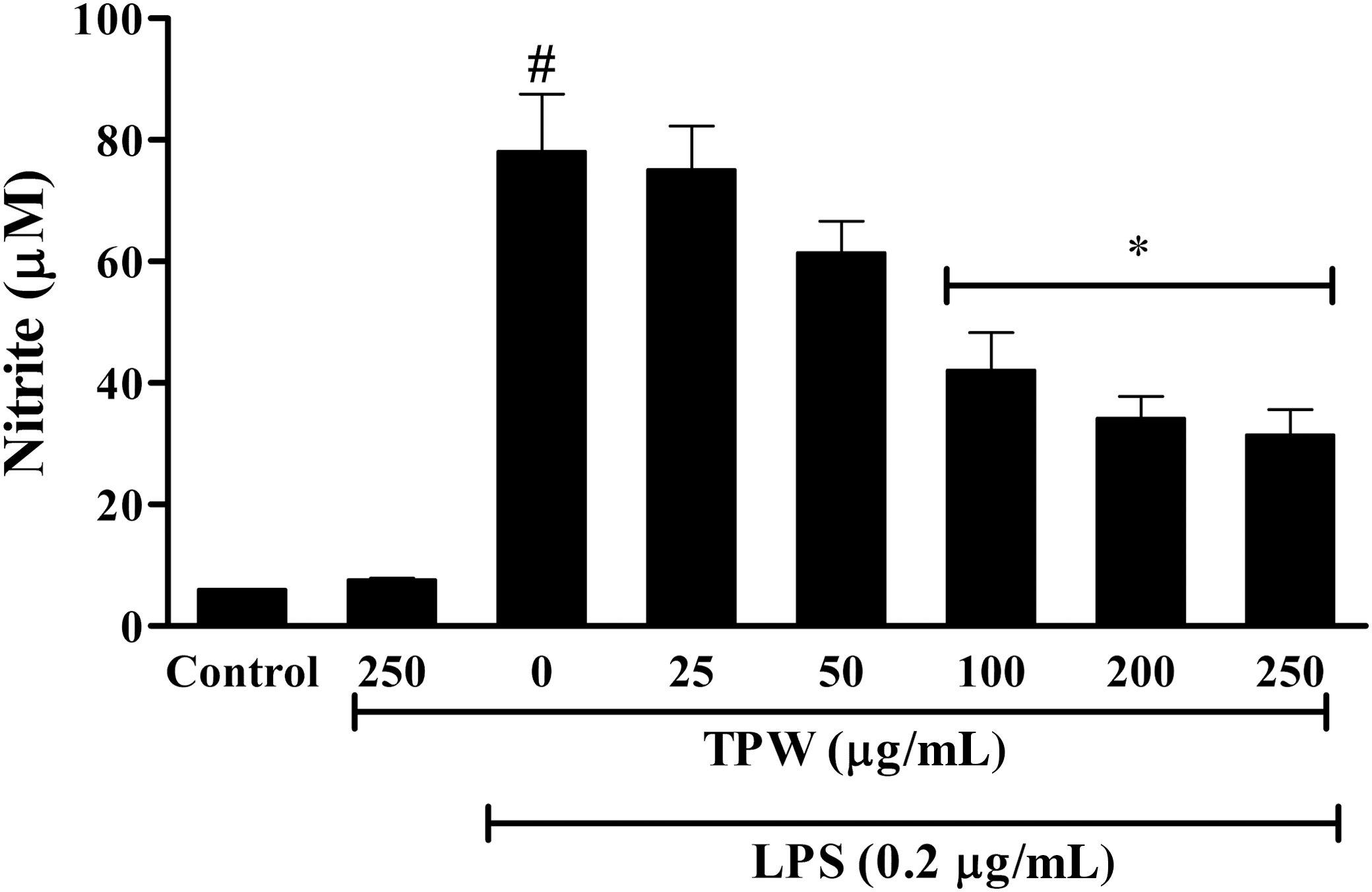
Protective effect of TPW against LPS-generated nitric oxide (NO) by nitrite assay in RAW 264.7 cells. LPS remarkably increased the level of NO, compared with control, but TPW decreased it in a dose-dependent manner. All the values were expressed as mean±S.E.M. (n=3) analyzed by one-way ANOVA followed by post hoc Dunnett's test. # P<.05 when compared with normal control, *P<.05 when compared with LPS control.
Effect of the TPW on cytokine release
To analyze the anti-inflammatory mechanisms of TPW, we determined its effect on LPS-induced production of pro-inflammatory cytokines, including TNF-α and IL-6. Figure 5 demonstrates that unstimulated RAW 264.7 cells cultured for 24 h produced a negligible quantity of cytokines. However, the levels of TNF-α and IL-6 were increased in the culture supernatant of the LPS-stimulated cells, whereas pretreatment with TPW resulted in a decrease in cytokine production.
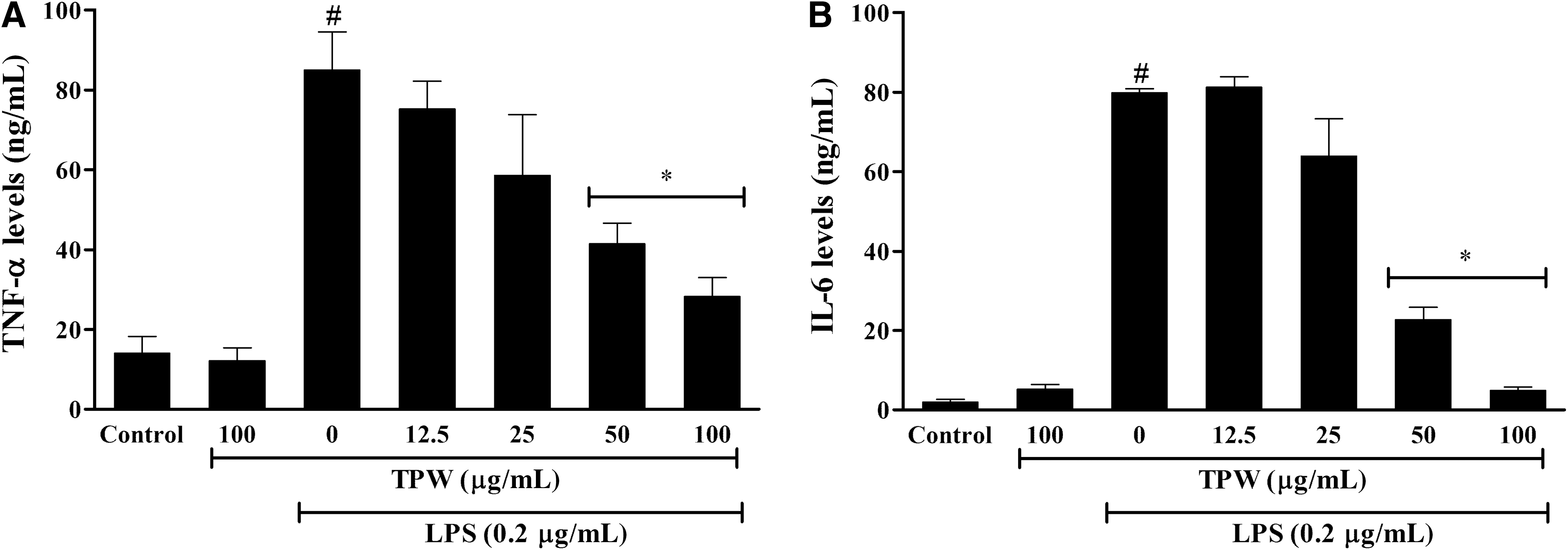
Effect of TPW on cytokine release
In vivo analgesic activity
Reduction in AA-induced writhing
After oral administration of TPW (100–400 mg/kg), a significant inhibition (when compared with vehicle-treated group) in the number of writhes was observed (Fig. 6). In addition, TPW administration induced a dose-dependent decrease (100 mg/kg, 11.2% inhibition; 200 mg/kg, 30% inhibition) in maximum inhibition of writhes at 400 mg/kg (41% inhibition) as compared with vehicle control. Positive control (diclofenac) also significantly inhibited the writhing response (63% inhibition).
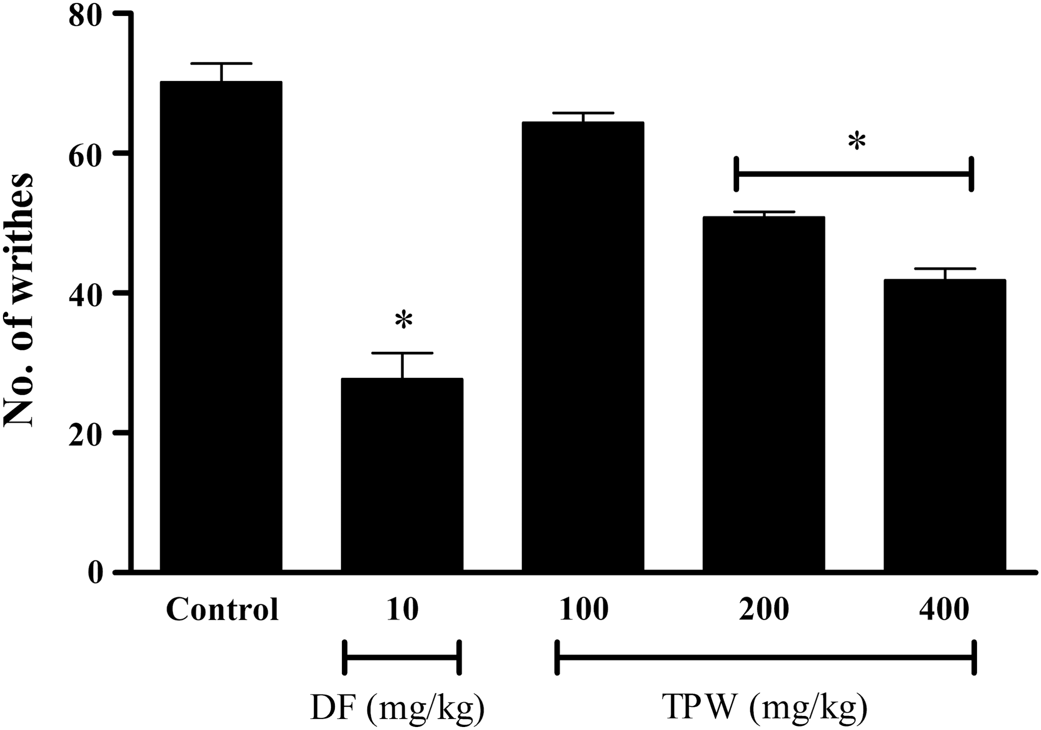
Effect of TPW on acetic acid-induced abdominal writhing in Swiss albino mice. Animals were pretreated by oral administration of different doses of TPW. Control groups were composed by vehicle and diclofenac (10 mg/kg, orally). Results are presented as mean±S.E.M. (n=6) of total writhings. Statistical significance was calculated by ANOVA followed by Dunnett's test. *P<.05 when comparing diclofenac or TPW-treated mice with vehicle-treated group.
Hot-plate test
The hot-plate test is a suitable model for testing centrally acting analgesics 27 and was employed to test the antinociceptive effect of extract. TPW (400 mg/kg) significantly increased the MPE as observed from the latency periods, in mice placed over a heated plate at 55°C (Fig. 7). This indicated attenuation of pain, which may be attributed to the inhibition of free radical generation by TPW at peripheral levels as well as central levels.
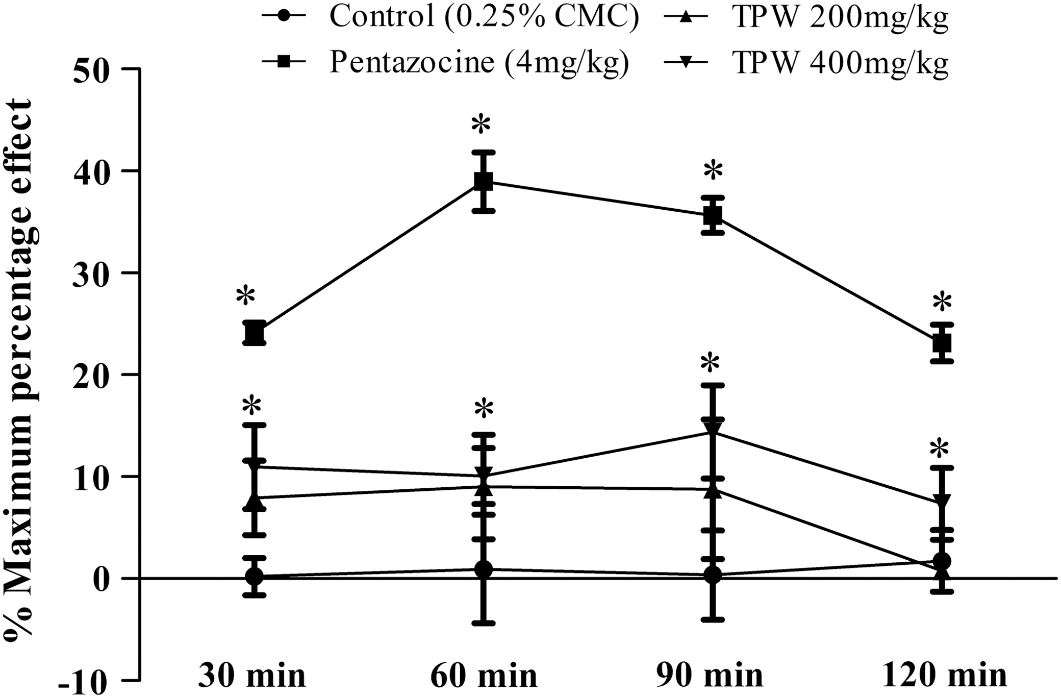
Effect of TPW on hot plate test in Swiss albino mice. Animals were pretreated by oral administration of different doses of TPW. Control groups were composed by vehicle and pentazocine (4 mg/kg, intraperitoneally). The results are presented as mean±S.E.M. (n=6) of % maximum percentage effect. Statistical significance was calculated by ANOVA followed by Dunnett's test. *P<.05 when comparing pentazocine or TPW-treated mice with vehicle-treated group.
Discussion
Polyphenolic compounds such as tannins (gallic acid and ellagic acid), caffeic acid, and flavonoids (quercetin and rutin) are reported to possess analgesic activity in various models of inflammatory pain and arthritis along with antioxidant properties. 28 –31 These polyphenols act through hydrogen or electron-donating and metal ion-chelating properties. 32 The presence of polyphenols such as gallic and ellagic acid 16,33 and flavonoids in Terminalia species with reported antioxidant properties 34 directed our interest to correlate its antioxidant and analgesic action.
In order to evaluate the antioxidant activity of a natural product, it is crucial to implement more than one antioxidant method, taking into consideration the various oxidation aspects in the systems under scrutiny. The TPW (12.5–400 μg/mL) exhibited DPPH and ABTS2− scavenging effects in a dose-dependent manner when compared with ascorbic acid. DPPH moiety is considered a model for a lipophilic radical (generated by lipid autoxidation). 35 However, ABTS2− radicals are more reactive than DPPH radicals and unlike the reactions with DPPH radical, which involve H-atom transfer, the reactions with ABTS2− radicals involve an electron transfer process. 36,37 Thus, TPW was found to inhibit both H-atom and electron transfer-mediated generation of free radicals. The pro-inflammatory role of nitric oxide (NO) has been reported in pain and inflammation. 30,38 In inflammatory conditions, free oxygen radicals react readily with NO to generate nitrite and peroxynitrite anions, which act as free radicals in the body. In the present study, TPW competes with the oxygen radicals to inhibit the generation of nitrite anions.
The links between superoxide radical, lipid peroxidation, and induction of COX pathway have been described earlier. 39 In the present study, superoxide radical scavenging activity of TPW (400 μg/mL) was comparable to 100 μg of ascorbic acid, which suggests the possible role of TPW in the prevention of lipid peroxidation and subsequent COX enzyme induction through superoxide scavenging activity. Furthermore, TPW interfered with the formation of complex between O-phenanthroline with Fe2+, indicating its metal-chelating ability. 40
The FRAP assay is widely used to evaluate the reducing potential of an antioxidant. In this assay, an easily reducible oxidant Fe(III) is used in excess. The reduction of Fe(III)–TPTZ complex by antioxidant, Fe(II)–TPTZ is formed, which can be measured spectrophotometrically at 595 nm. 41 Our results showed that TPW acts as a hydrogen donor to inhibit the formation of free radicals. The reducing ability (FRAP) of TPW was found in the range of 4.5±0.25 μg Fe(II)/g.
After confirmation of in vitro antioxidant activity of TPW in various chemically mediated oxidative reactions, it was subjected to test against in vitro inflammatory models, as up-regulation of pro-inflammatory mediators such as cytokines, ROS, and NO 42 is associated with inflammatory pain. Tissue macrophages are the first line of the body's defense against exogenous endotoxins such as pathogen-associated molecular pattern. 43 Identification of these patterns by toll-like receptors (TLRs) such as TLR-4 and TLR-5 present in macrophages, in turn, up-regulates several pro-inflammatory mediators, including cytokines such as IL-6 and TNF-α. 44 In the present study, inhibition of ROS, NO, IL-6, and TNF-α generation by TPW in LPS-stimulated RAW 264.7 macrophage cells suggests the involvement of TPW in modulating inflammation and associated pain through these pathways. Furthermore, the role of TPW in the modulation of TLRs has to be investigated.
It is well appreciated that, during tissue injury and inflammation, hyperalgesia results in a persistent state of peripheral afferent sensitization. The mechanism(s) is complex in nature and involves peripherally and spinally formed inflammatory mediators, such as peptides, prostanoids, NO, cytokines, and especially free radicals. 45 Superoxide formed during this process plays a major role in the development of pain through direct peripheral sensitization or through activation of lipid peroxidation, finally leading to induction of COX-2 pathway. 46
The involvement of free radical pathways in a particular setting can be better understood in a pharmacological setting; by using agents that remove or scavenge superoxide or NO. Inhibition of AA-induced writhing by TPW strongly suggests its peripheral analgesic activity. Therefore, the possible mechanism of analgesia could be the inhibition of free radicals at peripheral levels (Fig. 6), and the inhibition of subsequent lipid peroxidation-mediated activation of COX-2. 47
Conclusion
In conclusion, the TPW was found to possess analgesic activity in association with the scavenging of DPPH, ABTS, NO, superoxide anion, metal-chelating ability, and inhibition of IL-6 and TNF-α. This cumulative effect of TPW extract highlights the potential of natural sources with medicinal properties and suggests the potential to develop the extract as a nutraceutical.
Footnotes
Acknowledgments
The authors wish to thank the Department of Pharmacology, Manipal College of Pharmaceutical Sciences, Manipal University, for providing the funds and facilities to carry out this work. The authors would also like to acknowledge the financial support offered by the Board of Research in Nuclear Sciences, the Department of Atomic Energy (Government of India) in purchase of the Shimadzu HPLC system with PDA detector used in the present study.
Author Disclosure Statement
The authors declare that they have no conflict of interest.