
Abstract
Diabetic nephropathy (DN) is the major cause of end-stage renal disease. Resveratrol has been shown to ameliorate hyperglycemia in diabetic rats. However, the effects of resveratrol on DN remain unknown. The aim of the present study is to investigate the effects of resveratrol on early-stage DN. Diabetes was induced by streptozotocin injection in male Wistar rats. The diabetic rats were treated with resveratrol at a dose of 20 mg/kg body weight for 8 weeks. Plasma glucose, creatinine, kidney/body weight ratio, and 24-h urinary protein were determined. The renal pathological changes were examined with periodic acid Schiff staining, and renal mesangial cells were cultured in high glucose concentrations with indicated concentrations of resveratrol (2.5, 5.0, and 10.0 μmol/L). The proliferation of mesangial cells was evaluated by methylthiazoletetrazolium assay. Expressions of glutathione S-transferases Mu (GSTM) and nuclear factor erythroid 2-related factor 2 (Nrf2) were detected by western blot, and apoptosis was analyzed using a flow cytometer. Resveratrol reduced plasma glucose, creatinine, and urinary protein excretion, and attenuated renal hypertrophy. Moreover, resveratrol also reduced the expression of GSTM in diabetic rats. In vitro, resveratrol inhibited the proliferation of mesangial cells caused by high glucose and down-regulated GSTM and Nrf2 expressions in a dose-dependent manner. These findings suggest that resveratrol help prevent the progression of DN. The renoprotection by resveratrol is in part mediated through the inhibition of high glucose-induced rat mesangial cell proliferation and downregulation of GSTM expression.
Introduction
A
Resveratrol (trans-3,5,4′-trihydroxystilbene) is a naturally occurring polyphenol found in more than 70 plant species, such as grapes, berries, peanuts, and other traditional medicines. 5 Resveratrol has been shown to possess anticancer, antioxidant, anti-inflammatory, and cardioprotective effects in various experimental models, 6 –9 and to mediate its effects through the modulation of many different pathways. 10 Recently, resveratrol has been reported to have numerous beneficial effects in animal models of diabetes mellitus, including improving insulin sensitivity and lowering plasma glucose. 11,12 Nevertheless, the effects of resveratrol on DN still remain incompletely clear. In the present study, we investigated the effects of resveratrol on early DN in streptozotocin (STZ)-induced diabetic rats and explored the possible underlying mechanisms.
Materials and Methods
Animals, induction of diabetes, and resveratrol treatment
Male Wistar rats 10 weeks of age weighing 200–220 g were purchased from the Laboratory of Animal Center of Shandong University (Shandong, China). All of the procedures were approved by the Animal Ethics Committee of Shandong University. The animals were acclimated for 1 week before the experiments on a 12/12-h light/dark cycle and allowed food and water ad libitum. Diabetes was induced by a single tail vein injection of STZ (Sigma, Northbrook, IL, USA) diluted in citrate buffer (pH 4.5) at a dose of 55 mg/kg body weight. Age-matched control rats received an equal volume of citrate buffer. At day 5 after STZ injection, blood glucose levels were tested using One Touch II Glucose Analyzer (Johnson and Johnson). Only rats with fasted blood glucose higher than 16.7 mmol/L were considered diabetic. All experiments were carried out 1 week after diabetes induction.
STZ-induced diabetic rats were randomly divided into two groups (n=8 in each group) and treated with or without resveratrol. Resveratrol (more than 98% pure, lot no. 0810018-22) was provided by JF-NATURAL (Tianjin, China) and administered through gastric intubation at a dose of 20 mg/kg body weight/day for 8 weeks. The age-matched normal rats were fed an equal volume of normal saline. During the experiment, blood glucose levels, body weight, and blood pressure were measured weekly. Before sacrifice, after 8 weeks of therapy, the rats were placed in metabolic cages for 24-h urine collection for the measurement of urine protein concentrations. Blood samples were obtained by heart puncture. Serum was separated by centrifugation (4000 g, 10 min), and the samples were stored at −20°C until processed. Plasma urea and creatinine concentrations were measured by DVI-1650 Automatic Biochemistry and Analysis Instrument (Bayer, Leverkusen, Germany). Total urinary protein was measured by sulfosalicylic acid methods (722N).
Histological analysis
The kidneys were excised, and slices fixed in 10% formaldehyde, embedded in paraffin, and cut into 3-μm-thick sections for light microscopy. They were stained with periodic acid Schiff (PAS) under a light microscope at a total magnification of 400.
Western blot analysis
Equal amounts of proteins from renal tissues were separated by electrophoresis in a 12% sodium dodecyl sulfate (SDS)-polyacrylamide gel. After being transferred onto a polyvinylidene difluoride membrane (Millipore, Bedford, MA, USA), the blot was blocked with 5% nonfat milk in Tris-buffer saline and 0.05% Tween-20 for 1 h at room temperature and then probed with anti-glutathione S-transferases Mu (GSTM; 1:1000; Abcam, Cambridge, MA, USA) polyclonal antibody and anti-GAPDH (1:500), followed by incubation with the secondary antibody horseradish peroxidase-conjugated affinity anti-rabbit IgG (1:7500; Santa Cruz Biotechnology, Dallas, Texas, USA) for 1 h. Signal detection was performed by exposing the blots to enhanced DAB color reagents for 5 min. Quantification of the luminosity of each identified protein band was performed using a densitometric analysis (Digital Protein DNA Imagineware, Huntington Station, NY, USA).
Cell culture
Rat mesangial cells (RMCs) were obtained from Institute of Biochemistry and Cell Biology (Shanghai, China), and cultured in RPMI 1640 medium (Gibco, Grand Island, NY, USA) containing 10% fetal bovine serum (Front, Beijing, China) at 37°C in 95% air/5% CO2. RMCs were made quiescent by serum deprivation for 24 h before treatment. The medium contained 5.6 mmol/L glucose in the normal control group, and the final concentration in the high-glucose group was 30 mmol/L glucose. The cells were incubated with the indicated concentrations of resveratrol (2.5, 5.0, and 10.0 μmol/L) for 1 h before adding glucose.
Methylthiazoletetrazolium assay
Cell viability was determined by methylthiazoletetrazolium (MTT) assay. RMCs were cultured in 96-well culture plates for 48 h with 1 mg/mL MTT added to each well and incubated for 4 h at 37°C. After the experimental period, 10% SDS was added to the wells, which then were further incubated overnight at 37°C. Optical density was measured at a wave-length of 570 nm. The optical density of the control cells was assigned a relative value of 100. The experiments were performed in triplicate.
Apoptosis analysis with annexin V-propidium iodide dual staining
Cells were collected, and the annexin V-propidium iodide (PI) dual-staining assay was performed using a kit according to the manufacturer's instructions (Jingmei Biotech, Shenzhen, China). Collected cells were briefly washed with cold phosphate-buffered saline twice and resuspended in 500 μL of 1× binding buffer containing 5 μL annexin V and 5 μL PI for 10 min at room temperature in the dark. After incubation, the cells were analyzed using a flow cytometer (BD FACSCalibur, Franklin Lakes, NJ, USA).
Western blot analysis
The samples were treated with buffer, heated at 100°C for 5 min, and electrophoresed in a 12% SDS-polyacrylamide gel. After being transferred to a nitrocellulose membrane (Pall, Port Washington, NY, USA), the membrane was incubated in blocking buffer (0.05% Tween-20 and 5% nonfat milk) for 1 h at room temperature, followed by 1 h of incubation at room temperature in a dilution of polyclonal antibodies to GSTM, nuclear factor erythroid 2-related factor 2 (Nrf2), or β-actin (Abcam, Cambridge, MA, USA), and then incubated with horseradish peroxidase-linked secondary antibody for 1 h. The membrane was visualized with a chemiluminescent agent (Denville Scientific, Metuchen, NJ, USA), and the band densities were measured using Image J software.
Statistical analysis
All values are expressed as means±SE. Statistical analysis was performed using the statistical package SPSS 10.0, and the results were analyzed using the nonparametric test for multiple comparisons. P values<.05 were considered statistically significant.
Results
Hyperglycemia and urinary protein excretion were attenuated by resveratrol
At the end of the experimental period, plasma glucose and creatinine were significantly higher in diabetic rats than in control rats. Resveratrol-treated rats showed decreased plasma glucose and creatinine as compared with vehicle-treated diabetic rats. The kidney/body weight ratios in the resveratrol group were lower than those in the diabetic group. To evaluate the effects of resveratrol on functional abnormalities in diabetic rats, we measured the urinary protein excretion. Resveratrol treatment significantly reduced urinary protein excretion (Table 1).
P<.01 vs. C group; * P<.05 vs. DM group; # P<.01 vs. DM group.
DM, diabetic group; RSV, resveratrol-treated group; FPG, fasting plasma glucose; SCr, serum creatinine; K
Glomerular hypertrophy in diabetic rats was suppressed by resveratrol
In the kidneys of diabetic rats, glomerular hypertrophy, hypercellularity, and mesangial cell proliferation were observed. For ECM accumulation, the percentage of the PAS-positive area was analyzed using Leica QWin V3 image analysis software, and was shown to be higher in diabetic than in normal rats. Moreover, treatment with resveratrol significantly suppressed the mesangial matrix expansion and mesangial cell hyperplasia in diabetic rats (Fig. 1).
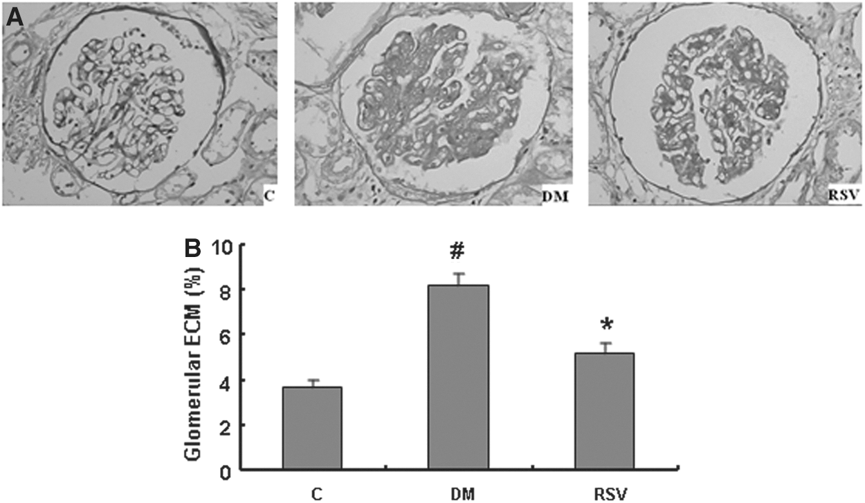
Effect of resveratrol on histologic changes in kidneys.
GSTM expression in diabetic kidneys was down-regulated by resveratrol
GSTM expression was evaluated by western blot analysis. Expression of GSTM was increased in diabetic rats. Notably, resveratrol treatment decreased GSTM expression (Fig. 2).
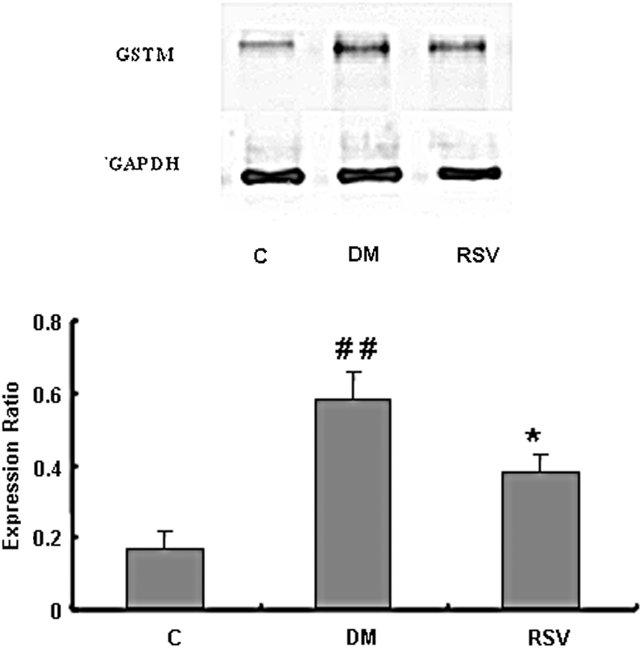
Glutathione S-transferases Mu (GSTM) expression in kidneys of diabetic rats. Data represent GSTM/GAPDH and are given as mean±SD of three experiments. ## P<.01, compared with C; *P<.05, compared with DM.
High glucose-induced RMC proliferation was inhibited by resveratrol
We examined whether resveratrol affects high glucose-induced proliferation of RMCs. The cells were treated with resveratrol (2.5, 5.0, and 10.0 μmol/L) for 48 h, and the viability was evaluated by MTT assay. High glucose stimulated the proliferation of RMCs. However, resveratrol at concentrations of up to 10 μmol/L inhibited the proliferation caused by high glucose in a dose-dependent manner (Fig. 3). The inhibition rate was 8.9% at 5 μmol/L and 16.1% at 10 μmol/L, respectively.
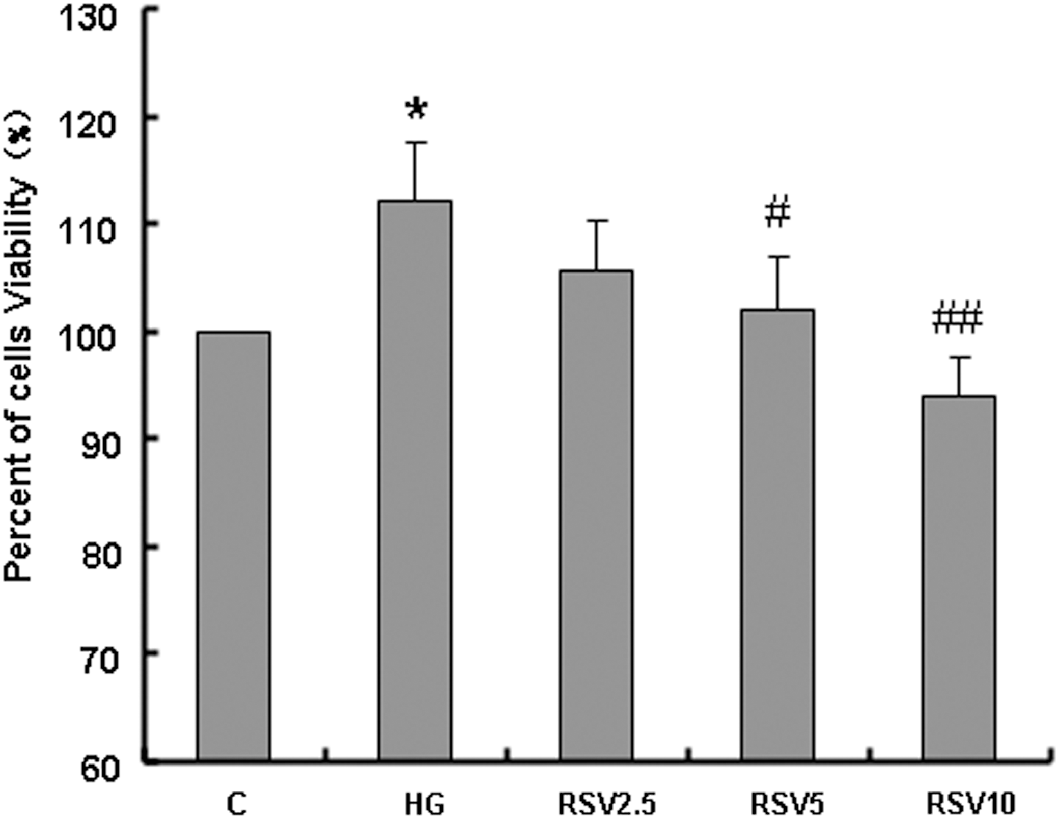
Effects of resveratrol (2.5, 5.0, and 10.0 μmol/L) on viability of rat mesangial cells (RMCs) as assessed by MTT. Results represent percentages of untreated cells (100%) and are given as mean±SD of three independent experiments. *P<.01, compared with C; # P<.05, ## P<.01, compared with HG. HG, high-glucose group.
Effect of resveratrol on apoptosis in high glucose-induced RMCs
The fluorophores annexin V and PI were used to analyze the effects of resveratrol on apoptosis in glucose-induced RMCs. Control, untreated glucose-induced cells showed rare presence of annexin V, indicating steady apoptosis in these cells. However, after resveratrol treatment, the apoptosis rate in RMCs slightly increased in a dose-dependent manner compared with that of glucose-induced RMCs. Significantly increased apoptosis was noticed at a dose of 5 μmol/L resveratrol, indicating the onset of apoptotic phenomena. Increasing concentrations of resveratrol (5 and 10 μmol/L) induced 2.2% and 4.53% apoptosis, respectively (Fig. 4).
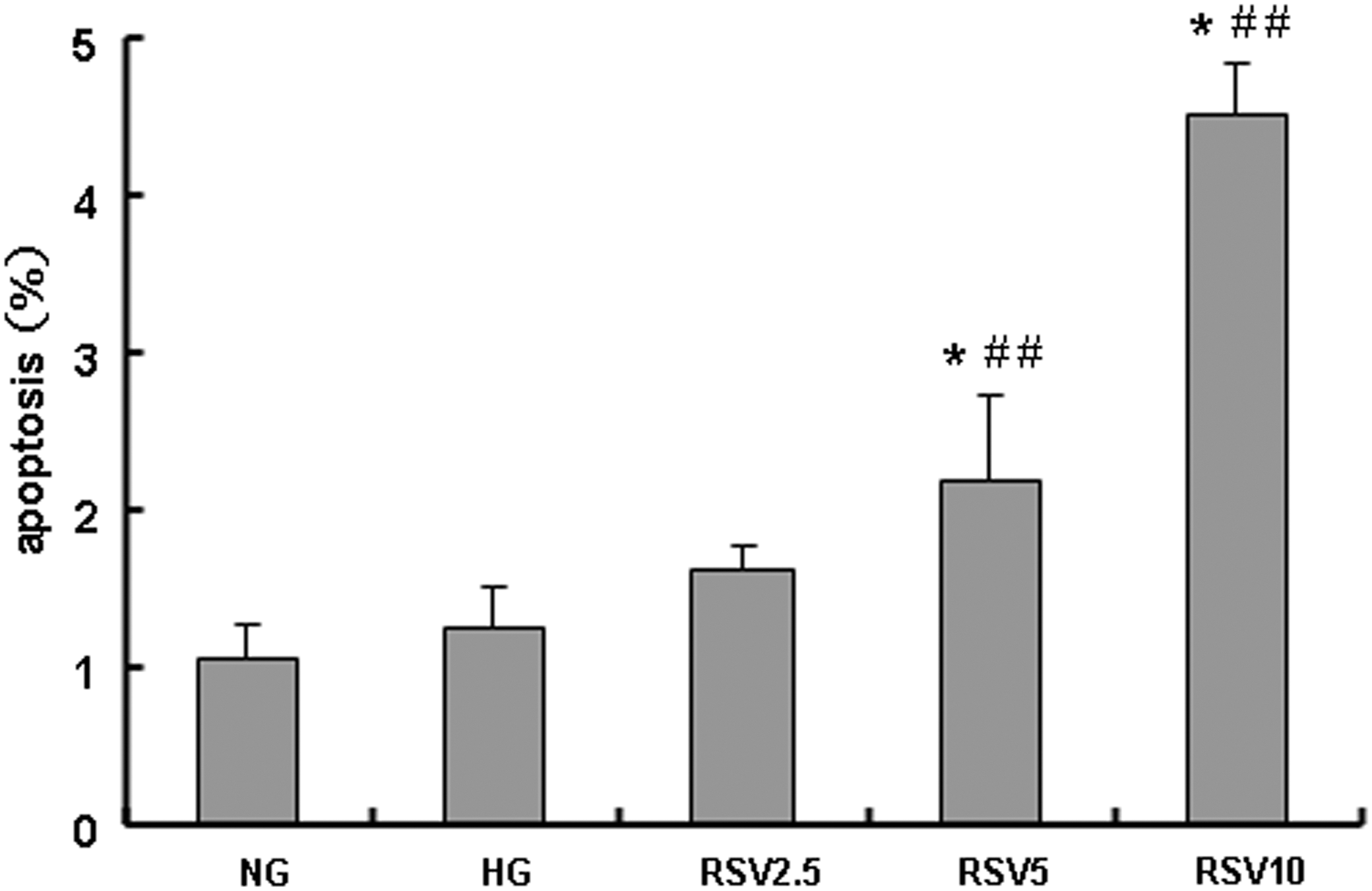
Effect of resveratrol (2.5, 5.0, and 10.0 μmol/L) on apoptosis of RMCs. Data are given as mean±SD of three experiments. *P<.01, compared with C; ## P<.01, compared with HG.
GSTM expression in high glucose-induced RMCs was down-regulated by resveratrol
In mesangial cells, high glucose significantly increased the expression of GSTM. A diminished GSTM expression was observed in cells treated with resveratrol for 48 h. Resveratrol dose-dependently inhibited GSTM expression. Resveratrol inhibited Nrf2 expression in a dose-dependent manner which was increased in the high-glucose conditions (Fig. 5). Nrf2 is one of the most important cellular defense mechanisms for coping with oxidative stress. It regulates intracellular antioxidants, phase II detoxifying enzymes, and the expression of many other proteins. Our findings indicate that resveratrol decreases GSTM expression induced by high glucose through the regulation of Nrf2 expression.
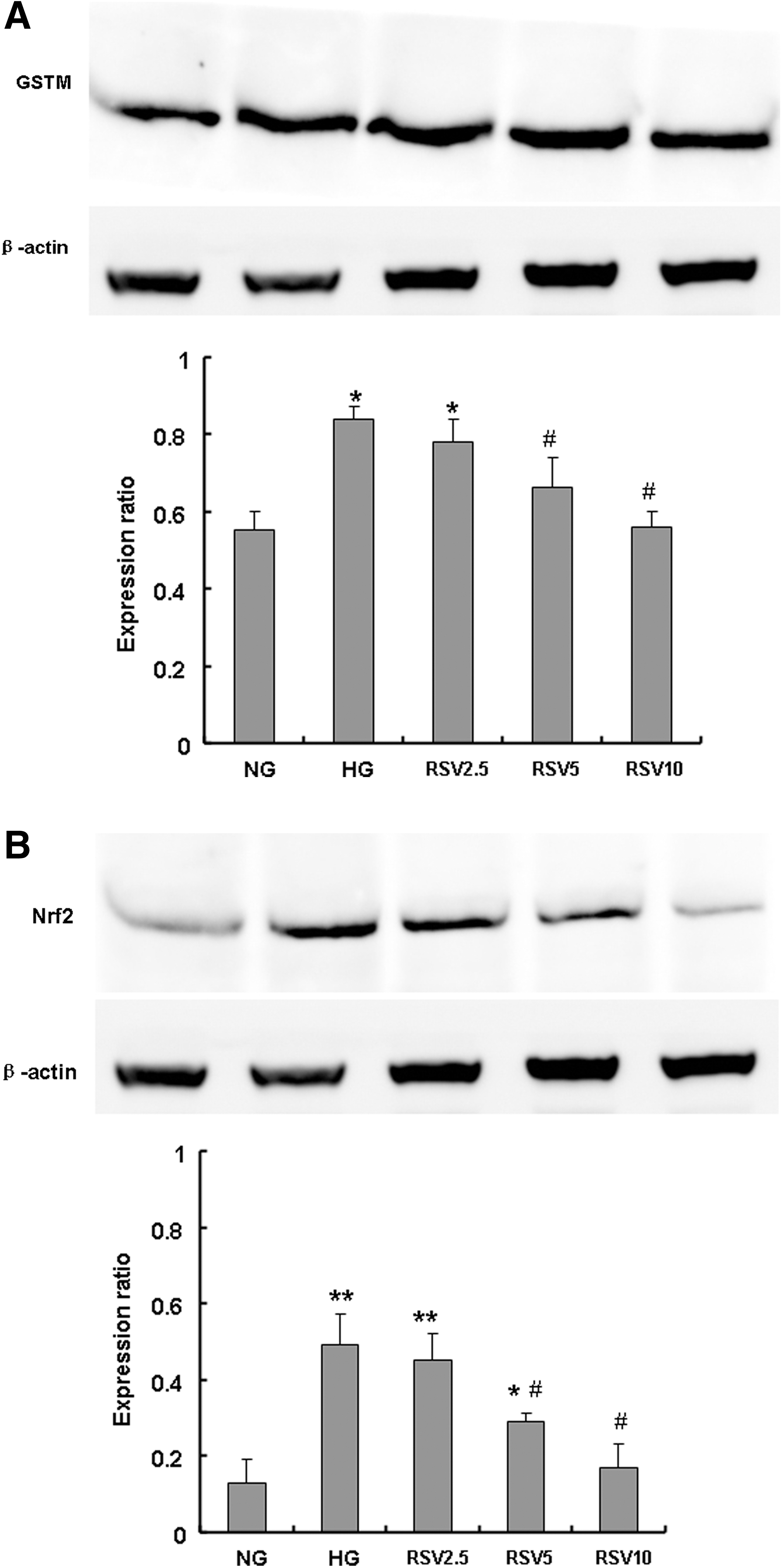
Effect of resveratrol (2.5, 5.0, and 10.0 μmol/L) on expressions of GSTM and nuclear factor erythroid 2-related factor 2 (Nrf2) in RMCs.
Discussion
DN is the leading cause of end-stage renal disease, and the STZ-induced diabetic rat model has been widely used to study early diabetic renal changes. 13 In our study, serum glucose concentrations in STZ-induced diabetic rats were significantly higher than those in the control group. The urinary protein excretion also showed a dramatic increase, and renal changes including glomerular hypertrophy and mesangial expansion were observed in diabetic rats. Thus, all these data were consistent with early-stage DN, suggesting the successful establishment of the STZ-induced DN model. In the present study, resveratrol-treated diabetic rats showed significant reductions in protein output levels compared with untreated diabetic rats. Consistent with previous studies, 13 –15 resveratrol treatment ameliorated glomerular hypertrophy and ECM accumulation in our study. All these results suggest that resveratrol has potential preventive effects on the progression of DN.
As shown in Table 1, serum glucose was only mildly reduced in treated diabetic rats. There might be other mechanisms involved in the protective role of resveratrol against the development of DN which were independent of its ability to decrease glucose level. In a recent study, resveratrol has been shown to attenuate early DN associated with the suppression of TGF-β/Smad and ERK1/2 signaling in STZ-induced diabetic rats. 13 Furthermore, resveratrol ameliorated glomerular hypertrophy and inhibited mesangial cell proliferation by activating AMPK. 14 –16 In our study, the results showed that the expression of GSTM in diabetic rats was significantly higher than in control rats. After resveratrol treatment, GSTM expression returned to normal levels, which was consistent with the result of our previous study. 17 Fujita et al. revealed that GSTM also showed increased expression in Akita mice compared to control mice. 18 The upregulation of GSTM in diabetic kidneys probably was in response to oxidative stress triggered by hyperglycemia or other toxic effects of glucose. However, it has been reported that the presence of the GSTM1 gene was associated with a susceptibility to type 1 diabetes mellitus. 19 These data suggest that the upregulation of GSTM might be involved in the development of DN. Moreover, downregulated GSTM expression might be one of the renoprotective mechanisms of resveratrol.
In addition, we investigated the mechanisms underlying such change through high glucose-induced RMCs. Glomerulosclerosis in DN is caused by an accumulation of ECM proteins in the mesangial interstitial space. 20 One of the important causes of ECM protein accumulation is the increased synthesis by mesangial cells. 21 Several studies have revealed significant mesangial cell proliferation in the early stages of DN, 22,23 which precedes the increases in the ECM proteins and glomerular sclerosis. In the present study, the proliferation of RMCs was increased by a high-glucose stimulus. Moreover, we investigated the effect of different concentrations of resveratrol on high glucose-induced mesangial cells. Our data suggest that resveratrol inhibits high glucose-induced mesangial cell proliferation in a dose-dependent manner. Furthermore, we investigated the effect of resveratrol on GSTM and Nrf2 expressions in mesangial cells. The results show that the expressions of GSTM as well as Nrf2 are increased in high glucose-induced RMCs, and resveratrol inhibits the expressions of GSTM and Nrf2. Apoptosis signal-regulating kinase 1 (ASK1), a mitogen-activated protein kinase, 24 is an important kinase in the intracellular signal transduction system leading to cell proliferation and ECM protein synthesis. It has been demonstrated that GSTM directly interacts with the N-terminal portion of ASK1 both in vivo and in vitro. This interaction caused a suppression of ASK1 activity as well as ASK1-dependent apoptotic cell death. 24,25 However, upregulation of GSTM expression in mesangial cells results in a suppression of ASK1-dependent apoptosis, which in turn leads to further mesangial cell proliferation. In our study, resveratrol downregulated the expression of GSTM; thus, the proliferation of RMCs is inhibited by resveratrol.
In conclusion, we investigated the effects of resveratrol on early-stage renal injury in diabetic rats and proliferation of high glucose-induced RMCs. As a result, we demonstrated that resveratrol reduces plasma glucose, creatinine, and urinary protein excretion, and attenuates renal hypertrophy in early-stage diabetic rats. These data suggest that resveratrol may prevent the progression of DN. The renoprotection of resveratrol is in part mediated through the inhibition of high glucose-induced RMC proliferation and downregulating GSTM expression. Therefore, this study suggests that resveratrol may have therapeutic potential in preventing and treating early-stage renal complications in diabetic patients.
Footnotes
Acknowledgments
This work was supported by grants from the National Natural Science Foundation of China (30873145), Shandong Provincial Natural Science Foundation, (Y2007C081) and Shandong University Foundation (2012ST204).
Author Disclosure Statement
No competing financial interests exist.