
Abstract
Mucuna pruriens, an underutilized native legume of South India has been reported to have high levels of L-Dopa, and used in the treatment of Parkinson's disease. Cellular damage arising from reactive oxygen and nitrogen species is said to cause neurodegenerative disorders. Antioxidants could assuage this oxidative damage of tissue directly and/or indirectly by enhancing natural defenses and also scavenging the free radicals. In this context, the antioxidative potential of different germplasm of Mucuna species was analyzed. Assays were performed to evaluate the enzymatic and nonenzymatic antioxidants in the extracts. Methanolic extracts of Mucuna (black germplasm) yielded high levels dietary antioxidants viz., flavonoids, alkaloids, saponins, steroids and phlobotannins qualitatively. Tannins, total phenols, flavanoids, and steroids accounted for 13.60±1.8 tannic acid equivalents, 58.47±3.19 gallic acid equivalents, 23.7±3.12 quercetin equivalents, and 20.3±1.01 mg per 100 mg β-sitosterol equivalents, respectively. Percentage of scavenging activity against hydroxyl, superoxide anion, nitric oxide, and hydrogen peroxide radicals were 39.12%; 57.1%; 41.26%, and 25.68%, respectively. Reducing capacity (17.74%) was seen to concurrently increase with extract concentration. Catalase, glutathione reductase, and polyphenol oxidase activities were found to be 30.15; 26.6 and 42.5 μmol/mg of protein, respectively. The methanolic extract yielded the most potent levels of dietary antioxidants and exhibited high free-radical–scavenging activity.
Introduction
T
Materials and Methods
Collection of germplasm seed samples
Different germplasm seed samples of Mucuna were collected from the Eastern and Western Ghats of South India.
Preparation of extract
Two hundred grams of each seed powder was extracted in sterile distilled water (5:l) with shaking (Stuart Scientific Orbital Shaker, S01, Essex, United Kingdom) for 48 h. The extract was filtered using a Buchner funnel and Whatman No.1 filter paper. The filtrate was quickly frozen at −40°C and dried for 48 h using a freeze dryer to give a yield of 15% dry extract. The resulting extract was reconstituted with sterile distilled water to the desired concentration and used throughout study.
Phytochemical screening of the seed extract
Solvent and aqueous extraction of phytochemical compounds
Phytochemical compounds were extracted using the following solvents: acetone, chloroform, methanol, n-hexane, and water. Soxhlet and flask extraction procedures were adopted for extraction. Ten grams of each powdered seed sample was packed in muslin cloth and used for extraction at a temperature lower than the boiling temperature of each solvent. Portions of the powdered samples were soaked in a conical flask containing individual solvents, wrapped with aluminium foil and placed in a shaker for 48 h at 120–130 rpm (∼0.805–0.945 g). All the extracts were filtered and pooled together and used for further studies.
Qualitative screening
10% of the dried extract was used for qualitative screening of phytochemical compounds (tannins, flavonoids, alkaloids, saponins, and steroids) in accordance with the standard methods. 15,16 Exactly 1.0 g of each seed extract was dissolved in10 mL of distilled water and filtered using Whatman No. 1 filter paper. A blue coloration, resulting from the addition of ferric chloride reagent to the filtrate, indicated the presence of tannins in the extract. Exactly 0.5 g of the plant extract was dissolved in 5 mL of 1% HCl on a steam bath. One milliliter of the filtrate was treated with a few drops of Dragendorff's reagent. Turbidity or precipitation was taken as indicative for the presence of alkaloid. About 0.2 g of the extract was dissolved in 2 mL of methanol and heated. A chip of magnesium metal was added to the mixture, followed by the addition of a few drops of concentrated HCl. The occurrence of a red or orange coloration was indicative of flavonoids. A freshly prepared 7% blood agar plate was prepared, wells were cut into it, and the crude extract dissolved in 10% methanol was used to fill the wells. Sodium nitroprusside was used as a positive control, while 10% methanol was used as a negative control. The plates were incubated at 35°C for 6 h and complete hemolysis of the blood around the extract was indicative of saponin. About 0.5 g of the extract was dissolved in 3 mL of chloroform and filtered. Acetic anhydride and concentrated H2SO4 were carefully added to the filtrate to form a lower layer. A reddish brown color at the interface was taken as positive for steroid ring.
Assay on nonenzymatic antioxidant activity
Total phenolic assay
Total phenolics were measured following the protocol of Shetty et al. 17 Phenolics were measured as gallic acid equivalents (GAE). We transferred 1 mL of seed extracts to a test tube along with 1 mL of 95% ethanol, 5 mL of distilled water, and 0.5 mL of Folin–Ciocalteu phenol reagent (Sigma Chemical Co., St. Louis, MO, USA). After incubation of ∼5 min, 1 mL of 5% Na2CO3 was added, mixed well, and the solution was kept in the dark for 1 h, after which the samples were vortexed and absorbance was measured at 725 nm.
Determination of total flavonoids
The total flavonoid content was determined using the Dowd method. 18 We mixed 5 mL of 2% aluminium trichloride (AlCl3) in methanol with the same volume of the extract solution (10 mg/mL). Absorption readings at 415 nm using a Perkin Elmer UV-VIS spectrophotometer were taken after 10 min against a blank consisting of extract solution with 5.0 mL methanol without AlCl3. Total flavonoid content was expressed as mg of quercetin equivalents (QE)/100 mg of sample.
Estimation of tannins
Colorimetric estimation of tannins was performed based on the measurement of blue color formed by the reduction of phosphotungstomolybdic acid by tannin like compounds in alkaline solution. 19 A known amount of extract was combined with 5.0 mL of Folin–Denis reagent and Na2CO3 solution and made up to 100 mL, mixed well, and absorbance was read at 760 nm after 30 min using a spectrophotometer. Total tannin content expressed as mg tannic acid equivalents (TAE)/100 mg of sample.
Determination of reducing power
The reducing power of the extract was evaluated according to the method described by Ranganna. 20 The mixture containing 2.5 mL of 0.2 M phosphate buffer (pH 6.6) and 2.5 mL of K3Fe(CN)6 (10 mg/mL) was added to 1.0 mL of the extract dissolved in distilled water. The resulting mixture was incubated at 50°C for 20 min, followed by the addition of 2.5 mL of TCA (100 mg/mL). The mixture was then centrifuged at 3000 rpm for 10 min to collect the upper layer of the solution (2.5 mL), further mixed with distilled water (2.5 mL) and 0.5 mL of FeCl3 (1 mg/mL). The absorbance was then measured at 700 nm against a blank.
DPPH free-radical–scavenging activity
The antioxidant activity of the samples was determined by the 1,1-diphenyl-2-picrylhydrazyl (DPPH) inhibition method.
21
To 3 mL of 60-mM DPPH, 100 mL of seed extracts were added, mixed well, and incubated at room temperature for 15 min. The absorbance was monitored at 517 nm. Antioxidant activity was reported as percentage inhibition:
Hydroxyl radical–scavenging assay
The hydroxyl radical–scavenging assay was performed by standard procedures.
22
Hydroxyl radical was generated by the Fenton reaction using a Fe3+-ascorbate-EDTA-H2O2 system (0.6 mL of 4 mM H2O2 and phosphate buffer pH 7.40). The assay quantifies the 2-deoxyribose degradation product by its condensation with TBA. All tests were carried out six times. Mannitol, a classical OH scavenger, was used as a standard compound. Percent inhibition was evaluated by the following equation:
where A0 was the absorbance of the control and A1 was the absorbance in the presence of the samples and standard.
Nitric oxide–scavenging activity
Determination of nitric oxide radical–scavenging activity of seed extract was performed as previously described.
23
Sodium nitroprusside in aqueous solution at physiological pH spontaneously generated nitric oxide which interacts with oxygen to produce nitrite ions, which was determined by the use of Griess reagents. 2 mL of 10 mM sodium nitroprusside dissolved in 0.5 mL phosphate buffer saline (pH 7.4) was mixed with 0.5 mL of seed extract at various concentrations. The mixture was incubated at 25°C. After 150 min, 0.5 mL of incubation solution was withdrawn and mixed with 0.5 mL of Griess reagent (1.0 mL sulfanilic acid reagent [0.33% in 20% glacial acetic acid at room temperature for 5 min with 1 mL of naphthylethylenediamine dichloride (1 mg/mL)]). The mixture was incubated at room temperature for 30 min. The absorbance was measured at 540 nm. The amount of nitric oxide radical was calculated following this equation:
where A0 is the absorbance before reaction and A1 is the absorbance after reaction has taken place.
Scavenging activity of superoxide anion
Superoxide anion–scavenging activity was determined by the method of Ruch et al. 24 The reaction mixture consists of seed extract (1 mg/mL), 1 mL of PMS (60 μM) prepared in phosphate buffer (0.1 M pH 7.4), and 1 mL of NADH (phosphate buffer), which was incubated at 25°C for 5 min. The absorbance was read at 560 nm against blank.
Assay of enzymatic antioxidants
Catalase (CAT) activity was measured by observation of the decrease in H2O2 concentration spectrophotometrically over time at 240 nm according to a previously described method. 25 Reduction in the glutathione level was measured spectrophotometrically at 412 nm by the method of Ellman. 26 The assay followed was a modified version developed was adopted for evaluating the peroxidase activity. 27 Superoxide dismutase (SOD) was assayed by measuring the inhibition of the formation of blue colored formazan at 560 nm according to the techniques of Arvouet-Grand et al. 28
Statistical analysis
All data were reported as the mean±standard deviation of three measurements. The statistical analysis was performed by Spearman correlation (Spearman Rank Correlation [v1.0.1] in Statistics Software [v1.1.23-r]).
Results
Phytochemical screening
Results on the phytochemical screening conducted on Mucuna germplasms were depicted in Table 1. The presence of tannins, phenols, alkaloids, steroids, triterpenoids, glycosides, saponins, anthroquinones, and phlobotannins was demonstrated. Quantitative analysis of phytochemical compounds of black-colored seed extract revealed the presence of higher levels of tannins, total phenols, and flavanoids than found in the white-colored seeds. The total phenol content of the methanolic extract of black beans was 58.47±3.19 mg/100 mg GAE, tannin contents was found to be 13.60±1.8 mg/100 mg TAE; flavanoid content was found to be 26.4±0.09 mg/100 mg QE, respectively (Table 2). The presence of phytochemical constituents of the Mucuna seed extracts may be related to their medicinal value and in turn are responsible for their dietary antioxidant defense mechanisms.
+, presence; −, absence; BS, black-colored seed; WS, white-colored seed.
Values are means of triplicate determinations. Data are presented as the mean±standard deviation.
GAE, gallic acid equivalent; TAE, tannic acid equivalent; QE, quercetin equivalent.
Free-radical–scavenging activity
Scavenging activities of methanolic extracts of the germplasms were investigated against both free radicals, such as DPPH, NO2, SOD, and hydroxy radicals. The inhibitory effect on individual radicals was given in Table 3. The black-colored germplasm registered higher levels of scavenging activity against the studied free radicals. The percentage of scavenging activity for black seeds were found to be 39.12% for DPPH, 57.14% for hydroxyl radical, 41.26±0.91 for NO2, and 25.68±0.48 for SOD, with reference to butylated hydroxy toluene (BHT).
Values are means of triplicate determinations. Data are presented as the mean±standard deviation.
Butylhydroxytoluence (BHT), chemically a derivative of phenol, is a lipophilic organic compound useful in assays for its antioxidant properties.
DPPH, 1, 1-diphenyl-2-picrylhydrazyl.
Correlation between phytochemicals and antioxidant activity
Scatter plot correlations between phytochemicals and antioxidant activities were carried out. The total phenol, tannins, and flavonoid content of the seed extract of both black- and white-colored germplasms significantly correlated with antioxidant activity (respectively: y=0.324x+18.41, R2=0.879; y=4.748x−24.87, R2=0.952; and y=1.850x−10.04, R2=0.990; Figs 1 –3). Specifically, tannins and flavonoids were highly correlated with radical-scavenging activity.
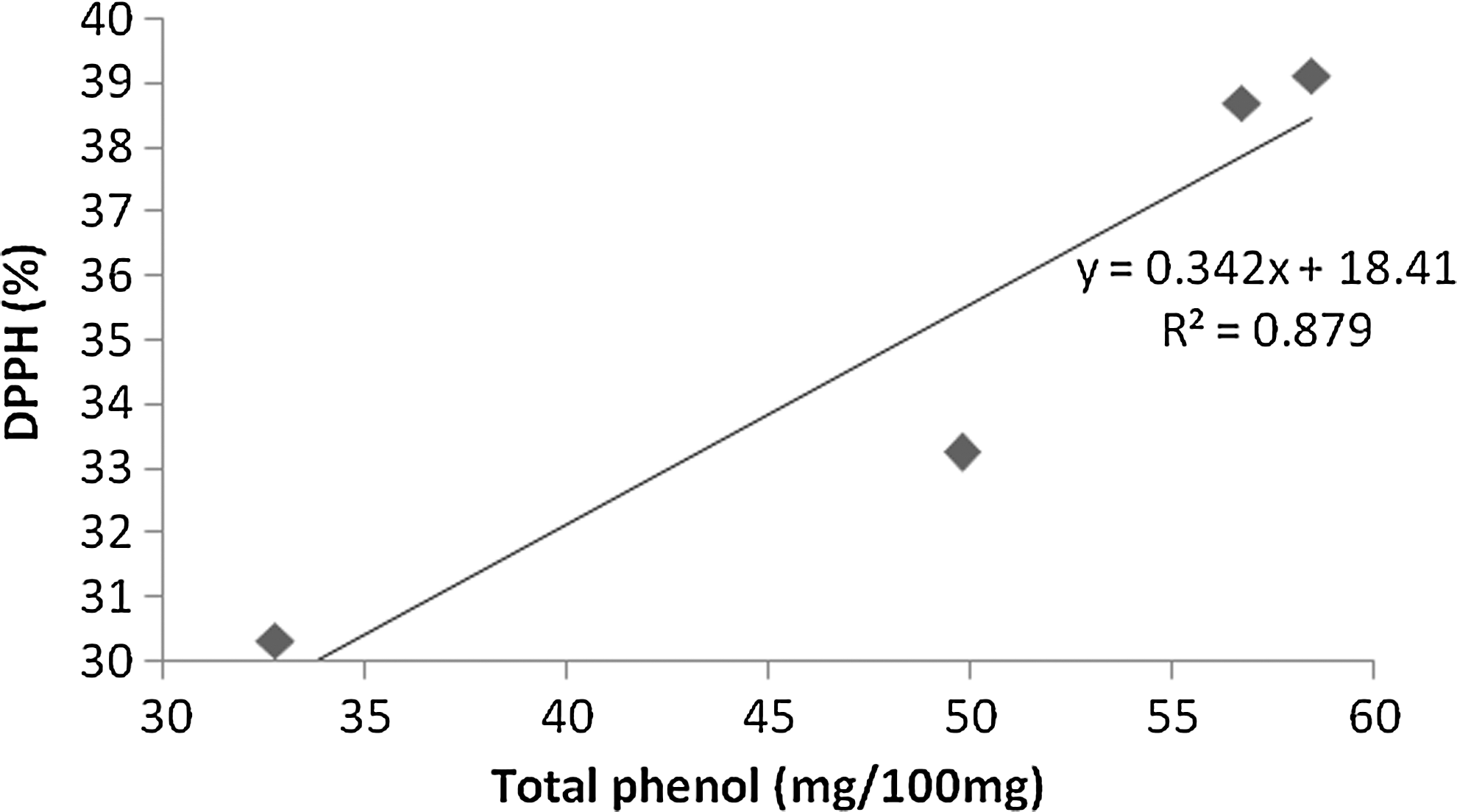
Scatter plot showing the correlation between DPPH scavenging activity (%) and total phenols (mg/100 mg) in black- and white- colored germplasms. DPPH, 1, 1-diphenyl-2-picrylhydrazyl.
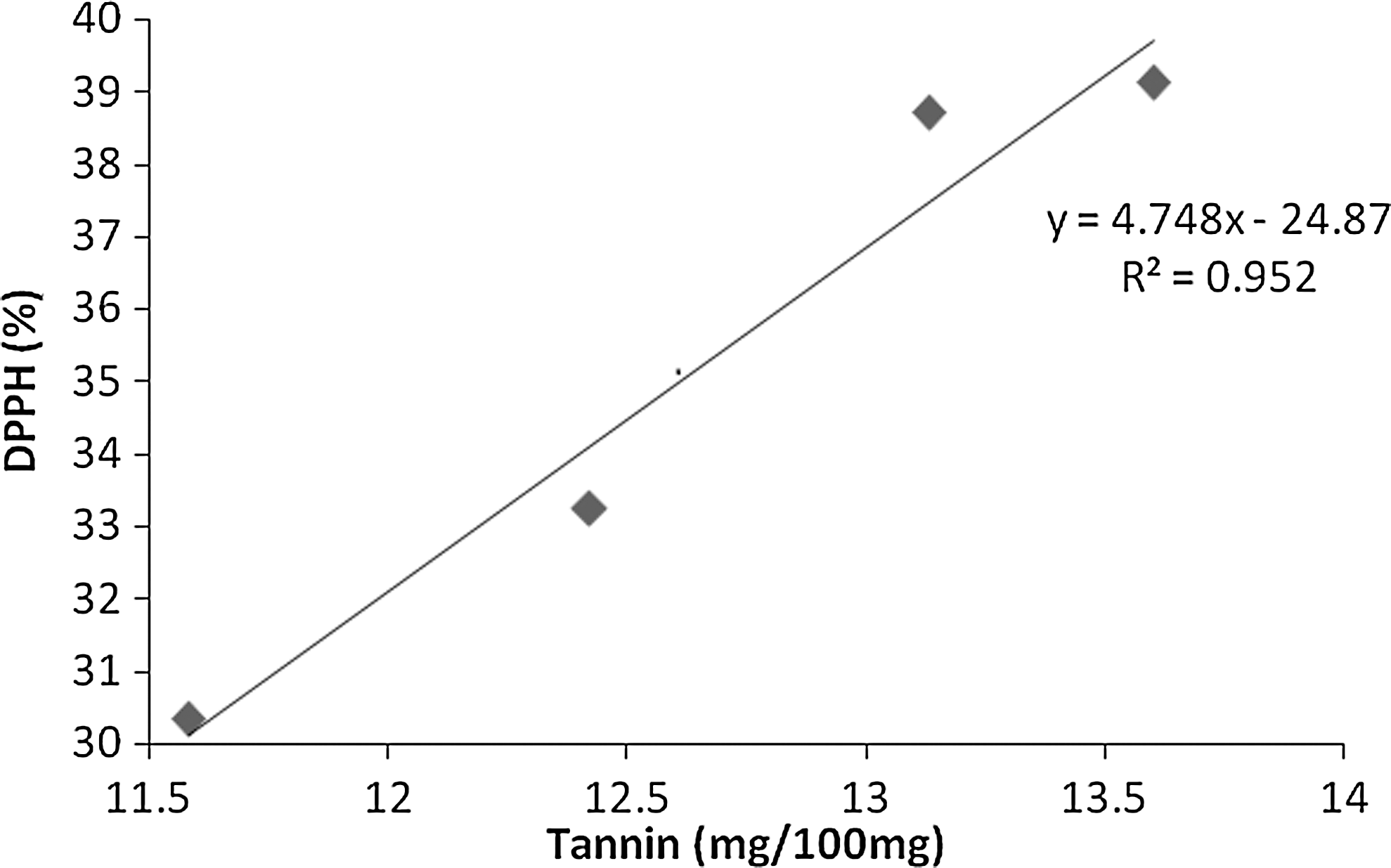
Scatter plot showing the correlation between DPPH scavenging activity (%) and tannin (mg/100 mg) in black- and white- colored germplasms.
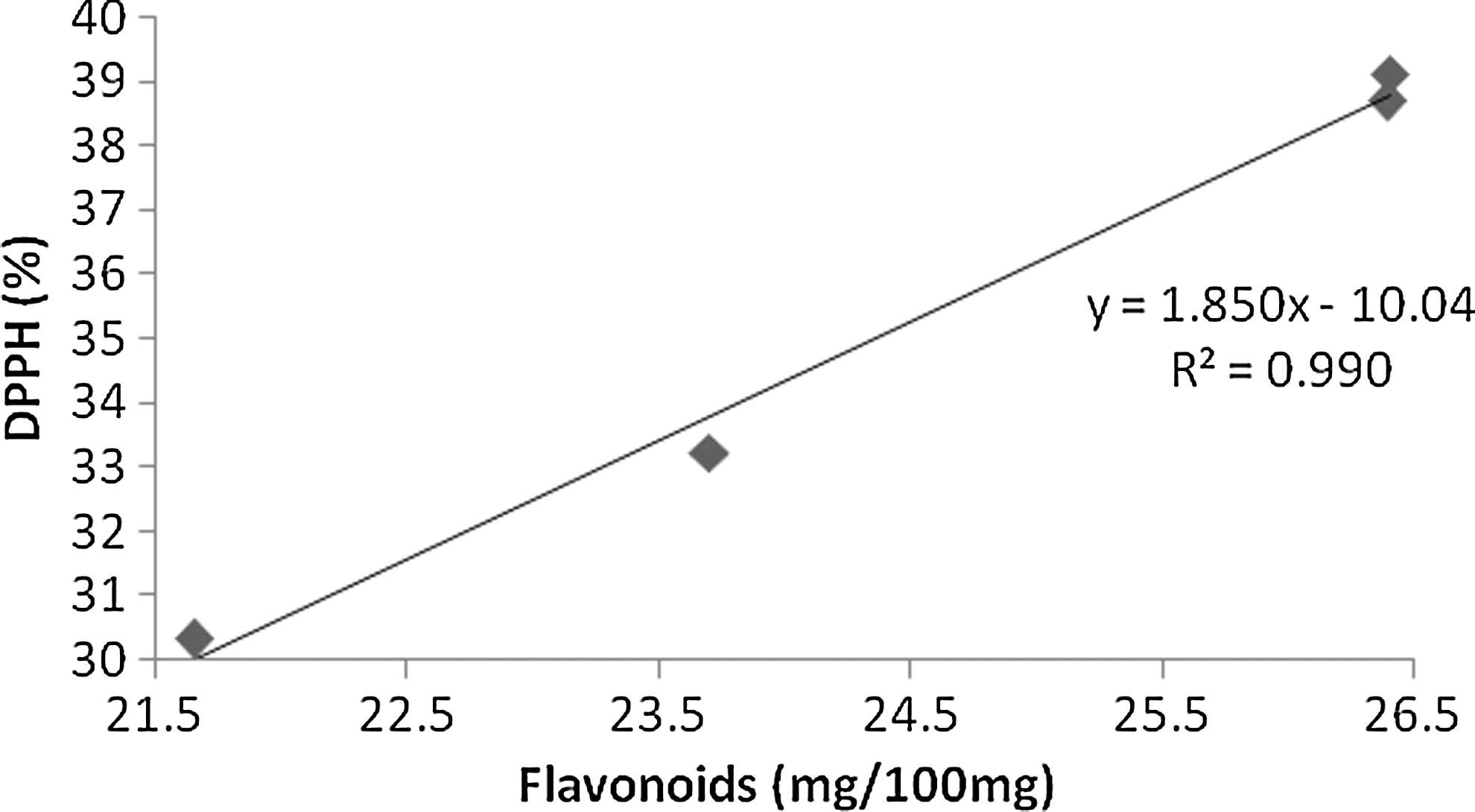
Scatter plot showing the correlation between DPPH scavenging activity (%) and flavonoids (mg/100 mg) in black- and white- colored germplasms.
Reducing capacity
The reducing capacity of an antioxidant is a measure of the sample to reduce Fe3+ to Fe2+ by donating an electron. The amount Fe2+ complex can then be monitored by measuring the formation of Perl's blue at 700 nm. The reducing capacity of the methanolic seed extract was evaluated in comparison with a standard BHT (Fig. 4). The pattern of reducing capacity of methanolic extracts of both germplasms followed a similar trend as the standard used in the present study. The reducing capacity was concomitantly increased with increasing concentrations of methanolic extract.
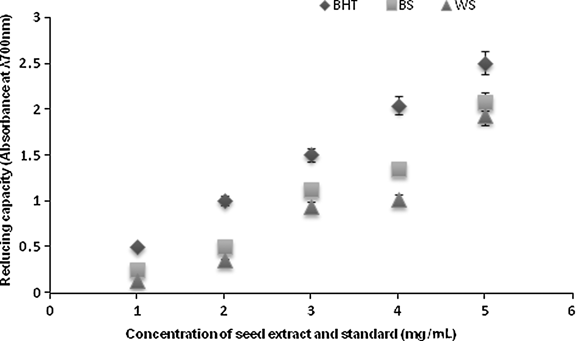
Reducing capacity of methanolic extracts of Mucuna in comparison with a standard (BHT) at λ 700 nm. BHT, butylated hydroxy toluene; BS, black-colored seed extract; WS, white-colored seed extract.
Antioxidant enzyme activity
Assays of the effects methanolic seed extracts on antioxidant enzymes CAT, glutathione reductase (GR), glutathione peroxidase (GPx), and SOD are depicted in Figure 5. In comparison with control (BHT), the activity of CAT and SOD were significant and reached a level of 34 and 33 units/min/mg of protein/μmoles, respectively, while GR and GPX exhibited somewhat lesser values. In general, between the two germplasms, blackcolored beans showed significantly greater antioxidant enzyme activities.
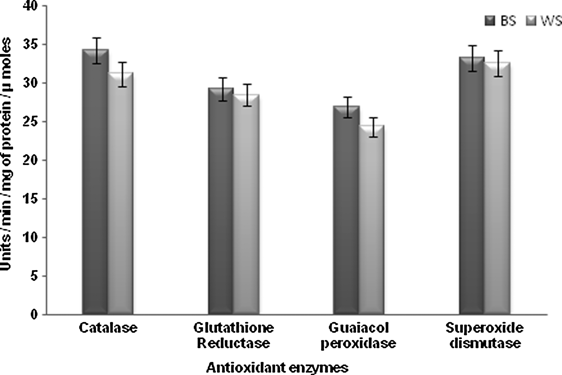
Enzymatic antioxidant activities of two different germplasms of Mucuna (black- and white-colored beans). CAT, catalase: 1 unit=amount of enzyme required to decrease the absorbance at 240 nm; GR, glutathione reductase: 1 unit=nanomoles of CDNB conjugated/min; GPx, glutathione peroxidase: 1 unit=changes in absorbance at 430 nm/min; SOD, superoxide dismutase: 1 unit=amount of enzyme (mg of protein) that is the amount that causes 50% reduction in NBT oxidation.
Discussion
Phytochemical compounds are known to be biologically active and exert bioactivity through different mechanisms. The phytochemical analysis conducted on Mucuna seed extracts revealed the presence of significant levels of tannins, phenol, glycosides, flavonoids, steroids, phlobotannins, anthroquinones, triterpenoids, and saponins. Phenolic compounds are secondary metabolites of plants and can act as antioxidants by many potential pathways, such as free-radical–scavenging, oxygen-radical absorbance, and chelation of metal ions. 29 Phytochemicals, especially plant phenolics, constitute a major group of compounds that act as primary antioxidants. 30 They have high redox potentials, allowing them to act as reducing agents, hydrogen donors, and singlet-oxygen quenchers. 31 Higher concentrations of phenolics are also present in the methanolic extract when compared to the other extracts. The preliminary phytochemical studies revealed the pronounced importance of the varied composition of secondary metabolites, for example in crude drugs. 32
The antioxidant activity of the Mucuna seed extract is dependent on the concentration of the extract. In this trial, the antioxidant assays selected are established methods with a history of evaluating the antioxidant potential of natural products. 33 Antioxidant compounds may function as free-radical scavengers, initiators of the complexes of pro-oxidant metals, reducing agents, and quenchers of singlet-oxygen formation. 34 Free radicals have been implicated in many disease conditions, especially superoxide radicals and hydroxy radicals. Herbal drugs containing free-radical scavengers are gaining importance in treating such diseases. 35,36 Earlier studies on Brazilian species of Mucuna demonstrated efficient antioxidative properties due to their phytoconstituents, including phenolics. 37 The reducing capacity of a compound acts as a significant indicator of its potential antioxidant activity. Antioxidant activity has been attributed to various mechanisms, including prevention of chain initiation, decomposition of peroxides, reducing capacity, and radical-scavenging activity. 38 Similar studies were conducted on Cyperus rotundus, suggesting that the reducing properties are generally associated with the presence of reductones, which have been shown to exert antioxidant action by breaking the free-radical chain through donation of a hydrogen atom. 35
The seed extracts of Mucuna were found to increase the activity of all of the antioxidant enzymes assayed, including CAT, GR, GPx, and SOD. These enzymes are known to constitute an essential defense system against oxygen toxicity. SOD catalyzes the breakdown of endogenous cytotoxic superoxide radicals to H2O2, which is further degraded by CAT. The marked decrease in GR and GPx activities seems to eliminate the ROS resulting from induced oxidative stress.
In conclusion, this study revealed in vitro antioxidant potential of Mucuna seed extract, with results comparable to those of the standard compounds, such as ascorbic acid and BHT. Black-colored germplasms registered higher levels of antioxidants. Methanolic extract yielded significant amounts of dietary antioxidants and exhibited high free-radical–scavenging activity. Studies of the neuroprotective effects of these germplasms are ongoing to validate their potential utilization and commercialization.
Footnotes
Acknowledgments
The authors express sincere gratitude toward Dr. C. Muthuamizhchelvan, Director (E&T), and Prof. M. Vairamani, Dean, of the School of Bioengineering, SRM University, for their support and continued encouragement. S.U. thanks Mr. R. Dinesh Kumar (Phathyusha Institute of Technology and Management, Thiruvallur, India) for his technical help in experiments.
Author Disclosure Statement
No competing financial interests exist.