
Abstract
The present study investigated the effects of sulfated polysaccharides from brown seaweed Sargassum wightii (Sw-SP) and seagrass Halophila ovalis (Ho-SP) in nociceptive and inflammatory models. In the formalin test, Sw-SP and Ho-SP significantly reduced licking time in both phases of the test at a dose of 10 mg/kg. In the hot plate test, the antinociceptive effect was observed only in animals treated with 10 mg/kg of Sw-SP and 5, 10 mg/kg of Ho-SP, suggesting that the analgesic effect occurs through a central action mechanism at the higher dose. Sw-SP and Ho-SP (10 mg/kg) significantly inhibited paw edema induced by carrageenan, especially at 3 h after treatment and potentially decreased neutrophil migration by 53% and 52%, respectively. In Freund's adjuvant-induced arthritic rats, there was a significant increase in the rat paw volume and decrease in body weight, but in Sw-SP- and Ho-SP-treated groups (10 mg/kg), a significant reduction in paw volume and a normal gain in body weight were observed. The present results indicate that Sw-SP and Ho-SP possess antinociceptive and anti-inflammatory effects and have potential usefulness for development as therapeutic agents.
Introduction
I
The formalin test is the most predictive model of tonic pain and is useful for the screening of novel compounds, since it encompasses inflammatory, neurogenic, and central mechanisms of nociception. Injection of 1% formalin into the paw is a biphasic event. The first phase (neurogenic pain) is caused by the direct chemical stimulation of nociceptors. The second phase (inflammatory pain) is triggered by a combination of stimuli, including inflammation of the peripheral tissues and mechanism of central sensitization. The hot plate test is used to screen the effects of compounds on the threshold for pain sensitivity.
Carrageenan-induced inflammation, a classical model of edema formation and hyperalgesia, has been widely used in pharmacological field to develop nonsteroidal anti-inflammatory drugs and selective COX-2 inhibitors. The edema induced by carrageenan is biphasic. The first phase starts with the release of a diverse set of mediators, such as histamine and serotonin during the 1st hour and release of kinins up to 2.5 h. In the second phase, prostaglandin-like substances are released over 2–3 h, and is characterized by an intense neutrophil infiltrate. 2 Neutrophil recruitment in the inflamed tissue contributes to vascular inflammatory events such as the increase in vascular permeability with protein exudation and vice versa. 3,4 It has been reported that selectin-adhesion molecules are responsible for mediating leukocytes rolling along the endothelial cells of blood vessels at sites of inflammation, and the neutrophils participate in the cascade of events leading to mechanical hypernociception. 5 Rheumatoid arthritis (RA), a destructive inflammatory polyarticular joint disease, is characterized by infiltration of inflammatory cells in the synovium, and hyperplasia of the synovial lining cellular components, leads to the formation of a very aggressive tissue called pannus, which invades and destroys the adjacent cartilage and bone. 6 The suppression of inflammatory responses by nonsteroidal anti-inflammatory drug administration is an important tool and has the ability to inhibit initial or later manifestations.
Seaweed polysaccharides received extensive attention due to their intriguing potential as novel anti-inflammatory and analgesic drugs. 7,8 Brown seaweeds contain large amounts of cell wall polysaccharides, most of which are sulfated polysaccharide fucoidans. Sargassum, a well-known brown seaweed belonging to the order Fucales, and class Phaeophyceae, is common to all oceans except Antarctica. In India, it is represented by 38 species. 9 Generally, chemical characteristics such as the molecular size, type of sugar, sulfate content, type of linkage, and molecular geometry have a great influence on their biological activities. 10 The structural and functional properties of sulfated polysaccharides derived from different Sargassum species have been imperatively emphazised. 11,12 The sulfated polysaccharides from S. vulgare, 13 S. hemiphyllum, 14 and alginic acid from S. wightii 15 have been explored for possible anti-inflammatory effects. The solvent extracts of S. swartzii, 16 S. micracanthum, 1 S. wightii, 17 S. fulvellum, and S. thunbergii 18 are reported to have an anti-inflammatory and analgesic activity. Seagrasses are marine vascular plants, which grow in sea water and survive the tides unscathed. The presence of sulfated polysaccharides was determined in three species, of which, the Ruppia maritima sulfated polysaccharide was purified and its structure was characterized. 19 To date, the sulfated polysaccharide of Halodule wrightii has been characterized for its antioxidant and anticoagulant activities. 19 We reported the existence of the anti-inflammatory activity in seagrass Halophila ovalis methanol extract. 20 However, there is no single report about the biological activities of sulfated polysaccharides from seagrass H. ovalis until now. Therefore, the present study was undertaken to determine the anti-inflammatory and analgesic activity of sulfated polysaccharides from brown seaweed S. wightii and seagrass H. ovalis in experimental animal models.
Materials and Methods
Animals
Male Wistar rats (180±20 g) were maintained in the Central Animal Facility of Pondicherry University in polypropylene cages in temperature-controlled rooms with a 12-h light/12-h dark cycle and had free access to standard feed pellets and water ad libitum. The study was approved by the Institutional Animal Ethics Committee (PU/IAEC/12/05) in accordance with CPCSEA guidelines.
Plant material
Samples of S. wightii were collected from the Gulf of Mannar, Tamilnadu, India, in August 2010. The seagrass H. ovalis was collected at low tide from the Chunnambar estuary (Puducherry, India) during September 2011. The samples were rinsed with sea water, and then successively with tap water and distilled water to remove the epiphytes and other wastes and shade dried. Voucher specimens were preserved in a 5% formalin solution and deposited at the School of Ecology and Environmental Sciences, Pondicherry University. Prof. N. Parthasarathy, Salim Ali school of Ecology, Pondicherry University, identified and authenticated the plant specimens.
Sulfated polysaccharides
Polysaccharide was extracted according to the method of Foley et al. 21 with slight modifications in triplicate. Briefly, the milled algal biomass (100 g) was treated with EtOH (80% v/v) at room temperature for 12 h and subsequentially at 70°C for 12 h to extract mannitol and some salts (S1 and S2) to leave a depigmented algal powder (DAP, 85 g). Extraction of depigmented algal powder (10 g) with Milli–Q H2O (0.1 mg/mL) at room temperature for 7 h, at 70°C for 7 h, and at 70°C for 4 h with constant stirring three times (S3, S4, and S5). All three fractions were pooled and treated with 2 M CaCl2 at room temperature for 5 h to precipitate alginates. The precipitated alginates were then removed by centrifugation at 16,000 g in SIGMA 6K 15 refregirated centrifuge for 30 min followed by filtration through a glass filter (G-3), yielding the polysaccharide in the supernatant. The residue was briefly washed with additional Milli–Q H2O and the wash was collected to maximize the polysaccharide recovery. Dialysis was carried out (MWCO 1000 Da, Spectrum, USA) at 4°C extensively against deionized water over a 48-h period to decrease salinity, and Milli–Q H2O was changed every 12 h and lyophilized. The recovered material was redissolved in water, and then precipitated with ethanol (4 vols.) overnight. This process was repeated twice for complete recovery of polysaccharides. The final pellet was dissolved in Milli–Q H2O and lyophilized to yield the water-extracted polysaccharide (S. wightii [Sw-SP], 1.11±0.1 g).
The dried whole plant H. ovalis (20 g) was ground to fine powder, suspended in 400 mL 0.1 M sodium acetate (pH 6.0), containing 2 g papain, 5 mM ethylenediamine tetra-acetic acid, and 5 mM cysteine. After 24-h incubation at 60°C, the mixture was centrifuged at 10,000 g for 10 min at 4°C and filtered through a glass filter (G-3) and the supernatant was collected. The sulfated polysaccharides in the solution were precipitated with 800 mL absolute ethanol. After 24 h, the precipitate formed was collected by centrifugation (2560 g for 20 min at 4°C). The final precipitate was dried at 60°C for 12 h. Approximately 1.3 g (dry weight) of crude polysaccharide (H. ovalis [Ho-SP]) was obtained after these procedures.
Antinociceptive activity of sulfated polysaccharides
Formalin test
The formalin test, which causes local tissue injury to the paw, was used as a model for tonic pain and localized inflammatory pain. 22 Wistar rats were injected with Sw-SP and Ho-SP (2.5, 5, or 10 mg/kg, intravenously [i.v.]) or sterile saline (0.9%, w/v, NaCl). After 30 min of administration, 20 μL of 1% aqueous formalin was injected intraperitoneally (i.p.) into the right hind paw and the licking time was then recorded from 0 to 5 min (phase 1, corresponding to the direct stimulation of nociceptors) and 20 to 25 min after formalin injection (phase 2, inflammatory). Indomethacin (5 mg/kg, subcutaneously [s.c.]) was also administered 30 min before formalin injection and used as a reference compound.
Hot plate test
The hot plate test was performed to measure the analgesic activity as described previously. 23 Each rat was placed twice on the heated plate (51°C±1°C), with a 30-min intertrial interval. The first trial familiarized the animal with the test procedure, and the second served as the control reaction time (licking the paw or jumping). Animals showing a reaction time >10 sec were excluded in subsequent analyses. Immediately after the second trial (control reaction time), a group of animals (n=6) received sterile saline (0.9%, w/v, NaCl), sulfated polysaccharide of S. wightii Sw-SP and H. ovalis Ho-SP (2.5, 5 or 10 mg/kg, i.v.), indomethacin (5 mg/kg, s.c.). The reaction times were measured at time zero (0 min) and 30, 60, and 90 min after drug administration with a cutoff time of 40 sec to avoid paw lesions.
Anti-inflammatory activity of sulfated polysaccharides
Carrageenan-induced rat paw edema
The rats (n=6 per group) were pretreated with Sw-SP and Ho-SP at doses of 2.5, 5, or 10 mg/kg (0.1 mL/100 g body weight, s.c.) or sterile saline (0.9%, w/v, s.c.) 1 h before carrageenan injection. Carrageenan (500 μg/paw, 100 μL, i.p.) was injected into the right hind paw. In a control experiment, diclofenac (1 mg/kg, s.c.), a synthetic glucocorticoid with potent anti-inflammatory and immunosuppressant properties was administered 1 h before carrageenan. 24 The paw volume was measured immediately before the stimulus (time zero) and at selected time intervals (1, 2, 3, and 4 h) using a plethysmometer (Panlab, Spain). The results are expressed as the variation in paw volume (mL), calculated as the difference from the basal volume (time zero) and the volume at each time interval.
Induction of peritoneal inflammation (Peritonitis model) in rats
Five groups of rats (n=6 per group) received sterile saline (0.9%, w/v, s.c.) or dexamethasone (1 mg/kg, s.c.), Sw-SP (10 mg/kg, s.c.), and Ho-SP (10 mg/kg, s.c.) 1 h before carrageenan (500 μg/paw, 100 μL, i.p.). The animals were sacrificed after 4 h, and the peritoneal cavity was washed with 10 mL saline containing heparin (5 IU/mL). The peritoneal fluid was collected, and the total leukocyte count was determined using the Neubauer chamber. To differentiate the leukocyte count, samples were stained by the hematoxylin–eosin method, and the cells were counted by microscopy. The data are expressed as the means±SEM of the number of cells ×103 mL of the peritoneal fluid. 25
Freund's adjuvant induced arthritis
Male Wistar albino rats weighing 180±20 g were divided into five groups of six animals each. The standard drug diclofenac sodium, Sw-SP, and Ho-SP were dissolved in water and administered immediately. Group I served as control (without treatment), Group II served as arthritic control (negative control), Group III was treated with diclofenac sodium at 5 mg/kg (positive control), Group IV received Sw-SP (10 mg/kg), and group V received Ho-SP (10 mg/kg). After 30 min of drug treatment, arthritis was induced by injecting a 0.1 mL (0.1% w/v) of Complete Freund's Adjuvant into the left hind paw. 26 The drug treatment was continued from the initial day, that is, from the day of adjuvant injection (0 day), and continued till the 14th day. The paw volume of all the groups was measured on days 0, 3, 7, and 14 by using a plethysmometer. The mean changes in injected paw edema with respect to initial paw volume, were calculated on respective days and % inhibition of paw edema with respect to the untreated group was calculated.
Statistical analysis
All results are expressed as mean±standard error of the mean. Statistical differences between the experimental groups were determined by one-way ANOVA, and differences were considered to be statistically significant if P<.05. The Tukey's post hoc test was performed for multiple group comparison between groups. All statistical analysis was performed using SPSS Version 7.0.
Results
Formalin test
There was no significant reduction of licking time observed during the first phase following 1% formalin administration (neurogenic) with 2.5 or 5 mg/kg of Sw-SP and Ho-SP, respectively. Whereas there was a considerable shorter licking time by 56.5% and 50.1% with 10 mg/kg of Sw-SP and Ho-SP (Figs. 1 and 2, panel A). In addition, the Sw-SP (2.5, 5, or 10 mg/kg; i.v.) injected 30 min before the formalin injection elicited a dose-dependent inhibition of the formalin response during the second phase (inflammatory) by 44.6%, 67.6%, and 78.1%, respectively (Fig. 1B). Similalry, Ho-SP caused does-dependent inhibition during the second phase (inflammatory) of 40.3%, 63.6%, and 75.3%, respectively (Fig. 2B). Indomethacin (5 mg/kg, s.c.) did not inhibit the first phase, but significantly decreased the licking time by 65.2% during the second phase compared to the control group.
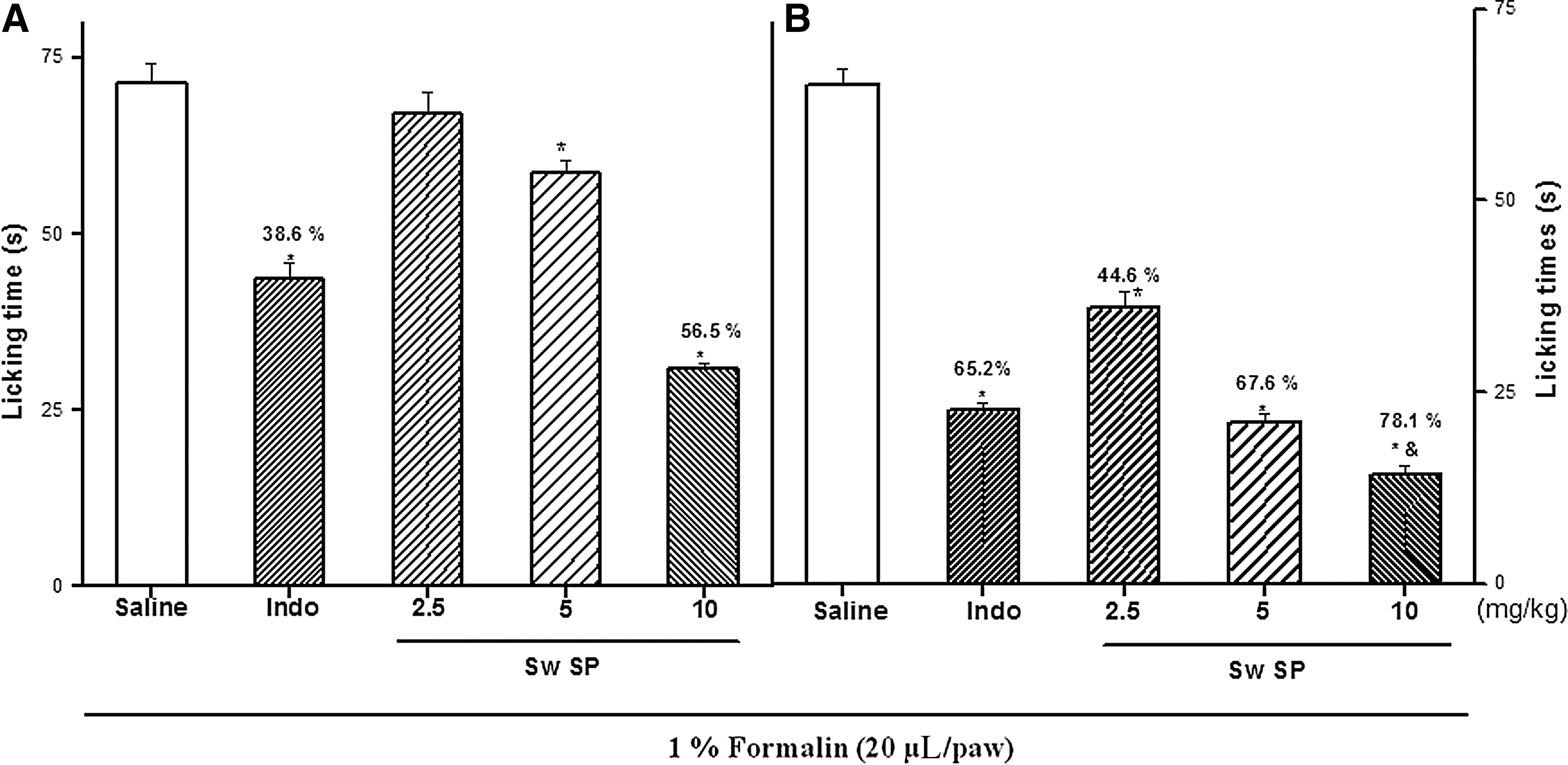
Effect of Sargassum wightii (Sw-SP) on the chemical stimuli (formalin test). Sw-SP (2.5, 5 or 10 mg/kg) or saline were given intravenously (i.v.) 30 min before the formalin and the licking time was determined during the first 5 min
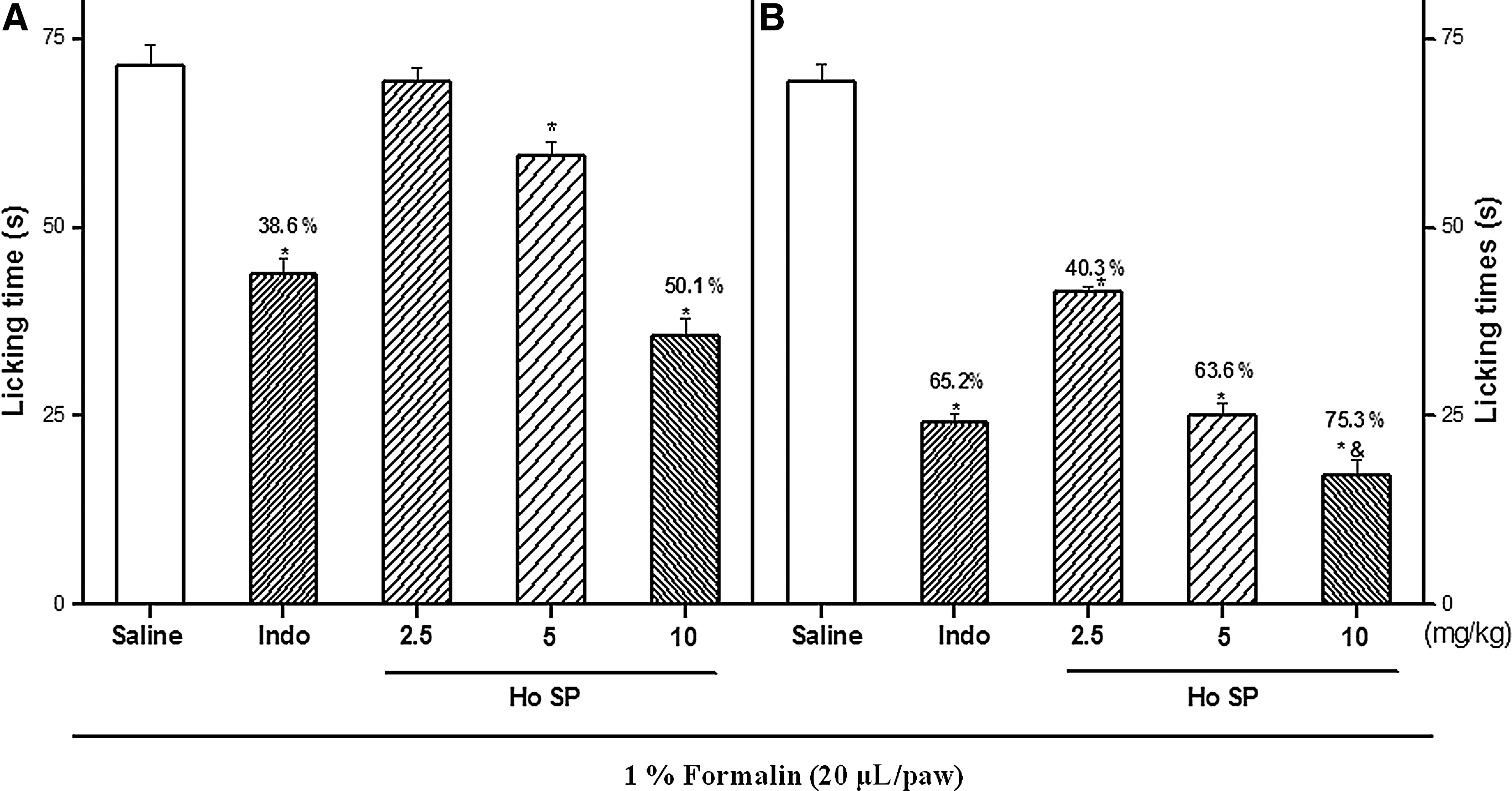
Effect of Halophila ovalis (Ho-SP) on the chemical stimuli (formalin test). Ho-SP (2.5, 5 or 10 mg/kg) or saline were given i.v. 30 min before the formalin and the licking time was determined during the first 5 min
Hot plate test
The treatment of rats with Sw-SP and Ho-SP (2.5, 5, or 10 mg/kg, i.v.) after the second hot plate trial produced significant antinociceptive effects, as measured by the reaction time during the 90-min observation (Fig. 3A, B). The systemic administration of Sw-SP and Ho-SP (10 mg/kg) significantly increased latency in the hot plate test at 30 (saline=5.60±0.13 s; Sw-SP=10.03±0.17 s; Ho-SP=9.99±0.19 s), 60 (saline=5.67±0.28 s; Sw-SP=15.42±0.38 s; Ho-SP=15.04±0.12 s), and 90 min (saline=5.70±0.38 s; Sw-SP=12.47±0.45 s; Ho-SP=12.69±0.30 s). At 10 mg/kg, the maximum latency to thermal stimuli was observed at 60 min after administration when compared to indomethacin-treated control. Sw-SP (5 mg/kg) increased hot plate latency at the 30-min interval compared to the indomethacin-treated group, but was ineffective at inducing antinociception at 60 and 90 min. However, Ho-SP (5 mg/kg) increased the latency period to thermal stimuli at all time intervals more than the indomethacin-treated group (Fig. 3B). The minimal doses of Sw-SP and Ho-SP (2.5 mg/kg) were ineffective at inducing antinociception at all time intervals.
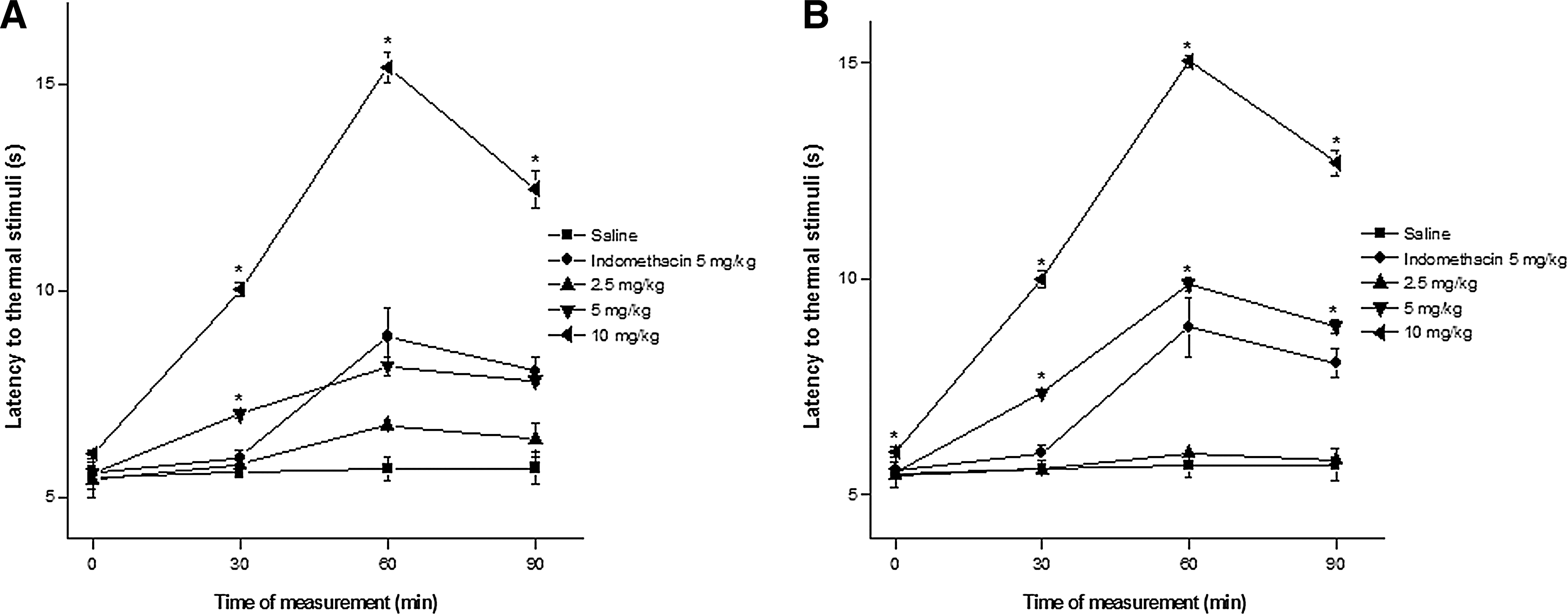
Effect of S. wightii and H. ovalis sulfated polysaccharides, indomethacin on the latency period to thermal stimuli (hot plate) induced in male Wistar rats. Animals received indomethacin (5 mg/kg, subcutaneously [s.c.]). Saline, Sw-SP, and Ho-SP (2.5, 5, or 10 mg/kg) were injected i.v. Data are expressed as mean±SEM of six rats per group. *P<.05 indicates significant difference from the saline group (ANOVA, Tukey's test).
Carrageenan-induced paw edema
The acute anti-inflammatory activity of Sw-SP and Ho-SP (2.5, 5 or 10 mg/kg) was evaluated by carrageenan-induced paw edema in rats (Fig. 4). Carrageenan (500 μg, i.p.) caused intense paw edema and reached a maximum level (0.59±0.03 mL) at 3 h after administration, and then decreased over the subsequent hour. Sw-SP (10 mg/kg) showed significantly inhibited carrageenan-induced acute paw edema after 1-h administration at all time intervals, first (0.13±0.01 mL), second (0.18±0.02 mL), third (0.19±0.01 mL), and fourth hour (0.15±0.01 mL) when compared to diclofenac-treated groups. However, there was no significant inhibition in the carrageenan-induced paw edema in Sw-SP 2.5 or 5 mg/kg Sw-SP-treated groups at any interval. Similarly, Ho-SP (10 mg/kg, but not 2.5 or 5 mg/kg) significantly inhibited the carrageenan-induced acute paw edema when it was administered 1 h before the local injection of carageenan at all time intervals, first (0.12±0.01 mL), second (0.15±0.008 mL), third (0.17±0.01 mL), and fourth hour (0.14±0.01 mL), in comparison to the diclofenac-treated group (Fig. 4).
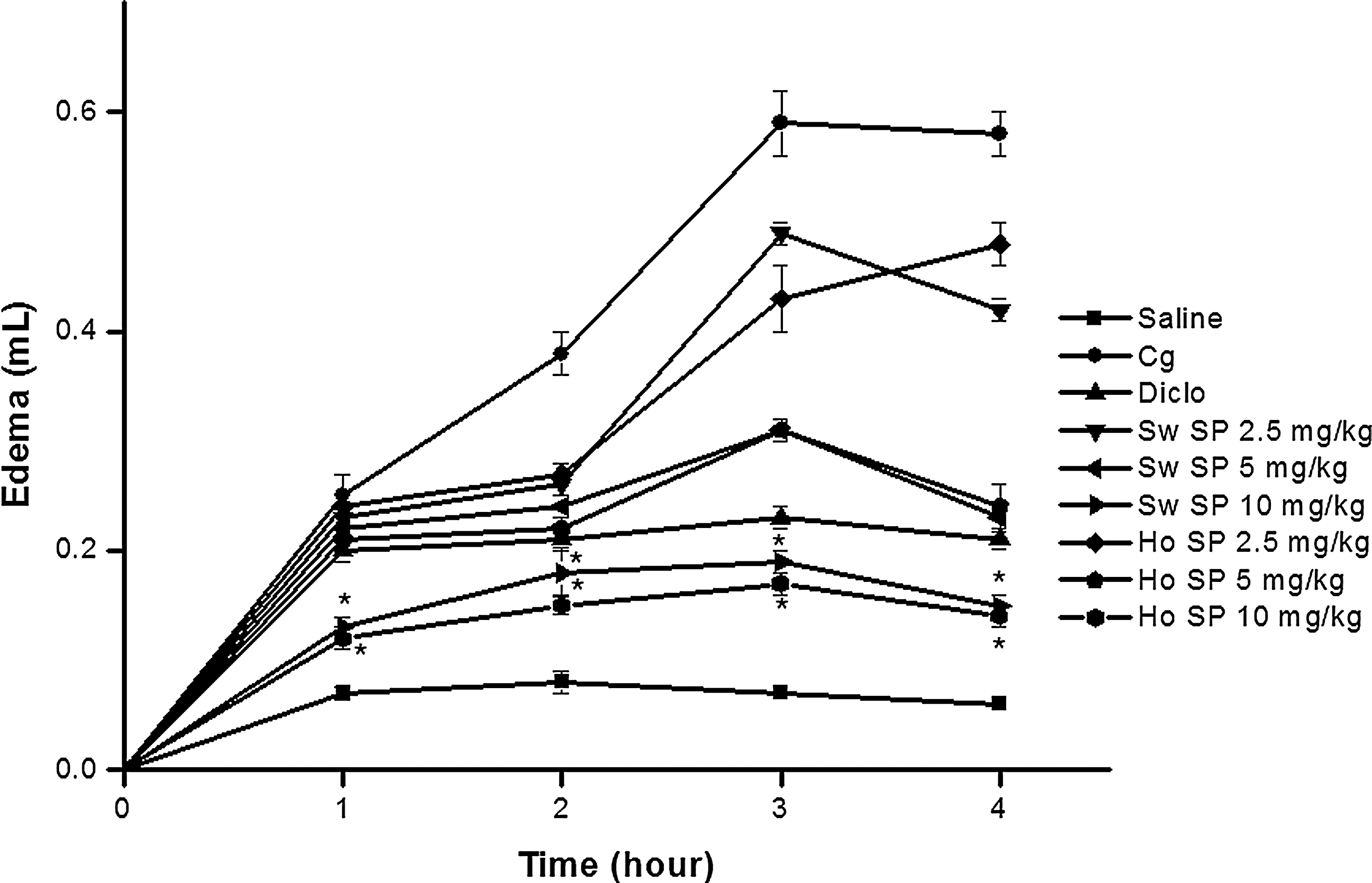
Effect of Sw-SP and Ho-SP on paw edema induced by carrageenan in rats. Before receiving a 0.1 mL injection of carrageenan (Cg, 500 μg/paw, intraperitoneally [i.p.]), rats received saline or Sw-SP or Ho-SP (2.5, 5 or 10 mg/kg). Diclofenac sodium (5 mg/kg) was injected s.c. Another group received only saline (s.c.) without Cg. Data are expressed as mean±SEM of six rats per group. *P<.05 indicates significant difference from the diclofenac-treated group (ANOVA, Tukey's test).
Peritonitis model
The anti-inflammatory activity of Sw-SP and Ho-SP (10 mg/kg) was evaluated in a carrageenan-induced peritonitis model. As expected, carrageenan (500 μg/cav, i.p.) induced significant leukocyte and neutrophil migrations when injected into the peritoneal cavity of rats. On the other hand, Sw-SP and Ho-SP (10 mg/kg) showed anti-inflammatory effects by decreasing leukocyte migration into the peritoneal cavity of these rats by 41% and 39%, respectively (Fig. 5A). The reference drug, dexamethasone (1 mg/kg, s.c.) significantly inhibited the leukocyte migration by 69% into the peritoneum of animals treated with carrageenan. Similarly, at the test dose of 10 mg/kg, Sw-SP and Ho-SP inhibited neutrophil migration by 53% and 52% (Fig. 5B). The reference drug dexamethasone (1 mg/kg, s.c.) decreased the carrageenan-induced neutrophil migration by 81%.
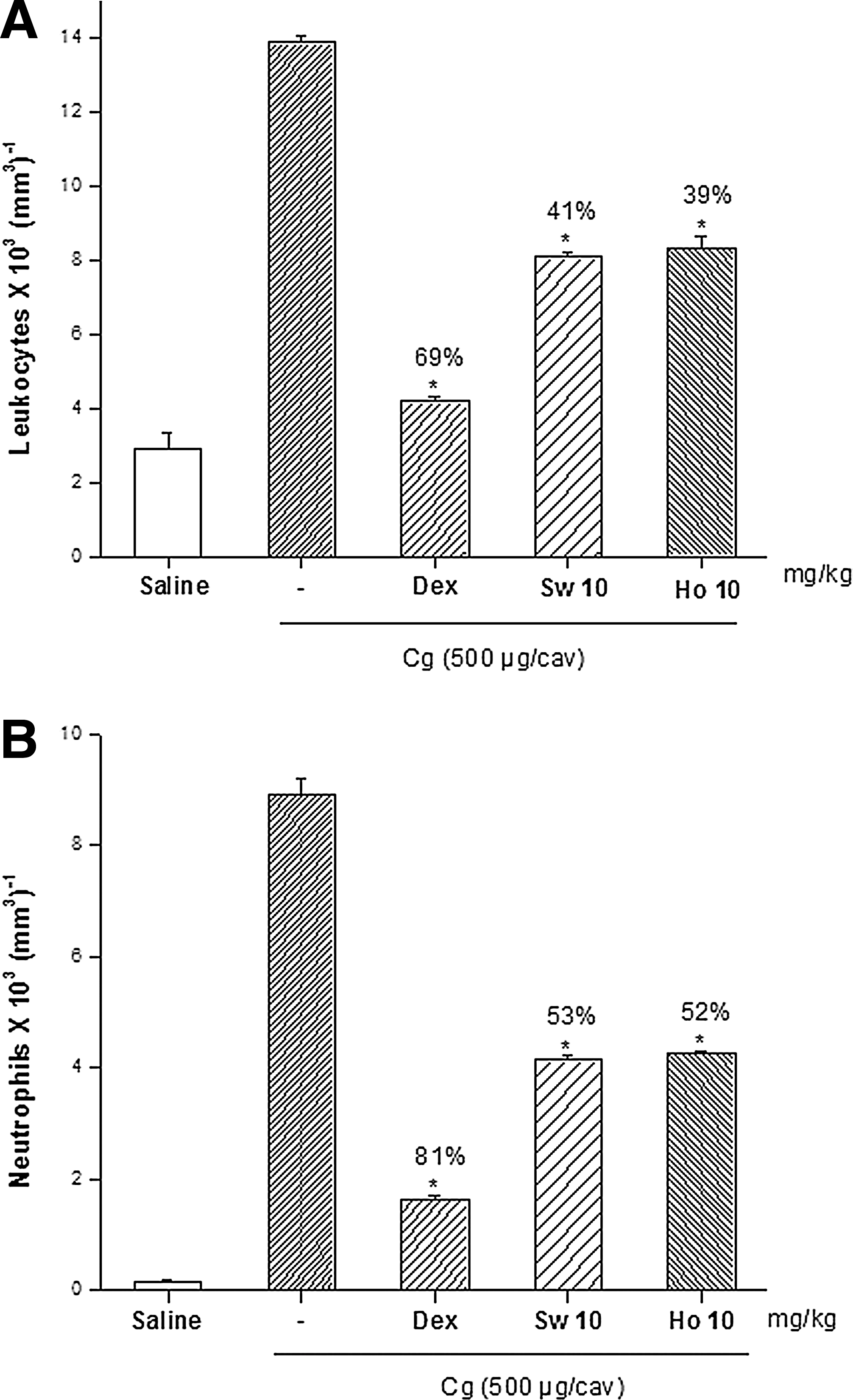
Effect of Sw-SP and Ho-SP on leukocyte
Freund's complete adjuvant–induced arthritis
The chronic anti-inflammatory activity of Sw-SP and Ho-SP (10 mg/kg) was evaluated by a Freund's complete adjuvant (FCA)–induced arthritis model in male Wistar albino rats during 0-, 3-, 7-, 11-, and 14-day intervals (Table 1). There was a significant increase in rat paw volume in FCA-injected arthritic control rats when compared to saline-treated rats at all day intervals. Whereas Sw-SP and Ho-SP (10 mg/kg) showed significant reductions in rat paw volume when compared with the arthritic group. On the 14th day, the % inhibitions of FCA-induced paw edema exhibited by Sw-SP and Ho-SP (10 mg/kg) were 46.4% and 47.7%, respectively. However, the inhibition was not significant when compared to diclofenac (5 mg/kg; 68.2% inhibition)-treated groups.
Values are mean±standard error of the mean, n=6 animals in each group. Values in parentheses indicate % inhibition of rat paw edema of sulfated polysaccharide-treated groups at 10 mg/kg versus the arthritic control group II.
P<.05 indicates significant difference from the arthritic control group.
Discussion
Indiscriminate use of commercially available analgesic and nonsteroidal anti-inflammatory drugs exerts adverse side effects and contributes to substantial mortality. 27 –29 Consequently, there is a strong interest in searching for new anti-inflammatory agents from natural products. 30 –32 Studies on the anti-inflammatory activity of solvent extracts of seaweeds and seagrass have been conducted previously. 16,20,33,34 However, very few studies have investigated the anti-inflammatory activity of sulfated polysaccharides obtained from seaweeds. 18,35,36 Currently, sulfated polysaccharides of different seaweed species provide new analgesic and anti-inflammatory agents. 8,37 To the best of our knowledge, no reports have been documented for the anti-inflammatory activity of sulfated polysaccharides extracted from seagrass species. This study demonstrated that sulfated polysaccharides obtained from the brown algae S. wightii and seagrass H. ovalis, produce antinociceptive and anti-inflammatory effects in models of nociception (formalin test and hot plate test), acute inflammation (carrageenan-induced paw edema test, peritonitis model), and chronic inflammation (FCA-induced arthritis test).
Formalin-induced nociception is considered the most predictive test of persistent pain, and is a mainstay for developing novel agents for postoperative pain treatment. 38 Substances that inhibit both phases are central analgesics, while those that inhibit only the second phase are peripheral analgesics, 39 suggesting that its nociceptive effect may be the result of inhibiting inflammatory mediators released in the effected tissue. 40 In the formalin test, administration of Sw-SP and Ho-SP (10 mg/kg) showed more potent inhibition of the second phase than of the first phase, suggesting that its antinociceptive effect is mainly against inflammatory pain. Similarly, SP-Sf of red seaweed Solierria filiformis, PII and carbohydrate fraction of Bryothamnion seaforthii seemed to act predominantly on the second phase of the response. 41,42
To discriminate between the central and peripheral antinociceptive action, we examined the effect of Sw-SP and Ho-SP using the hot plate test. This test is known to evaluate the possible specific central action in which, analgesic effects are exerted through opioid agents via supraspinal and spinal receptors. 43 In the present study, Sw-SP and Ho-SP showed a dose-dependent increase in the latency for jumping or licking when compared to indometacin, but the maximal antinociception effect was observed at 60 min. Red seaweed Gracilaria cornea GC-TSP (27 mg/kg) showed significant antinociceptive effects (at 60 min) compared to the other doses tested (3 and 9 mg/kg). 37 In contrast, SP sf from red seaweed S. filiformis (1 mg/kg) and Cc-SP2 of green seaweed Caulerpa cupressoides (9 mg/kg) showed more significant antinociceptive effects than higher concentrations tested. Therefore, this result indicate that the antinociceptive action of Sw-SP and Ho-SP could also occur via a central acting mechanism, similar to red seaweeds G. cornea and B. seaforthii. 37,42
There is a well-established link between the antinociceptive action and inflammatory pain. To evaluate this correlation, the anti-inflammatory activity of Sw-SP and Ho-SP was investigated in the paw edema model. Carrageenan has been used for decades to induce inflammation and to study the mediators of inflammation and the effectiveness of anti-inflammatory mediators. 44 From the present study, Sw-SP and Ho-SP at the higher doses of 10 mg/kg significantly inhibited the paw edema at all time intervals after the carrageenan administration. Similarly, oral administration of the crude polysaccharides of brown seaweed Turbinaria ornata significantly reduced the paw edema in a dose-dependent manner compared to the carrageenan-induced rats. 45 These results suggest that the anti-odematogenic response to Sw-SP and Ho-SP at the highest dose is related to inflammatory events involving the inhibition of osmotic edema through increased vascular permeability and may involve a diverse set of mediators.
Neutrophil migration into the peritoneal cavity of animals is P-selectin dependent. 46 Blockage of neutrophil migration to the center of inflammation is one way in which, the intensity of the inflammation process is decreased by sulfated polysaccharides. 47 In this model, Sw-SP and Ho-SP inhibited carrageenan-induced neutrophil migration into the rat peritoneal cavity. The inhibitory action of Sw-SP and Ho-SP on neutrophil migration is possibly due to its interaction with P-selectin 48 as supported by the inhibition of the chemotaxis of leukocytes into the peritoneal cavity of rats suffering from experimentally induced peritonitis. Similarly, Fucoidan from brown seaweed Laminaria saccharina and sulfated polysaccharides from Fucus vesiculosus inhibited leukocyte recruitment in the peritonitis model and blocked the interaction of P-selectin with its carbohydrate ligand. 49,50 In addition to that, sulfated polysaccharides of red algae Champia feldmannii were not anti-inflammatory and induced neutrophil migration. 51
Paw swelling is an index, employed to measure the antiarthritic activity of various drugs. Adjuvant-induced arthritis in rats is a classical model to study the chronic inflammatory disease, which is characterized by infiltration of the synovial membrane in association with destruction of joints resembling RA in humans. 52 In this study, Sw-SP and Ho-SP at the dose level of 10 mg/kg showed marked reductions in paw volume compared with the arthritic control group. Similarly, alginic acid at the dose level of 100 mg/kg extracted from S. wightii reduced paw edema in arthritic rats. 15 The ability of Sw-SP and Ho-SP to reduce edema formation in adjuvant-induced arthritis may be associated to its inhibition of prostaglandin synthesis. The loss of body weight has been used to assess the course of arthritis and the response to therapy of anti-inflammatory drugs. 53 Yoshikawa et al. 54 reported that there was a significant weight loss during the course of the experimental period, but thereafter continued to show normal weight gain in rats. Similarly, Somasundaran et al. 55 reported the loss of body weight during arthritic conditions on alterations in the metabolic activity of diseased rats. The present study indicates that there is a close relationship between the extent of inflammation and loss of body weight.
In conclusion, the present investigation demonstrated that sulfated polysaccharides isolated from brown seaweed S. wightii and seagrass H. ovalis possess antinociceptive and anti-inflammatory effects. However, the molecular mechanism of the Sw-SP and Ho-SP activity remains unknown. The action of Sw-SP and Ho-SP on proinflammatory mediators such as TNF, interleukins, and other mediators will be carried out in future to study its mechanism.
Footnotes
Acknowledgment
The authors thank the animal ethics committee, Pondicherry University, for animal work approval.
Author Disclosure Statement
The authors declare that they have no competing interests.