
Abstract
Cancer is a leading cause of death and is responsible for one in eight deaths worldwide. The use of herbs as complementary medicine for cancer, especially advanced cancer, has recently increased. The aim of this study was to evaluate in vitro, the antiproliferative effect of Origanum vulgare against human breast adenocarcinoma (MCF-7), and human colon adenocarcinoma (HT-29). The essential oil (EO) was extracted from a bought amount of O. vulgare dried leaves and analyzed in a gas chromatograph interfaced with a mass selective detector. The cytotoxicity test was performed by sulforhodamine B assay. The results show that the EO is composed mostly of 4-terpineol and induces a high cytotoxicity effect in HT-29. In the MCF-7 cell line the EO was less effective. In conclusion, this study showed that O. vulgare main component is 4-terpineol and was effective in inducing cancer cell growth inhibition.
Introduction
A
The use of herbs as complementary medicine for cancer, especially advanced cancer, has recently increased. 5,6 Questions concerning the safety of synthetic agents have increased the interest in the use of natural compounds and have encouraged more detailed studies of plant resources, which are a rich source of bionutrients or bioactive phytochemicals. 7 One new approach to cancer therapy focuses on anticancer and antimetastatic agents with little or no cytotoxic activity, such as the use of plant foods and their essential oils (EOs), which have a potential antioxidant effect. 8
EOs are volatile, natural complex compounds characterized by a strong odor and are formed by aromatic plants as secondary metabolites. 9 Origanum vulgare (oregano) is an annual, perennial, and shrubby herb native to the Mediterranean and has been used for many years as a medicinal plant with health-aiding properties such as its powerful antibacterial and antifungal properties. 8 Oregano belongs to the Lamiaceae family and it is well-known that Lamiaceae spices have potent antioxidant properties, mostly due to the polyphenolic compounds. 10,11 Phenolic phytochemicals are a large group of substances, which are found in significant quantities in vegetables, fruits, spices, and seeds, and are thought to promote optimal health partly through their scavenging effects in protecting cellular components against free radicals. 7
Recent findings revealed the antimicrobial, fungicidal, insecticidal, and antioxidant potential of the EO and extract of Origanum, which raised great pharmaceutical and industrial interest in oregano. 10 Natural antioxidants can protect the human body from free radicals and could retard the progress of many chronic diseases. 11 Oxidation of lipids is associated with cell membrane damage, aging, heart disease, and cancer in living organisms. 11 Studies have shown that components of oregano EOs, that is, carvacrol, thymol and protocatechuic acid, possesses strong antioxidant properties and can act like potent anticancer and antimelanogenic compounds. 12 –14 Modulation of carcinogenic and mutagenic effects by inhibitors from plant origin has been of crucial importance for the final outcome of some biological effects, particularly for cancer. 7
Therefore, the present study aimed to evaluate in vitro the antiproliferative effect of O. vulgare against human breast adenocarcinoma (MCF-7) and human colon adenocarcinoma (HT-29).
Materials and Methods
Material
Samples of oregano (dried leaves from Chile) were purchased from TecPharma Importação de Produtos Químicos e Farmacêuticos (TECPHARMA), a major manufacturer of chemical and pharmaceutical products. These samples were manually crushed and stored under nitrogen atmosphere. The analytical standards thymol, α-terpinene, and α-terpinene were purchased from Fluka® and the analytical standards α-terpineol, 4-terpineol, camphene, carvacrol, limonene, α-pinene, α-pinene, myrcene, p-cymene, 1,8-cineole, terpinolene, and linalool were purchased from Sigma-Aldrich®. Solutions of each standard were prepared (1000 mg/L) using dichloromethane (Merck; pa grade, bidistilled) and stored under refrigeration.
Extraction procedure
The EO was extracted by hydrodistillation over a period of 4 h using a Clevenger apparatus and the yield of oil was recorded at every 5 min. The density of the EO measured by the gravimetric method was 0.92 g/mL at room temperature (20°C). After hydrodistillation, water was removed by decantation and the EOs obtained were stored at 4°C in a dark-colored container to prevent light-sensitive decomposition.
Analytical methods
The EO was analyzed in a gas chromatograph interfaced with a mass selective detector—GC/MS (Shimadzu 5050A), using a capillary column DB-5 (30 m×0.25 mm×0.25 mm) and a flow rate of 1 mL/min, in electronic impact mode of 70 eV and in split mode (split ratio 1:50). The following temperature gradient was used: 40°C (0 min)—2°C/min—145°C—10°C/min—280°C (10 min). The interface temperature was maintained at 280°C. The identification of major compounds was accomplished by comparing their retention times with those of authentic standards, and by comparison of their mass spectra with those from the equipment library. Compositions were then expressed as percentages of normalized peak areas.
Cell lines
The cell lines under investigation were human breast adenocarcinoma (MCF-7) and human colon adenocarcinoma (HT-29). They were purchased from RJCB Collection (Rio de Janeiro Cell Bank). The cells were cultured in Dulbecco's modified Eagle's medium (Gibco®), supplemented with 10% fetal bovine serum (Gibco) and incubated at 37°C in a humidified atmosphere containing 5% CO2, as described previously. 15 –17
Cytotoxicity assay
According to the cells growth profile, cells were seeded with a density of 2×10⁴ cell/well and incubated at 37°C in a humidified atmosphere containing 5% CO2. After 24 h, the cells were treated with the EO, which was diluted in dimethylsulfoxide to produce nine concentrations, ranging from 10 to 500 mg/mL. Of each concentration, 100 μL/well was added to the plates in triplicates. The final dilution used for treating the cells contained not more than 1% of the initial solvent, this concentration being used in the solvent control wells. This concentration of dimethylsulfoxide did not significantly influence the proliferation rate as compared to media alone. The plates were incubated for 72 h. At the end of the exposure time, cell growth was analyzed using the sulforhodamine B (SRB) assay.
SRB assay
After incubation for 72 h, adherent cell cultures were fixed in situ by adding 50 μL of cold 40% (w/v) trichloroacetic acid and incubated for 60 min at 4°C. The supernatant was then discarded, and the plates were washed five times with deionized water and then dried. Of the SRB solution (0.4% w/v in 1% acetic acid), 50 μL was added to each well and incubated for 30 min at room temperature. Unbound SRB was removed by washing five times with 1% acetic acid. Then, the plates were air-dried and 100 μL of 10 mM Tris base pH 10.5 (Sigma®) was added to each well to solubilize the dye. The plates were shaken gently for 20 min on a plate shaker, and the optical density was determined in an ELISA multiplate reader (Thermo Plate TP-Reader; Thermo Fisher Scientific) using a wavelength filter of 492 nm. The cell growth inhibition was calculated as the percentage inhibition of cell growth and was determined as follows: inhibitory rate=(1 – Abs492treated cells/Abs492control cells)×100. All observations were validated by at least two independent experiments, and for each experiment, the analyses were performed in triplicate
Statistical analyses
Data sets from the SRB assay were analyzed using a two-way ANOVA followed by a Tukey test for multiple comparisons. Two factors were considered: the cell type and the concentration of the compound. Significance was considered at P<.05 in all analyses. The data are expressed as the mean±SEM.
Results
EO composition
The EO was obtained from O. vulgare and its composition is listed in Table 1. The table shows the retention indices calculated on the DB-5 column and the percentages of the detected compounds. Retention times of the sample components were calculated on the basis of homologous compounds under the same conditions, and the compounds were identified by injection of standards and/or by comparing the mass spectra with the equipment library.
Compounds identified by comparison with standards compounds; other compounds identified by literature data and Willey Library.
C, normalized peak areas without using the correction factors; ND, not detected (based in Rt of standards compounds); Rt, retention time.
The average extraction yield of oregano EO was determined to be 1.33±0.16 wt%, achieved after about 60-min extraction; this value is similar to that found by Rodrigues et al. 18 and Busatta et al. 20 The chromatogram of O. vulgare EO (Fig. 1) shows that 4-terpineol (peak 20 and percent area of 41.17) is the major component, followed by thymol (peak 28, percent area of 21.95), γ-terpinene (peak 12, percent area of 5.91), and carvacrol (peak 29, percent area of 4.71).
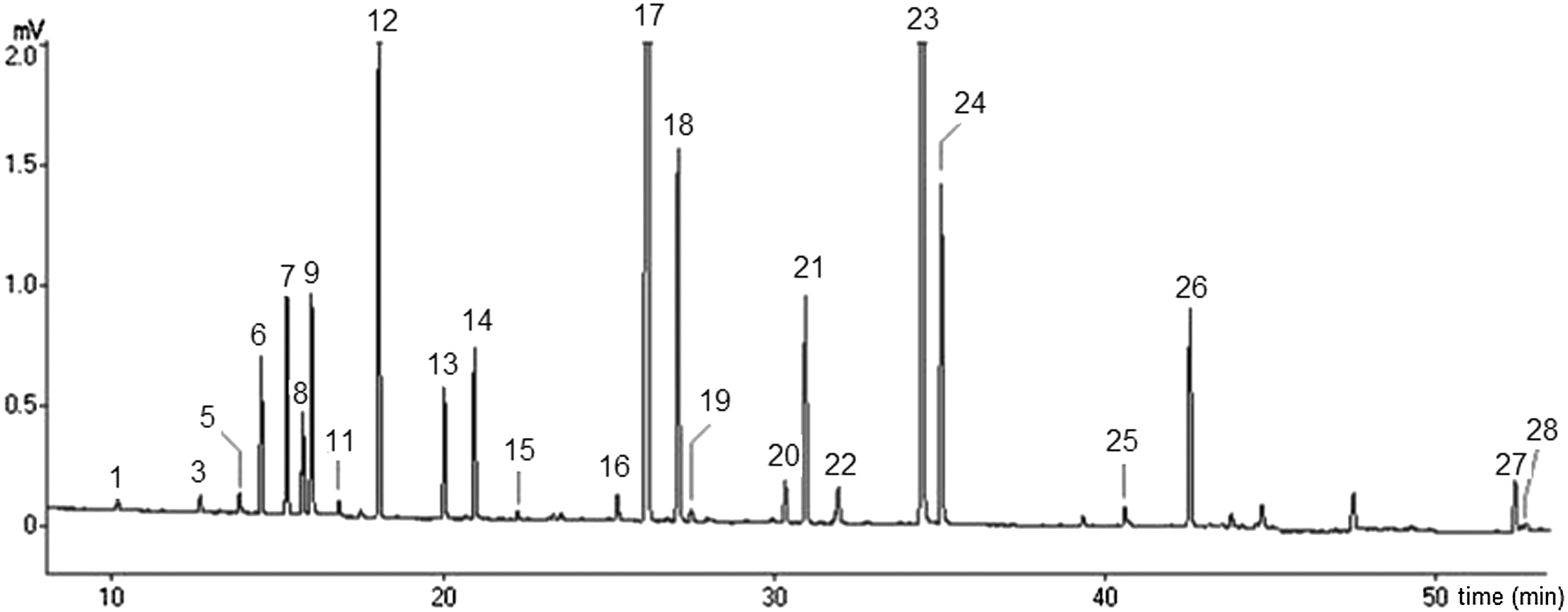
Chromatogram of oregano essential oil (EO) obtained by hydrodistillation. Peaks identification according to Table 1.
Antiproliferative activity
The cell growth inhibition of O. vulgare was determined by the cell proliferation assay using the MCF-7 and HT-29 cell lines. As shown in Figure 2, the EO was most effective in the HT-29 cell line when compared with MCF-7. In both cell types, the concentration of 10 mg/mL was not effective; in HT-29 cells, the percent of cell growth inhibition was 8.18% and in MCF-7 cells, this concentration induced cell proliferation. The most effective concentration for HT-29 and MCF-7 was 50 mg/mL: 60.8% and 48.9%, respectively; however, an increase in the EO concentration did not enhance the cell growth inhibition (Fig. 2).
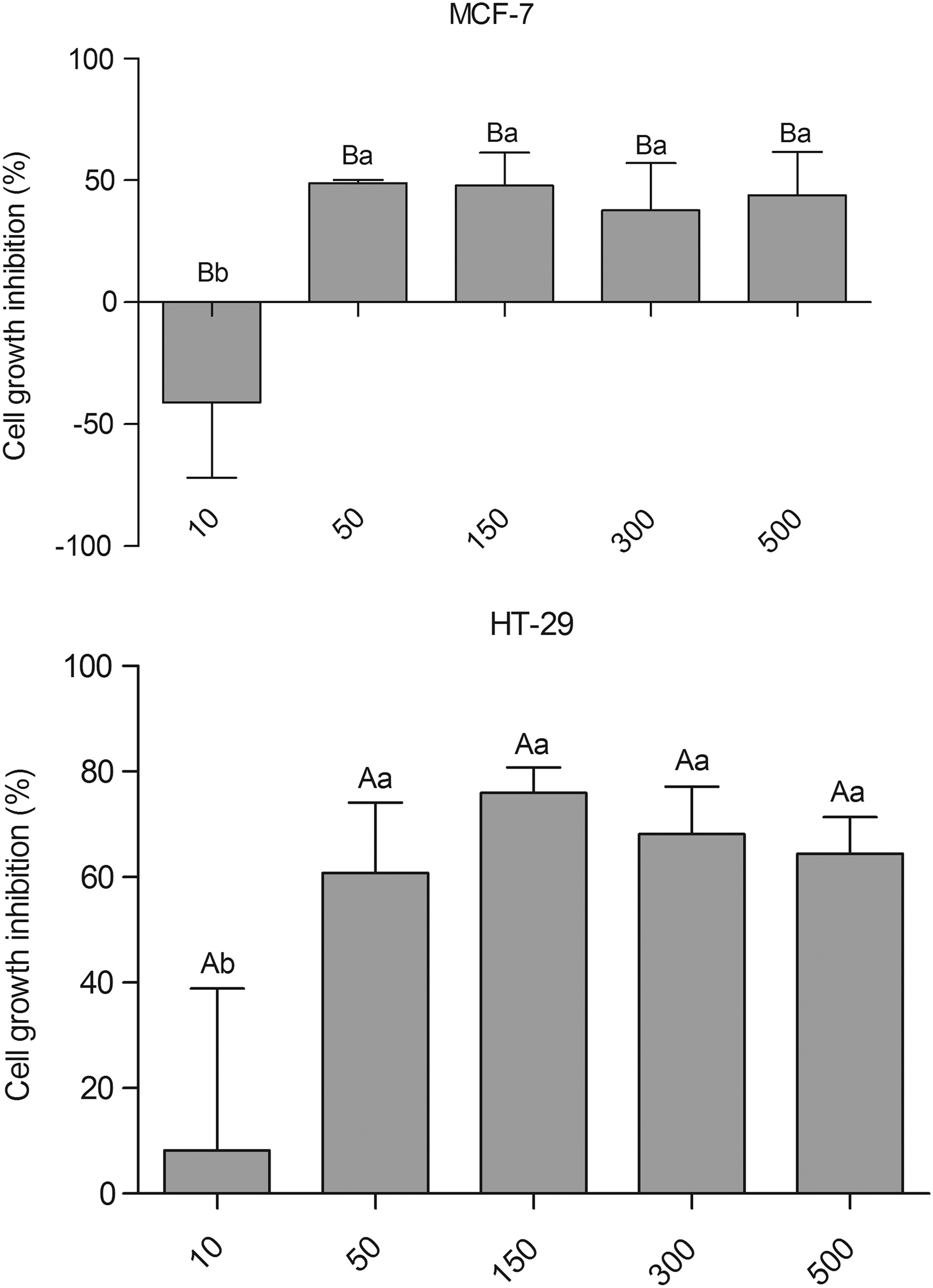
Cell growth inhibition of MCF-7 and HT-29 cell lines by Origanum vulgare EO. Uppercase letters indicate significant differences between tumor cell type and lowercase letters indicate significant differences in the concentration used. A P-value<.05 was considered significant (Tukey test).
Discussion
O. vulgare L. is the most variable species of the genus Origanum, and variations in the composition of the EO have been the topic in the reports of several researchers. 12,10 In the composition of the EO of oregano, great variations in the major and minor constituents were recorded. Several oregano species are characterized by the presence of two major components, thymol and carvacrol. Another intermediate type would contain a high content of two monoterpene hydrocarbons, γ-terpinene or p-cymene, which are biogenetic precursors of thymol and carvacrol. However, some species were found with high values of linalool and others with monoterpenes and sesquiterpenes. 12,18
The EO composition and yield of Origanum species vary with the origin of these plants and other factors, mainly the geographical and time of harvest. 19
Additionally, it is important to know the botanical origin of oregano as well as to do the investigation of the chemical constituents of this species. With regard to the taxonomic viewpoint, it is difficult to establish a correspondence between subspecies of O. vulgare and composition of its EO. In this study, 4-terpineol was the main component of Origanum EO, followed by thymol, γ-terpinene, and carvacrol. This chemical profile is very similar to that found by Busatta et al. 20 where 4-terpineol, γ-terpinene, carvacrol, and thymol were the major components. Comparison of the data produced herein with previous reports showed that the chemotype variability has a straight relationship with the geographical region where the species is found. 8,10,11 Reports 2,21,22 have described that oregano grown in a Mediterranean climate contains higher amount of phenols, whereas oregano from the inland contains a higher amount of terpene alcohols. In a study of Russo et al., 23 the authors evaluated the chemical composition of the EO of 24 samples of O. vulgare, hirtum variety that grows in Southern Italy by GC/MS. A total of 56 compounds were identified, the major compounds were carvacrol (0.12–56.63%) and thymol (7.91–53.62%). This same species, hirtum variety, grown in Northern Italy, was analyzed by Bocchini and coworkers 24 and they found a group of oregano with high content of thymol, carvacrol, and linalool, other with a large variation among the sesquiterpenes, and a third group with loads of sesquiterpenes.
Furthermore, the technique of obtaining the EO may influence the quality of the extract obtained and the amount of extracted aromatics. Parameters that affect the distribution coefficients of the main components present in the EO of O. vulgare, as the fraction of monoterpene hydrocarbons, depend on the equilibrium time, temperature, and density of the solvent used in the extraction process. 19 Comparing the extraction process with those obtained from the supercritical CO2 extract indicated that the extraction method plays an important role in the final extract's composition, 18 especially with regard to the hydrocarbon terpene fraction.
MCF-7 is an adenocarcinoma cell line recommended as one of the models for breast cancer tissue by the National Cancer Institute (NCI). The cell line expresses estrogen receptors and has been well studied and documented. 22 In the present study, the Origanum EO was less effective in the MCF-7 cell type and was able to induce cytotoxicity in a concentration of 50 mg/mL (cell growth inhibition of 48.9%). The increase in the EO concentration did not enhance the cell growth inhibition. In the HT-29 cell line, the EO was significantly more effective (cell growth inhibition of 60.8%) and also presented the same characteristic of inducing cytotoxicity at 50 mg/mL and the nonenhancement of cell growth inhibition values with the increase of EO concentration. These results suggest that the EO could have a selective activity and therefore offer an opportunity to investigate its use as a therapeutic agent.
The cytotoxic activity of oregano oil can be attributed to the action of its principal phenolic components, carvacrol, and thymol, which exhibit significant anticancer and antimutagenic activity when tested separately. 7,13 Arunasree 13 demonstrated the anticancer effects of carvacrol in MDA-MB 231, a human metastatic breast cancer cell line. These authors showed that carvacrol-treated cells exhibited prominent morphological changes like cell shrinkage with rounding of cells and formation of membrane blebs characteristic of apoptosis. Other studies have suggested that carvacrol might be potentially useful in counteracting free radical-mediated injuries and in DNA damage by the ability to enhance the levels of antioxidants along with its antilipid peroxidative activity, 25,26 what can be a beneficial action of carvacrol against pathological alterations like melanogenesis and cancer.
In conclusion, it was found that the O. vulgare EO main component is 4-terpineol and that the oil presents a significant effect on the cancer cell line tested, which could be associated with the major components of the extracted oil.
Footnotes
Acknowledgment
This study was made possible by an undergraduate fellowship provided by the National Council of Scientific and Technology Development of Brazil (CNPq).
Author Disclosure Statement
The authors declare that they have no conflicts of interest concerning this article.