




Abstract
The bioactive composition and cytotoxic and antioxidative/prooxidative effects of four medicinal plants: yarrow (Achillea millefolium L.), hawthorn (Crataegus oxyacantha L.), ground ivy (Glechoma hederacea L.), and olive (Olea europea L.) on human laryngeal carcinoma cell line (HEp2) were investigated. Water extracts of these plants obtained by infusion, maceration, and decoction were characterized for their polyphenol content and antioxidant capacity. Based on the extraction efficiency of polyphenols, the final extracts were obtained whose polyphenolic profile, polysaccharides, mineral content, and cytoprotective activities were determined. The overall highest content of polyphenols and antioxidant capacity was determined in hawthorn, followed by yarrow and ground ivy, and the lowest in olive leaves extract. Phytochemical screening revealed the presence of phenolic acids, as the most abundant bioactive compounds, followed by flavonoids, flavons, and flavonols. All examined medicinal plants reduced the cell viability and reactive oxygen species formation in a dose- and time-dependent manner. Ground ivy and yarrow containing a high content of phenolic acids and polysaccharides were more efficient to decrease the cell survival when compared to olive leaf and hawthorn. Experiments confirmed the importance of polyphenolic composition rather than content of investigated plants and revealed a relationship between the polyphenolic and polysaccharide contents and antioxidant/prooxidant characters of medicinal plants.
Introduction
T
Antioxidants have been studied extensively for decades aiming to find compounds that exhibit protective effects against a number of diseases attributed to free radical-induced damage and oxidative stress. A large number of studies involving screening for plant antioxidants are now being questioned with respect to their in vivo relevance. 5 Medicinal plants typically contain mixtures of different chemical compounds that may act individually, additively, or in synergy to improve health. Thus, the use of naturally occurring antioxidants from medicinal plants either as a single chemical entity or as a multicomponent extract may have a real tangible benefit. Therefore, only with the exact knowledge of the bioactive composition of medicinal and other plants, and their impact on biological systems, it will be possible to develop a new generation of standardized, effect-optimized mono- and multi-extract preparations, which fulfill today's standards for quality, safety, and efficiency of medicinal preparations. 6
Extraction techniques have been widely investigated to obtain such valuable natural compounds from plants for commercialization. 7 The choice of solvent for the extraction is influenced by what is intended with the extract. Since for food purposes, the end product will contain traces of residual solvent, the solvent should be nontoxic, while the extraction technique should be simple and economically feasible. 8,9 Medicinal plants have been extensively studied for the presence of natural antioxidants, but most emphasis has been given to essential oils or to hexane, acetone, ethanol (or methanol), and carbon dioxide extracts. As the result of growing interest in innovative extraction techniques developed and employed recently for the extraction of bioactives from different medicinal plants, conventional extraction techniques that use water as the extraction media have been less-favored in recent years. Despite the fact that in household preparation, medicinal plants are still most commonly consumed as infusions, decoctions, or macerates, these extraction procedures have been neglegted in terms of comparing their bioactive compound contents and bioactivity. Since tea and herbal infusions are generally prepared by steeping the leaves and herbal parts in hot water, a more comprehensive and thorough survey of the composition of water herbal extracts is necessary.
Laryngeal carcinoma is the most common type of head and neck cancer and one of the most aggressive human malignancies, with a tendency toward an increasing occurrence of new cases and deaths annually. In 2008, over 150,000 new cases of laryngeal cancer were estimated worlwide, with around 82,000 deaths casued by this type of cancer. 10 The high incidence of larynx cancer is primarily attributable to the habit of tobacco, betel quid chewing, and alcohol consumption. Over the past two decades, the survival rate of the patients has not considerably improved, despite the multimodal therapeutic strategies, especially chemotherapy. 11 However, a large number of chemotherapeutic agents are highly toxic and have a low survival profile. 12 Thus, novel effective chemotherapeutic agents and less harmful therapies are needed in the treatment of laryngeal carcinoma. Additionally, larynx cells are useful as a biological test system in experiments where biologically active compounds are investigated, since these cells are in direct contact with nonmetabolized compounds present in the food.
The aim of this study was to develop water-based extracts of polyphenols from four medicinal plants (ground ivy, hawthorn, yarrow, and olive leaf) and to obtain and characterize extracts that could be used in food and pharmacological purposes. These plants are widely spread in Croatia and south-eastern European region and are consumed very often due to their similar traditional uses (Table 1), 13 –20 but come from distant botanical families, and their current positions in phytotherapy are also different. To avoid the application of organic solvents for the extraction of polpyhenols and to ensure a simple extraction technique with low economical costs, infusions, macerates, and decoctions of medicinal plants were prepared and the extracts examined for their polyphenolic content and profile. To highlight the relationship between their bioactive profile and antioxidant potential, the aim of this study was to determine the polyphenolic profile, the content of soluble polysaccharides and mineral elements, cytotoxic and antioxidative/prooxidative activities of their water extracts on human laryngeal carcinoma cells, as well as to evaluate whether a correlation between the bioactive composition and biological activity of these medicinal plants can be established.
Materials and Methods
Chemicals
Analytical grade of Folin–Ciocalteu, formic acid, potassium peroxodisulfate, sodium carbonate, formaldehyde, n-hexane, methanol (HPLC grade), and hydrochloric acid were supplied by Kemika (Zagreb, Croatia). 2,2-diphenyl-1-picrylhydrazyl (DPPH) was supplied by Fluka (Buchs, Switzerland). Vanillin, 6-hydroxy-2,5,7,8-tetramethylchromane-2-carboxylic acid (Trolox), 2,2-azino-bis(3-ethylbenzthiazoline-6-sulphonic acid)diammonium salt (ABTS) as well as (−)-epicatechin (EC), procyanidin B2, gallic acid (GA), caffeic acid (CA), chlorogenic acid (ChlA), rosmarinic acid (RA), luteoline-7-glucoside (L-7-G), apigenine, quercetine and oleuropein, neutral red (NR), and 2′7′-dichlorofluorescein diacetate were obtained from Sigma-Aldrich (Steinheim, Germany).
Preparation of different medicinal plant extracts
Dried plant materials of hawthorn (Crataegus oxyacantha L.), yarrow (Achillea millefolium L.), olive leaf (Olea europaea L.), and ground ivy (Glechoma hederacea L.) were purchased from a local medicinal plant manufacturer. All plant materials were collected in April 2012, in the Žumberak area around Zagreb, except for olive leaf, which was obtained from the dalmatian coastal area (Zadar) of Croatia. Infusions [2.0 g of plant sample was extracted in 200 mL of distilled water (80°C) and stirred with a glass rod for 10 min], macerates [2.0 g of sample was extracted in 200 mL of distilled water (room temperature) during 72 h with occasional stirring], or decoctions (2.0 g of plant was subjected to boiling in 200 mL of distilled water during 20 min) of dried plant samples were prepared. The final extract used for cytotoxicity and reactive oxygen species (ROS) formation was prepared by pouring 200 mL of boiling water over 10 g of plant sample and extracted for 30 min, without maintaining the water temperature. After extraction, all extracts were filtered through a tea strainer and analyzed.
To determine the biological effects of medicinal plants, the previously prepared extracts were evaporated to dryness and re-dissolved (and concentrated) in 10 mL of DMSO. Different concentrations of extracts (ranging from 0.1× to 2.5×) were prepared in the growth medium (Dulbecco's modified Eagle's medium [DMEM]). In household preparation of herbal infusions for consumption as warm beverages, the ratio of 2 g of plant sample and 200 mL of hot water is used. The concentration of bioactive compounds present in thus prepared infusion is marked as 1×. The lowest concentration contained 50% of bioactive compounds that are present in ordinary prepared water infusion, and the highest concentration contained 2.5 times higher concentration of all bioactive compounds in comparison to those present in water extract prepared for consumption. According to the measurement of dry matter content in the final extracts obtained for the experiments on HEp2 cells (10 g of plant material in 200 mL of water), the final concentrations of each extract expressed as the extraction yield amounted to the following range for each sample (concentrations provided for the range of 0.1–2.5×): yarrow, 4–95 g/L; ground ivy, 9–217 g/L; hawthorn, 6–145 g/L; olive leaf, 7–180 g/L. All concentrations of diluted extracts used in the experiment were checked for the formation of microscopically apparent crystals to be sure that all components are in soluble form after dilution of stock solutions in DMEM. 21
Determination of polyphenolic compounds of medicinal plant extracts
Total phenol content (TPC) of medicinal plant extracts was determined spectrophotometrically according to a modified method of Lachman et al. 22 To determine flavonoids, the formaldehyde precipitation was used and the flavonoid content calculated as the difference between total phenol and nonflavonoid contents. All determinations were carried out in triplicates and the results were expressed as mg gallic acid equivalents/g of dry weight (dw) of plant. The content of flavan-3-ols was determined using the vanillin assay and the reaction with p-dimethylaminocinnamaldehyde as described by Di Stefano et al. 23 The content of flavan-3-ols was expressed as mg (+)-catechin/g dw. Proanthocyanidins (i.e., condensed tannins) were analyzed by the procedure described by Porter et al., 24 with some modifications. The quantity of condensed tannins was determined from a standard curve of cyanidin chloride treated with BuOH-HCl-FeIII mixture, and expressed as mg cyanidin chloride equivalents (CyE)/g dw. The content of tannins was determined according to a procedure described by Schneider 25 and expressed as percentage toward the mass of dry plant material. Individual polyphenolic compounds of the final extract used for cytotoxicity evaluation were identified and quantified according to the HPLC method described by Belščak-Cvitanović et al. 26
Mineral analysis of medicinal plants
Mineral content of the plants was determined using a procedure described by Belščak-Cvitanović et al., 26 with a Prodigy High Dispersive ICP spectrometer (Teledyne Leeman, Hudson, NH) working in a simultaneous mode. The instrument was operated with optimal parameters: a radiofrequency power of 1.1 kW; plasma gas flow rate (Ar) of 18 L/min, and auxillary gas flow rate (Ar) of 0.8 L/min. For the analysis, ∼0.25 g of the ground plant was weighted into Teflon reaction vessels (in triplicate), and 5 mL of nitric acid, (50:50 v/v) was added to each sample. For the microwave-assisted digestion of plants, MWS-2 Microwave System Speedwere BERGHOF was used, and the digestion procedure was conducted in three steps, (1) 150°C/10 min, (2) 160°C/10 min, and (3) 190°C/20 min. The resulting clear solutions of digestion procedure were then brought to 25 mL with ultrapure water. The system was calibrated using aqueous mixed standards. Calibration ranges were modified according to the expected concentration ranges of the elements of interest. All determinations were performed in triplicate. All standard deviations (SDs) were based on measurements in triplicate and amounted to less than 10%.
Determination of soluble polysaccharides
The content of soluble polysaccharides was determined according to a modified method of Wei et al. 27 Briefly, plant material (10 g) was ground in a blender to obtain a fine powder and then extracted with 50 mL of distilled water at 100°C for 2 h. The extracted soluble polysaccharides were separated from the insoluble crude residue by centrifugation (15,600 g for 10 min, at 4°C) and concentrated by vacuum evaporation. The obtained concentrate was precipitated by the addition of three volumes of ethanol to a final concentration of 75% (v/v). The precipitates collected by centrifugation (15,600 g for 15 min, at 20°C) were solubilized in deionized water and lyophilized to get the crude polysaccharides. The polysaccharides yield was calculated as the polysaccharide content of extraction divided by dried sample weight (10 g).
Determination of antioxidant capacity
Ferric reducing/antioxidant power
The ferric reducing/antioxidant power assay was carried out according to a standard procedure by Benzie and Strain. 28 All measurements were performed in triplicate. Aqueous solutions of FeSO4×7H2O (100–1000 μM) were used for the calibration, and the results are expressed as mmol Fe(II)/L.
ABTS radical scavenging assay
The Trolox equivalent antioxidant capacity was estimated by the ABTS radical cation decolorization assay. 29 The results, obtained from triplicate analyses, are expressed as Trolox equivalents and derived from a calibration curve determined for Trolox (100–1000 μM).
Determination of biological activity in vitro
Human cell lines
Human laryngeal carcinoma (HEp2) cells were provided as a gift by the Rudjer Boskovic Institute, Zagreb, Croatia. Cells were grown as monolayer cultures in the DMEM (Gibco, Grand Island, NY, USA), supplemented with 10% of the fetal bovine serum (Gibco), 4500 mg/L of glucose and 1% of penicillin/streptomycin. HEp2 cells were seeded in wells (2×10⁴/well) of 96-well plates.
Cytotoxicity assay
Cytotoxicity of medicinal plant extract was determined by NR assay, as described in Durgo et al.
30
Briefly, after 24-h incubation, cells were treated with extracts in different concentration ranges (0.5–5× of concentration present in originally prepared medicinal plant extract) for 1 and 2 h, with and without subsequent recovery period that lasted for 24 h. After treatment and recovery, the NR assay was carried out, as described by Babich and Borenfreund.
31
Cell viability was calculated using the following equation:
where ABSsample is the absorbance of cells treated with extracts and ABSCtrl is the absorbance of corresponding vehicle control (growth medium+0.25% DMSO).
ROS determination
ROS formation in the cells after the treatment with plant extracts was determined by dichlorohydrofluorescein assay using microplate reader,
32,33
as described by Durgo et al.
30
Cells were cultured to confluence and subsequently treated with different concentrations of extract for 1 and 2 h, with and without subsequent recovery period of 24 h. Each extract concentration was tested in quadruplicate, and each experiment was repeated three times. Data are reported as fluorescence intensity in relation to cell survival (data obtained according to the equation):
where ABSsample is the absorbance of cells treated with extracts and ABSCtrl is the absorbance of corresponding vehicle control (growth medium+0.25% DMSO).
Statistical analyses
Statistical analyses were performed using the SPSS v. 8.0 (SPSS, Inc., Chicago, IL, USA). The one-way analysis of variance (ANOVA) was employed to determine whether the means obtained with various groups differ significantly from each other. The significance was established using the Tukey and Dunnett post hoc test. The probability level of P<.05 was considered significant. All data are expressed as mean±SD of the values obtained by three independent measurements.
Results and Discussion
Extraction efficiency of polyphenols using different water-based extraction techniques
Using conventional extraction procedures such as infusion, maceration, and decoction for the extraction of polyphenols from four medicinal plants resulted with a high variation in total and specific subclasses of polyphenolic compounds. Since for the preparation of infusions warm water (80°C) was used for a short period of time (10 min), maceration is performed with cold water in a closed container for a longer period (72 h) and decoction is done by boiling the crude plant in water for a short time (20 min), the differences observed are not surprising.
As can be seen in Table 2, the extraction efficiency of every type of extract depends on the nature of plant (plant part used), as well as on the chemical nature of plant, the extraction method employed, solvent, temperature, and extraction time. TPC of all plants prepared by infusion is higher when compared to their macerated extracts, except for olive leaf macerate, which exhibits twofold higher TPC than olive leaf infusion. Similarly, nonsignificant (P>.05) TPC was obtained by maceration and decoction of olive leaf since this plant is characterized with thick rigid leaves, which require more intensive extraction and penetration of solvent to extract the required bioactives. The TPC of ground ivy decoction was significantly (P<.05) lower when compared to infusion and macerate, which indicates that high temperature used during boiling of thin ground ivy leaves may have led to degradation of phenolic compounds.
Values with matching superscript letters are not significantly different (P>.05).
TPC, total phenol content; GAE, gallic acid equivalents; dw, dry weight; CyE, cyanidin chloride equivalents; FRAP, ferric reducing/antioxidant power.
Although it may be expected that the ranking of specific subclasses of polyphenolic compounds follows the ranking of TPC, each class of compounds exhibits a different content depending on the extract type. Ground ivy decoction, with the lowest TPC, is characterized with the highest flavan-3-ols. Similar is observed also for the other three plants, whose decoctions exhibit the highest flavan-3-ol and proanthocyanidin contents. The contents of tannin, which impart astringency to the extract, are also extracted during the preparation of water extracts, so their content should be minimized. According to the obtained results, the highest tannin content was obtained after decoction of ground ivy and olive leaf, while for yarrow and hawthorn, tannins were mostly abundant in their infusions. Generally, maceration of plants did not yield a high content of tannins, when compared to the other two applied extraction procedures. Similarly, the antioxidant capacity was again dependent on the extraction procedure. Among the prepared extracts, infusions of ground ivy and hawthorn and decoctions of yarrow and olive leaf were the most efficient radical scavengers. Katsube et al. 34 determined that polyphenolic compounds and their antioxidant capacity in plant substrates are stable up to temperatures of 70–80°C, whereas above temperatures of 80°C, they start to deteriorate and consequently loose their activity. According to our results, room temperature is not sufficiently effective to extract the polyphenolic compounds, whereas decoction procedure at 100°C leads to thermal degradation of polyphenols in some plants. However, it has been reported that the phenolic compounds in grapes, extracted using PLE at high temperature (>100°C), are quite stable even for most oxidizable phenolics. 32 The most probable cause for a higher polyphenolic content at high temperature is the breakage of bonds between various phenolics (analytes) and the plant matrix. 35 Therefore, based on the obtained results, the combination of high temperature (100°C) and prolonged extraction (30 min) time (without maintaining the temperature) of medicinal plants was selected as the best extraction technique for obtaining the highest content of polyphenolic antioxidants. To increase the content of polyphenols in the final extract, the authors used a higher plant/solvent ratio and obtained a final extract, which was characterized in terms of polyphenolic profile and bioactive properties.
According to the obtained polyphenolic content and antioxidant capacity of final extracts of four evaluated medicinal plants (Table 2), hawthorn is characterized with the highest TPC, followed by yarrow and ground ivy with similar TPC, while olive leaf contains the lowest TPC, almost threefold lower than hawthorn. The content of specific subclasses of polyphenolic compounds differentiates depending on the plant species. The most abundant was hawthorn, in which total flavonoids, flavan-3-ols, and proanthocyanidins predominated when compared to other plants. Flavonoids (TFC) of hawthorn, ground ivy, and yarrow constituted more than half of total phenols (58.6%, 57.7%, and 52.2% of TPC), while in olive leaf, flavonoids represent the most abundant polyphenolic group (64.9% of TPC). Flavan-3-ols and proanthocyanidins (determined by both vanillin index and the reaction with p-DAC reagent) prevailed in hawthorn final extract, where significant differences (P<.05) were observed when compared to other plants. The antioxidant capacity of medicinal plants is consistent with their TPC ranking.
Individual polyphenolic compounds were distributed among the different plants in various contents. The most abundant and representative polyphenolic compounds of each medicinal plant final extracts are provided in Table 3. Among the analyzed plants, CA and ChlA are the most widely distributed phenolic compounds, with the highest content of CA determined in the extract of hawthorn (1.97 mg/g dw) and of ChlA in the extract of yarrow (6.23 mg/g dw). The presence of RA (α-O-caffeoyl-3,4-dihydroxyphenyllactic acid) found in ground ivy is the characteristic for this plant, hence this compound is often regarded as biomarker of this plant species. The Lamiaceae family is distinguished by a high content of RA as the main phenolic compound as well as by numerous flavonoid glycosides, so the high content of RA in ground ivy extract (3.24 mg/g dw) is not surprising. As for the RA, epicatechin [(−)-E], and procyanidin B2 are the phenolic compounds specific for hawthorn extract. 36 These flavan-3-ol compounds present the main active constituents of a number of plant derived and frequently consumed foods, especially tea and cocoa, whereas in the hawthorn extract, the content of these compounds amounted to 2.03 mg/g dw for (−)-E and 1.59 mg/g dw for procyanidin B2. Hawthorn extract also contained significant amounts of quercetin derivatives (1.85 mg/g dw). Among other flavonoid compounds, luteolin-7-glucoside was found to be the predominant flavonoid constituent of olive leaf extract (525.80 μg/g dw), which was also characterized with a high content of oleuropein (1.73 mg/g dw), a representative constituent of olive species. HPLC analysis also demonstrated substantial content of apigenin derivatives in hawthorn and yarrow.
Values are given in μg/g dw and are presented as mean±SD.
These values are not significantly different (P>.05).
CA, caffeic acid; ChlA, chlorogenic acid; RA, rosmarinic acid; EC, (−)-epicatechin; L-7-G, luteoline-7-glucoside; n.d., not detected.
Cytotoxicity
It is well documented that the stability of numerous bioactive compounds varies with the cell culture conditions (pH 7.0–7.4). Under these conditions, half-life of polyphenols is less than 2 h in the presence of cells. 37 Long and Halliwell 37 demonstrated a high instability of polyphenols, such as RA or hydroxytyrosol in commercially used growth media, such as DMEM or RPMI 1640. These compounds undergo rapid oxidation and hydrogen peroxide is formed. Therefore, the effect of plant extracts subsequent to 1 and 2 h cell treatment with or without recovery period of 24 h was examined in this study. The investigated plant extract concentrations were chosen according to the expected quantities of biologically active compounds that can be introduced into organism by periodical and/or everyday consumption of herbal infusions and beverages prepared from these plants. According to that, the range of investigated concentrations was 0.1–2.5×.
As can be seen on Figure 1a, all medicinal plants decreased the cell survival in dependence of concentration and time of exposure. Among the examined extracts, ground ivy, yarrow (Fig. 1a), and hawthorn (Fig. 1b) caused 50% of cell death. All extracts caused a dose–response decrease (with some fluctuations) in cell survival during 1 h of incubation, whereas prolonged incubation (2 h) with plant extracts caused a further decrease in cell survival. The highest extracts concentration (2.5×) proved to be toxic to HEp2 cells regardless of the time of exposure, whereas the cell survival after 2 h of exposure to the highest extracts concentration (2.5×) reduced for in average 40.7% when compared to the initial analyzed concentration (0.1×).
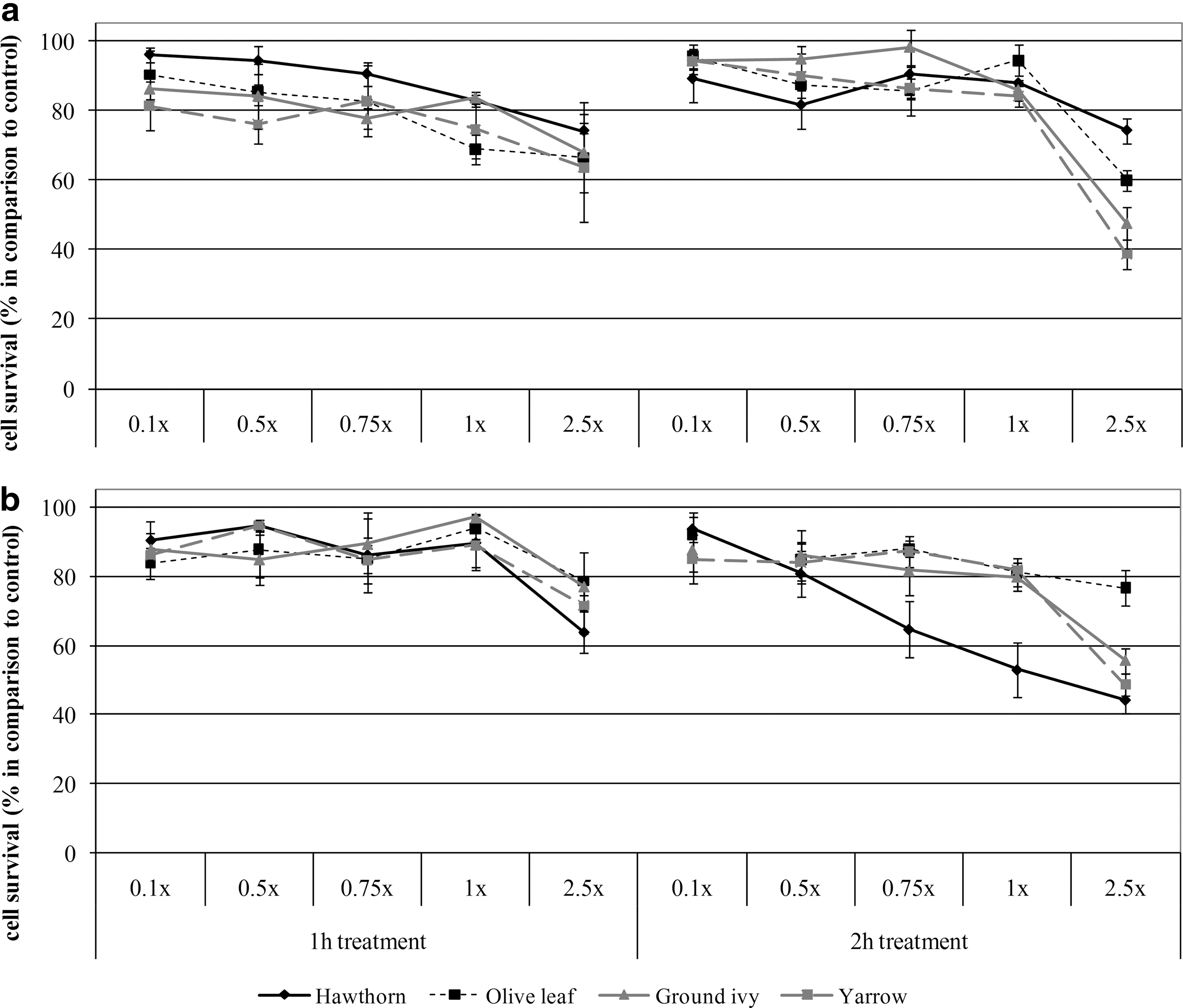
Survival of human laryngeal carcinoma (HEp2) cells following
Interestingly, after the subsequent recovery period that lasted 24 h (Fig. 1b), cytotoxic effect of plant extracts becomes even more pronounced. Prolonged exposure (2 h) to the highest concentrations (2.5×) of hawthorn and yarrow extracts leads to a 50% growth inhibition of HEp2 cells. Again, the highest concentrations of all plant extracts exhibit the most potent cytotoxic effect, although hawthorn extract markedly decreases the cell viability already at the concentration of 0.75×. These findings point out that during the treatment with plant extracts, irreversible events take place and consequences (meaning cytotoxic effect of higher concentrations) become obvious after 24 h, that is, after a period of one cell cycle.
As can be seen from Table 2 and Figure 1a and b, yarrow does not exhibit the highest TPC but induces the strongest cytotoxicity on HEp2 cells, followed closely by hawthorn and ground ivy, whereas olive leaf exhibits different cytotoxic effect despite the low TPC. Cytotoxic effect of olive leaf is the most observable in case of 2 h incubation without the subsequent recovery period (Fig. 1a), indicating that high level of flavonoids plays a crucial role in its prolonged expression of toxicity, which is caused by irreversible events in the first few hours of incubation. Since the results of our study revealed different effects on cytotoxicity induced by different plants, a potential connection between the cytotoxic activity and polyphenolic content and composition of medicinal plants was observed. Namely, the high cytotoxic activity may be attributed to some polyphenolic constituents of these plants. According to Yamamoto et al., 38 ROS are formed during cellular metabolism of higher concentrations of catechins and are often responsible for their cytotoxic effects in tumour cell lines. Lee et al. 39 showed that micromolar concentrations of phenolic acids and their phenethyl esters caused cytotoxic effect on oral cancer cells through G2/M phase arrest. Maurya and Devasagayam 40 also revealed the concentration-dependent antioxidant effects of caffeic and ferulic acids in terms of inhibition of lipid peroxidation and ROS-scavenging after 2,2′-azobis-amidinopropane dihydrochloride-induced damage in mouse liver microsomes and splenic lymphocytes.
Since in ground ivy and yarrow phenolic acids were the predominant polyphenolic compounds, as opposed to hawthorn, which was characterized by a higher variability of polyphenolic constituents, the results imply on a more prominent effect of polyphenolic composition rather than content of investigated plants.
Previous studies indicate that antimicrobial activity 41 and cytotoxic activity 42 might be mediated by the flavonoid-rich yarrow extracts, especially the presence of dicaffeoylquinic acids and flavonic compounds. A water-soluble fraction from a hydro-alcoholic extract of yarrow showed antiproliferative effect on B16 mouse melanoma cells after 2 days of growth. A lyophilized decoction of yarrow was evaluated for antihepatoma activity (cytotoxicity) on five human liver cancer cell lines; at 2 mg/mL the average inhibition of proliferation was 55% on nonhepatitis B virus cell lines and 20% on hepatitis B virus cell lines. 43 Similarly, Bouallagui et al. 44 determined significant antiproliferative activity of olive leaves extract in a dose-dependent manner on MCF-7 human breast cancer cells. However, this effect was prescribed to hydroxytyrosol, which was not identified in our study. It has been well established that extracts obtained from the original material or substrate often exhibit better biological activity than the purified individual substances due to the synergistic effect of all bioactive compounds in the mixture. 45 Synergistic effects have been previously observed in a RA/CA mixture by 2,2′-azobis(2-amidinopropane) dihydrochloride-induced oxidation. 46
Antioxidant/prooxidant activity of medicinal plant extracts
Experiments in this study conducted on human cell line revealed a strong relationship between the type of product/concentration/time of exposure and antioxidant/prooxidant character of medicinal plant extracts. According to our results, noncytotoxic concentrations of ground ivy and hawthorn extract slightly increased ROS formation after 1 h of incubation in dose-dependent manner (Fig. 2), and this effect was more obvious in the case of 2 h of incubation for hawthorn. In spite of the increase of ROS levels for ground ivy and hawthorn, it can be observed that higher concentrations of all examined extracts resulted with a decrease in ROS formation when compared to control. Prolonged incubation (Fig. 2) resulted with even more pronounced decrease of ROS formation, so the highest concentrations of all extracts further decreased the levels of ROS. Olive leaf extract showed antioxidative effect, decreasing the basal level of free radicals determined in the control cells. In case when the highest toxicity of olive leaf extract was obtained (2 h of incubation without recovery period), it was noticed that nontoxic concentration (0.1×) caused a prooxidative effect, indicating possible cause of increased cell death (Fig. 2.).
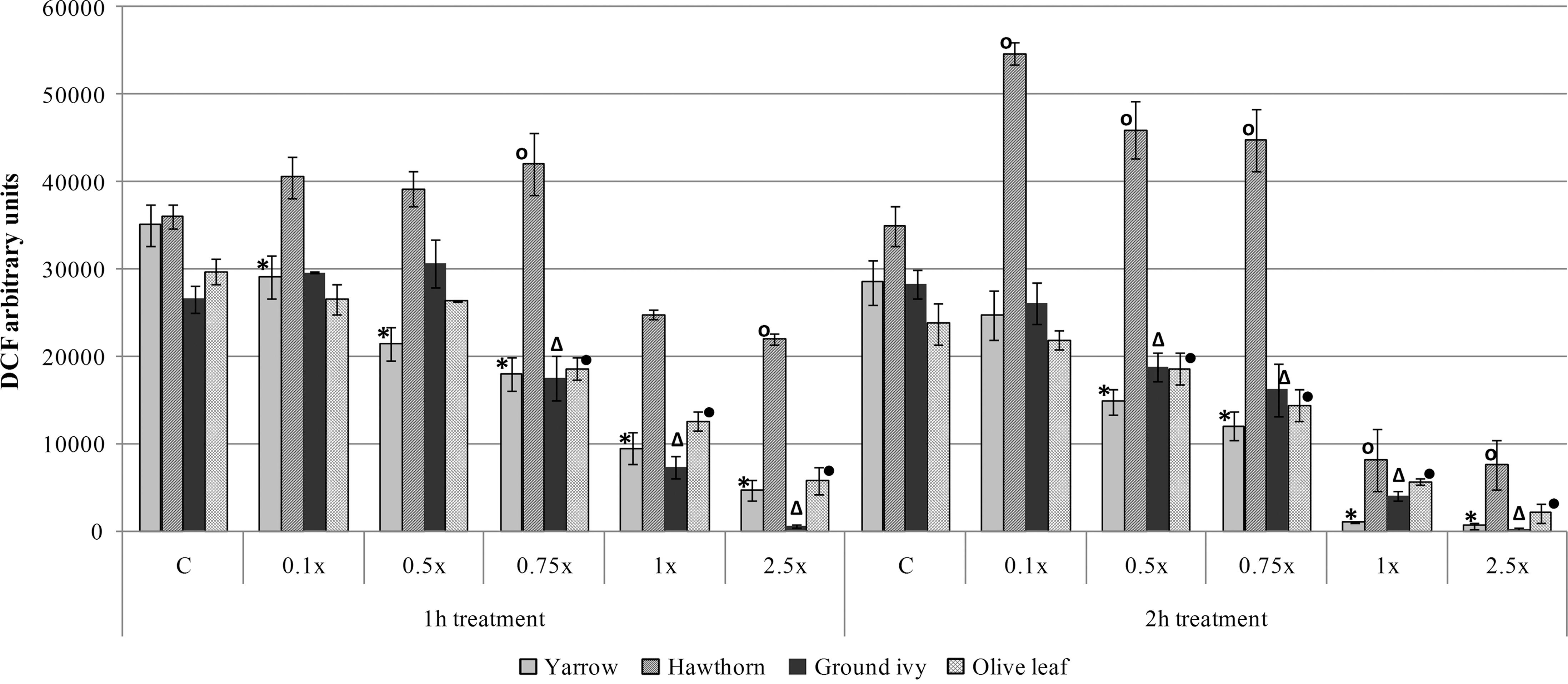
Comparison of reactive oxygen species (ROS) formation in HEp2 cells treated with medicinal plant extracts for 1 and 2 h. Pooled data obtained from three experiments (the mean at the point±standard deviation [SD]). Values superscripted with the same mark (*, o, Δ, •) are significantly different (P<.05) compared to control (C).
As can be seen on Figure 3, ROS formation in the HEp2 cells treated with medicinal plant extracts after the subsequent recovery period of 24 h is also affected by their concentration. In general, the treatment of HEp2 cells with higher concentrations of all medicinal plants resulted in a decrease in ROS formation when compared to control. The prolonged incubation time did not have any influence on these events. During the recovery period (Fig. 3), it can be seen that lower concentrations of hawthorn extract after 2 h treatment induced a significant (P<.05) increase in ROS, whereas the highest concentration caused a decrease in ROS formation. This was also observed for olive leaf (Fig. 3). Since prolonged incubation of HEp2 cells with hawthorn (Figs. 2 and 3) and olive leaf (Fig. 3) extracts caused an increase in the ROS formation, one of the possible explanation for such an effect could be a cytotoxic effect of phenoxyl radicals and hydrogen peroxide formed during cell incubation with the plant extracts.

Comparison of ROS formation in HEp2 cells treated with medicinal plant extracts for 1 and 2 h after subsequent recovery period (24 h). Pooled data obtained from three experiments (the mean at the point±SD). Values superscripted with the same symbol (*, ○, ▵, •) are significantly different (P<.05) compared to control (C).
Correlation between cytotoxicity and ROS formation induced by medicinal plants
The coefficients obtained by establishing a linear correlation between the cytotoxicity and ROS formation in the cells treated with plant extracts ranged from r=0.751–0.954 after 1 h treatment and r=0.509–0.704 after 2 h treatment. The recovery period resulted with lower correlation, varying between r=0.128–0.719 after 1 h treatment and r=0.667–0.972 after 2 h treatment. The generation of ROS is generally considered to be a major contributor to substrates toxicity and their formation, by exceeding the cellular defensive capacity and causing oxidative damage to biomolecules. 47 However, since our results demonstrate a high correlation between cytotoxicity and ROS levels, which is attributed to high cytotoxicity and decrease of ROS formation after the treatment with four analyzed medicinal plants, the results suggest the antioxidative activity of medicinal plants and induction of different cellular responses, depending on the chemical properties of plants. Sakihama et al. 48 proposed that polyphenols can act as antioxidants by donating electrons to different enzymatic and nonenzymatic systems in the cell for the detoxification of hydrogen peroxide produced under stress conditions. Also, the effect of the mixture of strong bioactives may be attributed for the observed antioxidative effects of plant extracts. Taking into account the chemical composition of medicinal plant extracts examined in this work, some other bioactive constituents of these medicinal plants, beside polyphenols may have contributed to exerting such biological responses.
It has frequently been suggested that metal impurities can contribute to cell toxicity, by triggering ROS production by Fenton's reaction. Dietz et al. 49 and Sahw et al. 50 reported that heavy metals induce oxidative stress in cells and tissues by transfering electrons directly in single-electron reactions, which generate free radicals. The so-called transition metals (Fe, Cu, Mn, etc.), which have unpaired electrons in their orbitals, accept and donate single electrons, thus promoting monoelectron transfers to O2 and generally ROS interconversion and oxidoreduction phenomena. In our study, a high variability of toxic metal impurities in four medicinal plants was found using ICP-AES assays. As can be seen in Table 4, all medicinal plants contain different content and composition of transition elements, which would imply on a potential for elevating and inducing ROS formation in HEp2 cells after the treatment with the plant extracts. However, since our study revealed a decrease in ROS formation, this effect cannot be attributed to the presence of metal impurities in medicinal plants indicating that some other constituents of medicinal plants may have influence the cell response.
Excerpt from Belščak-Cvitanović et al. 26
<LOD, below level of detection.
In recent years, bioactive polysaccharides isolated from natural sources have attracted much attention in the field of biochemistry and pharmacology. Polysaccharides isolated from different medicinal mushrooms have been shown to display inhibitory effects on various tumor cells, but the effect of polysaccharides from various medicinal plants is much less known. Stimpel et al. 51 reported that purified polysaccharides (EPS) prepared from Echinacea purpurea are shown to strongly activate macrophages. Macrophages activated with these substances develop pronounced extracellular cytotoxicity against tumor targets. According to the results of our study, ground ivy and yarrow are characterized by a higher content of soluble polysaccharides, in comparison to hawthorn and olive leaf, which is in agreement with the observed higher cytotoxicity exerted by these two extracts on HEp2 cells. A plausible explanation of our findings is that both polyphenols and polysaccharides block the formation of the ROS that result from the metal-mediated reduction of hydrogen peroxide. Our experiments indicate that bioactive compounds from water extracts of these four medicinal plants may act as antioxidants in Fenton-type reactions, although the chemical mechanism is unclear. Supported by these facts, it seems that a mixture of all bioactive compounds from the four investigated medicinal plants exhibits beneficial cytotoxic activity on HEp2 cells, but the contribution of each individual component must be further clarified.
Conclusions
This study provides the relationship between the bioactive content and composition of four traditional medicinal plants and their effect on cytotoxic and antioxidant/prooxidant activity on HEp2 cells. Comparison of values linked to differently prepared plant extracts points to the highest polyphenolic yield measured in extracts prepared at higher temperature and longer extraction time. The results of experimental analyses revealed a relationship between the type of product/concentration/time of exposure and cytotoxic and antioxidant/prooxidant characters of plant extracts. All examined plants exhibited potent cytotoxic effect, as well as antioxidant properties at higher concentrations (decrease of ROS formation), thus obtaining a high linear correlation between these two biological responses. Due to a high variability and presence of different metals in the examined plants, the high cytotoxicity and no generation of ROS may be prescribed to the effect of polyphenolic compounds or soluble polysaccharides, which have been known to exert similar effects in previous studies.
Footnotes
Acknowledgments
This work was supported by the Ministry of Science, Education and Sports, Republic of Croatia projects no. 058-0000000-3470 and 058-0582261-2246, as well as Croatia-Serbia Bilateral agreement 2011–2012.
Author Disclosure Statement
The authors declare that they have no conflict of interest nor competing financial interests to disclose.