
Abstract
This study was aimed at assessing the potential of essential oil from the leaf of Hoslundia opposita in the treatment of diabetes. Forty-eight rats (Rattus norvegicus) were randomized into two groups; nondiabetic and diabetic groups, each with four subgroups. Animals in the diabetic group were induced with diabetes using a single dose of alloxan monohydrate, 160 mg/kg body weight (b. wt.). The rats were treated with 110 and 220 mg/kg b. wt. of the essential oil. All treatments were administered, intraperitoneally, once a day for 4 days. In the nondiabetic condition, there was no effect of the oil on fasting blood glucose (FBG) levels in rats. In diabetic rats, the oil caused a significant reduction in FBG levels. Treatment with 110 mg/kg b. wt. of the oil reduced FBG almost to the normoglycemic level by day 4 and the overall glucose excursion during a 3-h intraperitoneal glucose tolerance test approached the baseline level at 120 min. Also, hepatic glycogen was significantly higher, while the glucose concentrations were lower in the diabetic-treated group when compared with the diabetic untreated group. Histological examinations revealed a mildly distorted architecture of the pancreatic islets β-cells of diabetic rats treated with the oil, while those of the untreated rats were severely degenerated. Overall, the in vivo antihyperglycemic activity of the essential oil may prove to be of clinical importance in the management of type 2 diabetes.
Introduction
D
Conventionally, T1DM is treated with exogenous insulin and T2DM with synthetic oral hypoglycemic agents like sulfonylureas and biguanides. 8,9 However, the hormone fails as a curative agent for complications of diabetes and synthetic oral drugs produce adverse health effects. 9,10 Due to the high cost of management and profiles of adverse side effects associated with various available synthetic antihyperglycemic agents, there is a need to develop improved alternatives with less side effects for the management of DM by exploring potentials offered by traditional phytotherapies. The oral hypoglycemic agent metformin was derived from the flowering plant, Galega officinalis (goat's rue or French lilac), which was a common traditional remedy for diabetes. 11
Hoslundia opposita Vahl. (Lamiaceae) is a herbaceous perennial shrub native to Africa and grows as a wild plant in Nigeria where it is commonly known as “Oke ota” by the Igbos and “Efirin odan” by the Yorubas ethnic groups. 12 Infusions of its leaves are widely used in African traditional medicine as a purgative, diuretic, febrifuge, antibiotic, and antiseptic, as well as antidiabetic. 13,14 In African traditional medicine, extracts from the leaves of H. opposita are used in the management of diabetes. 12 Numerous reported biological activities of the extracts from H. opposita, such as the antimalarial, anticonvulsant, and antimicrobial activities, confirm their use in folk medicine. 15 –17 Bioactive compounds such as of alkaloids, tannins, flavonoids, cardiac glucosides, and essential oils have been identified as phytoconstituents responsible for pharmacological actions. 13,18
Essential oils are natural multicomponent systems composed mainly of terpenes and other nonterpene components. 19 They are valuable natural products used as raw materials in many fields, including perfumes, cosmetics, aromatherapy, phytotherapy, spices, and nutrition. 20 The bioactivities of aromatic plants are partly attributed to the presence of essential oils. The possible role and mode of action of these natural products with regard to their bioactivities have been investigated by numerous researchers. 19 Studies in our laboratory had established the antidyslipidemic and lipid-lowering effect of the 1,8-cineole (eucalyptol) chemotype essential oil extracted from the leaves of H. opposita in experimental rats. 21 –23 A few antihyperglycemic effects of essential oils from plants have been reported. 24,25 However, the mechanisms of antidiabetic action of such phytochemicals are scanty in literature. Thus, this study was designed to evaluate the antidiabetic potentials and possible mechanisms of the antihyperglycemic activity of leaf essential oil of H. opposita in rats.
Materials and Methods
Chemicals and apparatus
The assay kit for glucose was obtained from Randox Laboratories (Antrim, United Kingdom). Alloxan monohydrate was a product of Sigma-Aldrich (St. Louis, MO, USA). Dimethyl sulfoxide and all other reagents used were of analytical grade and supplied by BDH Laboratories Ltd. (Poole, United Kingdom). Accu-Chek Active Glucometer and strips from Roche diagnostic (Mannheim, Germany). UV-Vis spectrophotometer (Lab-kits, Hunan Province, China) and OHAUS analytical balance (Ohaus Corporation, Parsippany, NJ, USA).
Collection of plant material
Fresh leaves of H. opposita were obtained from the Parks and Gardens Unit of the University of Ilorin, Nigeria. Plant identification was carried out at the herbarium of the Forestry Research Institute of Nigeria (FRIN), Ibadan, Oyo State, where a voucher specimen was deposited.
Oil isolation and standardization
Pulverized leaves of H. opposita (500 g) was hydrodistilled for 3 h in a Clevenger-type apparatus according to the British Pharmacopeia specification (1980). The resulting oils were reconstituted in 5% and 10% saline solution of dimethyl sulfoxide (DMSO). 26
Animal grouping
Forty-eight albino rats with an average weight of 132.50±14.65 g were obtained from the animal house of the Department of Biochemistry, University of Ilorin, Nigeria. All rats were maintained under standard laboratory conditions (12-h light–12-h dark cycle, 25°C±2°C). Before experimentation, the rats were acclimatized to laboratory conditions for a week. The animals were randomized into two groups and labeled as the nondiabetic and diabetic group with four subgroups under each group. This study was carried out following approval from the Ethics Committee on the Use and Care of Laboratory Animals of the Department of Biochemistry, University of Ilorin, Nigeria. The research also adhered strictly to the Principles of Laboratory Animal Care (NIH Publication, No. 85-23).
Induction of experimental diabetes
Animals were subjected to an 18-h fast, and rats in the diabetic groups were given a single dose of alloxan monohydrate (160 mg/kg body weight [b. wt.]) intraperitoneally in an ice-cold 0.9% NaCl solution. Rats in the nondiabetic groups were injected with normal saline. Fasting blood glucose (FBG) was determined after 48 h, using a glucose oxidase-based commercial glucometer (AccuChek active, Roche Diagnostic). Rat showing FBG above 250 mg/dL glucose were considered diabetic. 27
Dose determination
Literature search and preliminary studies were carried out to determine the therapeutic doses. From literature, 1, 8-cineole (eucalyptol), the major constituent of H. opposita leaf essential oil (HOLEO), has been found to possess an LD50 of 2480. 28 Various groups of hyperglycemic rats (n=6 in each; ≥250 mg/dL glucose) were treated with intraperitoneal administration of the vehicle DMSO and 55, 110, and 220 mg/kg b. wt. of HOLEO (equivalent of 62.5, 125, and 250 μL/kg b. wt of HOLEO; oil density 0.88 g/mL) once a day for 4 days and FBG levels were determined on day 4. The results revealed that the 55 mg/kg b. wt. and DMSO failed to show an antihyperglycemic effect, while the 110 and 220 mg/kg b. wt. treatment group showed significant antihyperglycemia. Therefore, 110 and 220 mg/kg b. wt were selected as antihyperglycemic doses for this study.
Administration of oil
All treatments were administered intraperitoneally to rats once a day for 4 days as shown below:
Nondiabetic groups
• Untreated nondiabetic rats
• Nondiabetic rats treated with 220 mg/kg b. wt. of the vehicle; DMSO
• Nondiabetic rats treated with 110 mg/kg b. wt. of essential oil
• Nondiabetic rats treated with 220 mg/kg b. wt. of essential oil
Diabetic groups
• Untreated diabetic rats
• Diabetic rats treated with 14.2 mg/kg b. wt. of standard drug; metformin
• Diabetic rats treated with 110 mg/kg b. wt. of essential oil
• Diabetic rats treated with 220 mg/kg b. wt. of essential oil
Determination of blood glucose
The method of Ortiz-Andrade et al. 29 was used with slight modifications. The animals were subjected to a 12-h overnight fast with free access to water (feeds were withdrawn from 7 pm to 7 am). The rats were treated as shown above. On day 1, blood samples were obtained from their caudal vein, FBG levels were determined at 0 h before treatment and 3 and 6 h after treatment. Feeds were returned and left ad libitum until 7 pm. FBG levels were also determined at 24 and 96 h after treatment.
Sugar tolerance test
This was performed on day 3 of administration. All test animals were fasted for 4 h (from 8 am) and then administered with DMSO, essential oils, or metformin. After 30 min, each rat was injected with 2 g/kg of sugar substrate (equal ratio of glucose and sucrose) solution, intraperitoneally. FBG levels were determined at 0 (before substrate administration), 1, 2, and 3 h after administration. 6,30
Preparation of tissue homogenates
The animals were starved overnight, sacrificed after 96 h of treatment, and quickly dissected; the tissues were excised and immersed in an ice-cold 0.25 M sucrose solution (to maintain the integrity of the tissues). Homogenates were prepared by cutting a known weight of the tissue finely with a clean scissors. The tissues were thereafter homogenized in ice-cold 0.25 M sucrose solution (1g to 5ml solution) using pestle and mortar. Triton x-100 was added to make a final concentration of 1% v/v. These processes were carried out at a temperature ≤4°C. The homogenates were labeled, stored in the refrigerator, and used for analysis within 24 h. 31
Hepatic glycogen and glucose
Glycogen measurement was carried out, as described by Passoneau and Lauderdale, 32 with slight modifications. Glucosyl units were determined by enzymatic oxidation.
Histological examination
Pancreatic tissue specimens were fixed in 10% (v/v) formaldehyde, dehydrated through ascending grades of ethanol, cleaned in xylene, and processed into paraffin blocks. Tissue sections (5 μm thick) were prepared according to the method described by Drury and Wallington 33 and stained with hematoxylin/eosin. Sections were examined using light microscopy (×100) for demonstration of pancreatic pathological changes.
Statistical analysis
All data are expressed as the mean of six replicates±standard error of mean. One way analysis of variance with Dunnett's post hoc test was used for multiple comparison of treatment groups using GraphPad prism version 5.02. Values were considered statistically significant at the 95% confidence level.
Results
The intraperitoneal (IP) administration of 110 mg/kg b. wt. of the oil caused significant reduction [F (5, 30)=194.8; P<.0001] in the glycemic level after 3 h, while 220 mg/kg b. wt of HOLEO produced a significant [F (5, 30)=101.1; P<.0001] reduction in hyperglycemia after 6 h (Table 1). The reduced FBG levels observed after the first day of treatment with both doses of the oil were maintained until the end of the treatment. The study revealed a normoglycemic value on day 4 (96 h) of treatment with IP administration of 110 mg/kg b. wt.
Values are expressed as mean of six replicates±SEM; values with the different superscript letters across a row are statistically different (P<.05).
HOLEO, Hoslundia opposita leaf essential oil; SEM, standard error of mean.
In nondiabetic rats, FBG levels were reduced during the acute experimental period (0–6 h), but returned to normal range after 24 h. Nondiabetic rats administered with 110 or 220 mg/kg b. wt. of HOLEO revealed no significant reduction in FBG levels, except on day 4, the FBG levels significantly [F (3, 20)=30.02; P<.0001] reduced in nondiabetic rats administered 110 mg/kg of HOLEO or 220 mg/kg b. wt. of DMSO, when compared with the normal rats not administered essential oil (Table 2)
All values are expressed as mean of six replicates±SEM; values with different superscript letters across a row are statistically different (P<.05). Values with * along a column are significantly different (P<.05) from each other.
DMSO, dimethyl sulfoxide.
The effect of HOLEO (110 and 220 mg/kg b. wt. treatment) on the glucose tolerance test in normoglycemic and diabetic rats evaluated on day 3 of treatment revealed normal fasting glycemia and increased basal hyperglycemia in the normoglycemic and diabetic groups, respectively, and this was aggravated by the IP sugar load. At 120 min, the glucose approached baseline levels in rats treated with 110 mg/kg b. wt. of HOLEO, indicating glucose tolerance (Tables 3 and 4).
Values are expressed as mean of six replicates±SEM; values with different superscript letters across a row are statistically different (P<.05).
Values are expressed as mean of six replicates±SEM; values with different superscript letters across a row are statistically different (P<.05).
In the nondiabetic state, hepatic glucose [F (3, 20)=5.892; P=.0047] and glycogen levels were not significantly altered on treatment with either 110 or 220 mg/kg b. wt. of HOLEO [F (3, 20)=2.001; P=.1463; Table 5]. However, glucose concentrations were significantly [F (3, 20)=27.10; P<.001] reduced and the glycogen content increased [F (3, 20)=1630; P<.0001] in diabetic rats treated with HOLEO (110 and 220 mg/kg b. wt.) or metformin compared with levels in the untreated diabetic group (Table 6).
Values are expressed as mean of six replicates±SEM; values with different superscript letters along a column are statistically different (P<.05).
Values are expressed as mean of six replicates±SEM; values with different superscript letters along a column are statistically different (P<.05).
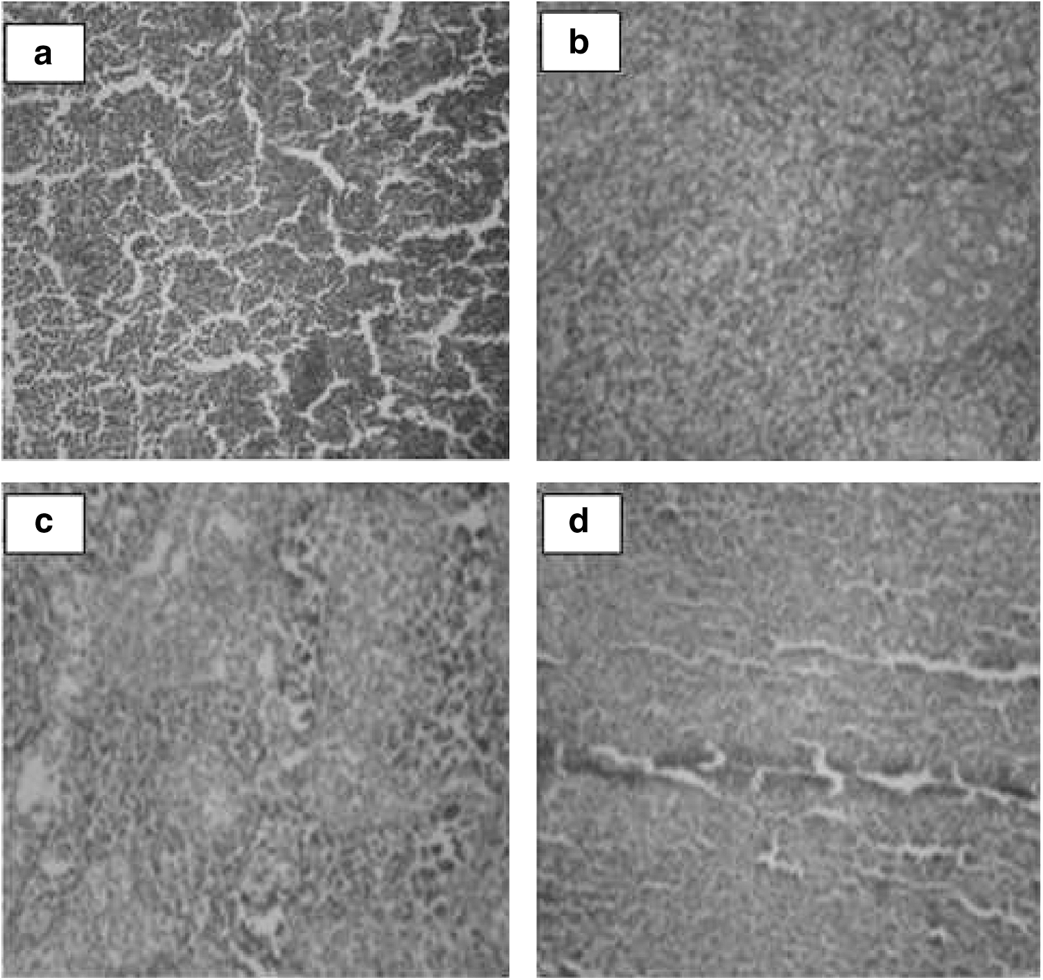
The pancreas from nondiabetic untreated rats display normal architecture with abundant compactly arranged islets of Langerhans of varied sizes, scattered unevenly in the tissue without inflammatory cells (Fig. 1a). The 110 and 220 mg/kg b. wt. of HOLEO-treated groups presented a mild disorganization of the exocrine but with preserved architecture (Fig. 1b, c).

Photomicrograph (×100) of the pancreas of untreated nondiabetic rat
On the contrary, the pancreatic tissue of the diabetic untreated rat was severely degenerated with distorted architecture (Fig. 2a). Diabetic rats on treatment with HOLEO extracts or metformin (Fig. 2b–d) revealed mildly or moderately degenerated pancreas with distorted architecture, but to a much lesser extent, when compared with the alloxan-induced diabetic untreated rats.
Photomicrograph (×100) of the pancreas of diabetic rat
Discussion
Alloxan has been reported to cause massive selective destruction and reduction of β cells of the islets of Langerhans mediated by the formation of reactive oxygen species, resulting in partial or complete loss of insulin synthesis and leading to the development of hyperglycemia. 34,35 Observed hyperglycemia in diabetic rats following alloxan induction may also be due to induced gluconeogenesis in the absence of insulin. 36 In this study, essential oil extracted from H. opposita reduced hyperglycemia after 3 and 6 h following a single dose IP administration. However, when studying a chronic disease such as diabetes, it is more pertinent to test for sustained low blood glucose levels with long-term treatment rather than acute antihyperglycemic effects after a single dose. 30 Subchronic investigation revealed that the essential oil of H. opposita progressively reduced blood glucose in alloxan-induced hyperglycemic rats to levels comparable to the standard drug metformin after 24 h and this effect was maintained until the end of treatment. The lower dosage of 110 mg/kg b. wt. of H. opposita leaf essential oil was the most effective dosage, reducing the blood glucose level almost to the normoglycemic level by day 4 of the administration. The lowering of blood glucose levels due to the administration of essential oil affirmed the claim of the use of H. opposita extract in traditional medicine for treatment of diabetes. 14
Although scanty literature is available on glycemic control effected by essential oils extracted from plants in experimental animals, a significant reduction in FBG levels have been reported in diabetic rats treated with essential oils. 24,25 In our previous investigation on the essential oil extracted from leaves of H. opposita, 20 compounds were identified. 21 The bulk of the oils were characterized by the abundance of oxygenated monoterpenes with 1, 8-cineole (72.3%) as the principal constituent Essential oils are complex mixtures of compounds. Each of these compounds is known to contribute to the beneficial or adverse effects of these oils. 20 From previous studies, 1,8-cineole has been found to exhibit an in vitro antidiabetic activity. 37
In addition, no remarkable hypoglycemia was observed in blood glucose levels of nondiabetic rats treated with both doses of HOLEO. Thus, HOLEO was more potent in diabetic than in nondiabetic rats. These findings suggest that the metabolic regulation of glucose by the extract may be similar to the approved antihyperglycemic drug metformin, which does not reduce blood glucose in normoglycemic patients unlike the effect exhibited by glibenclamide. 9 Metformin causes a higher glucose decrease in diabetic individuals by suppressing liver glucose production in the gluconeogenesis pathway. 6,38
Results from this study also suggest that the oil improved plasma glucose homeostasis. This may be either through peripheral uptake of glucose or hepatic glucose reduction as evident from the hepatic glycemic result or through stimulation of the undamaged or residual pancreas to secrete insulin. 29,30 The diabetic rats on treatment with HOLEO showed improved pancreatic architecture. This study correlates with other studies on histopathological amelioration of rat pancreas architectural degeneration by antidiabetic medicinal plant extracts. 39,40
In conclusion, data from this study revealed that intraperitoneal administration of the essential oil extracted from the leaf of H. opposita exhibits antihyperglycemic effects in diabetic rats without net hypoglycemic effects in the nondiabetic state.
Footnotes
Acknowledgments
The authors are grateful to the academic and research technologists of the Department of Biochemistry, University of Ilorin, Ilorin, Nigeria and the Biotechnology/Chemistry Advanced Laboratories, Sheda Science and Technology Complex, Abuja, Nigeria for their technical support.
Author Disclosure Statement
No competing financial interests exist.