




Abstract
Exposure to ultraviolet (UV) radiation induces various pathological changes, such as thickened skin and wrinkle formation. In particular, UVB irradiation increases matrix metalloproteinase (MMP)-1 production and collagen degradation, leading to premature aging, termed photoaging. The azuki bean (Vigna angularis; VA) has been widely used as a food product as well as a traditional medicine. However, its activity needs additional study to confirm its functional application in foods and cosmetics for protecting skin. In this study, hot-water extract from VA (VAE) and its active component, rutin, were investigated to determine their antiphotoaging effects. VAE was found to have antioxidant activity. In UVB-exposed normal human dermal fibroblasts cells with VAE and rutin treatments, MMP-1 production was significantly suppressed (90% and 47%, respectively). The effects of both topical and oral administration of VAE were tested in UVB-irradiated hairless mice. VAE suppressed wrinkle formation and skin thickness by promoting elastin, procollagen type I, and TGF-β1 expression (118%, 156%, and 136%, respectively) and by diminishing MMP-1 production. These results suggest that VAE may be effective for preventing skin photoaging accelerated by UVB radiation.
Introduction
S
Collagen and elastin are major components of ECM fibers, which support cells. In particular, type I collagen is known as a primary structural protein in skin. 6 It is released in precursor form as procollagen type I. 7 MMP-1 secretion and transforming growth factor β1 (TGF-β1) productions affect the synthesis of procollagen type I. 8 –10 Elastin provides skin elasticity and shape recovery. 11 Similar to collagen, it is influenced by MMP secretion and TGF-β expression, which activate the elastin promoter. 12,13
To protect skin against photoaging, it is important to maintain water balance in the SC of the epidermis. To maintain skin moisture, various hygroscopic molecules, such as natural moisturizing factors (NMFs), are present in the SC. 14 UV exposure causes alteration in the skin barrier, which influences trans-epidermal water loss (TEWL). TEWL is defined as the quantity of water lost from the epidermal layer through evaporation and diffusion. 15 TEWL is well known as a functional indicator of the SC barrier. 16
Azuki beans (Vigna angularis; VA) are one of the most important crops in Korea, China, and Japan. In Korea, VA has been consumed as a traditional food, often prepared as patjuk (red bean porridge), for many centuries. When made into foods, such as breads and confectionaries, VA is usually boiled and mashed. VA contains rutin, glyceroclycolipids, digalactosylononitol, kaempferolrutinoside, procyanidin dimer, protocatechuic acid, and azukisaponin. 17,18 Rutin, a polyphenolic bioflavonoid, has been studied extensively due to its promising pharmacological actions, such as antitumor, antibacterial, antiviral, and antiallergic effects. 19,20 Many researchers have reported that VA has antibacterial and antioxidative activities. 21,22 In particular, hot-water extract from VA (VAE) reduces serum cholesterol levels and has a hypoglycemic effect in diabetic mice. 23,24 In skin, VA stimulates melanogenesis and pigmentation while inhibiting adhesion, invasion, and metastasis of murine melanoma cells. 25,26 However, there are no studies that investigated the antiphotoaging effects of VA and its constituents. In this study, we investigated the protective effects of hot-water-extract VA and its active components against UVB-induced photoaging in normal human dermal fibroblasts (NHDF) cells in vitro and in hairless mice in vivo.
Materials and Methods
Materials
VA was obtained from Jeongseon Nong-hyup (Jeongseon-gun, Gangwon-do, Korea). 500 g of VA (dried materials) were extracted for 1 h in 5 L of hot water. The extract was purified by filtration (Whatman, Piscataway, NJ, USA), and the solvent was evaporated under vacuum.
Liquid chromatography–mass spectrometry analysis
Identification of VAE standard composition was determined by liquid chromatography–mass spectrometry (LC/MS; Technologies 6530 Q-TOF 6410 Triple Quad LC/MS; Agilent Technologies, Inc., Waldbronn, Germany). First, VAE was fractionalized using ethyl acetate and butanol solution to elevate detection sensitivity before LC/MS analysis. In this procedure, rutin was used as the VAE standard composition. LC/MS was conducted with solvent containing 0.1% formic acid in water and acetonitrile for gradient elution under a flow rate of 2 mL/min. Rutin with concentrations of 5, 10, 25, 50, and 100 ppm was used to detect the calibration curve. The ethyl acetate and butanol fractions of VAE at 2000 ppm were analyzed to compare compositions.
2,2-Diphenyl-1-picrylhdrazyl scavenging activity
Free-radical-scavenging activity was measured by 2,2-diphenyl-1-picrylhdrazyl (DPPH) assay. A 20-μL sample in distilled water was placed in a 96-well plate, and 180 μL of 2,2-diphenyl-1-picrylhdrazyl (0.2 mM) in methanol was also added. After 30-min reaction in dark conditions, the absorbance was recorded by a microplate reader (Molecular Devices E09090; San Francisco, CA, USA) at wavelength of 520 nm. VAE and arbutin were separately tested at various concentrations from 0.1 to 250 μg/mL.
Cell culture
NHDFs were obtained by a skin biopsy on a healthy, young male donor (MCTT Core, Inc., Seoul, Korea). NHDFs were cultured in DMEM supplemented with 10% heat-inactivated FBS and 1% penicillin-streptomycin at 37°C in atmosphere containing 5% CO2. Cells were cultivated in 100-mm culture dishes and seeded in 40-mm culture dishes (1.2×10⁵ cells/well) when they reached more than 80% confluence.
UV irradiation and VAE treatment
UVB irradiation and sample treatments were performed according to a method previously reported by Hwang et al. 27 The NHDFs were seeded in 40-mm tissue culture dishes (1.2×10⁵ cells). When cells reached over 80% confluence, they were rinsed twice with phosphate-buffered saline (PBS). All irradiations were performed under a thin layer of PBS. The cells were then exposed to UVB (144 mJ/cm2) with a thin layer of PBS (300 μL) using a UVB irradiation machine (Bio-Link BLX-312; Vilber Lourmat GmbH, Marne-La-Vallée, France). After UVB irradiation, cells were washed with PBS in triplicate and immediately treated with VAE at 1, 10, and 25 μg/mL in serum-free medium conditions. Nonirradiated control cells were maintained in the same culture conditions without UVB exposure.
Measurement of ROS generation
After 24 h of UVB irradiation, cells were stained with 30 μM 2′7′-dichlorofluorescein diacetate (DCFH-DA; Sigma-Aldrich, St. Louis, MO, USA) for 30 min at 37°C in a CO2 incubator. The cells were then analyzed by flow cytometry (FACSCalibur™; Becton-Dickinson, San Jose, CA, USA).
MTT assay
MTT assay is a colorimetric assay capable of measuring cell viability (CV) by reducing MTT to formazan dyes, producing a purple color. After 72 h of incubation, the volume of medium was reduced to 1 mL, and 100 μL of 1 mg/mL MTT was added to each well. Next, cells were incubated in the presence of 5% CO2 and 95% O2 at 37°C. After 2 h of incubation, cell medium was removed, and 1 mL of DMSO was added into each well to dissolve the formazan crystals. Absorbance was determined on a microplate reader (Molecular Devices E09090) at a wavelength of 570 nm. Blank group means cells and sample free; it was measured by using spectrophotometer. CV for a well was calculated by the following equation: CV=(the OD value of treated well/the OD value of nontreated control well)×100%. 27
Measurement of MMP-1 production
After 72 h of incubation, the cell medium was collected from each well. MMP-1 production was analyzed from cell medium using commercially available ELISA kits (Human total MMP-1 kit; R&D Systems, Minneapolis, MN, USA) in accordance with the manufacturer's instructions. Each sample was determined in triplicate.
Experimental animals
Six-week-old male hairless mice (SKH:HR-1) (29–34 g; n=48) were obtained from Central Lab Animals, Inc. (Seoul, Korea). The animals were randomly divided into eight groups of six mice per cage (three topical application groups and five dietary groups) and housed in conditions of 22°C±1°C, 60%±5% humidity, and 12-h light/dark cycles. Animals were acclimatized for 1 week before the study. The experimental protocol [KHUASP(SU)-12-09] was approved by the Institutional Animal Care and Use Committee of Kyung Hee University.
UVB irradiation
Five Sankyo Denki sunlamps with a peak irradiance at 310 nm were used for UVB irradiation (Kanagawa, Japan). Irradiance was measured using an IL1700 Research Radiometer (International Light, Inc., Newburyport, MA, USA). Bulbs were positioned 15 cm above mice. In the topical application group, mice were exposed to 100 mJ/cm2 UVB radiation (1 minimal erythematal dose=100 mJ/cm2) seven times per week for the first week, and to 200 mJ/cm2 three times a week for 5 weeks thereafter. In the dietary group, mice were exposed to 100 mJ/cm2 UVB irradiation seven times per week for the first week, and three times a week for 10 weeks thereafter.
Topical application
Thirty hairless mice were randomly divided into five groups of six mice per cage: (1) no UVB exposure (Normal), (2) UVB irradiation alone (UVBcont), (3) UVB irradiation with 1% RP treatment (RP 1%), (4) UVB irradiation with 1% VAE treatment (VAE 1%), and (5) UVB irradiation with 5% VAE treatment (VAE 5%). For the first week, all mice were exposed to UVB without any other intervention. Thereafter, hairless mice were treated with a vehicle (7:3 [v/v] propylene glycol:ethanol) or 1% and 5% VAE, and 1% RP mixture three times a week after 1 h of UVB irradiation until 4 weeks. However, the normal group was not exposed to UVB radiation. In this experiment, 1% RP was used as a positive control.
Diet, body weight, and food efficiency rate
Eighteen hairless mice were divided into three groups of six mice per cage: (1) Normal (control diet only), (2) UVBcont (UVB exposure+control diet), and (3) VAE 1% (UVB exposure+diet containing 1% VAE). All mice were fed a solid formula feed. During the experimental period, access to food and water was provided ad libitum. The compositions of the experimental diets given to each group are shown in Table 1. The amount of dietary fat (supplied as corn oil) was fixed at 10% of the dietary weight. To investigate the food efficiency ratio of each group, body weight and food intake values were measured once per week during the experimental period.
Group Normal: control diet only; group UVBcont: UVB irradiation+control diet; group VAE 1%: UVB irradiation+diet containing 1% VAE;× indicates nonirradiated hairless mice and ○ indicates UV-irradiated hairless mice. VA, Vigna angularis; VAE, hot-water extract from VA.
Wrinkle measurement
To determine skin wrinkles, skin replicas were made by SILFLO impression material (Flexico, Colchester, United Kingdom) from the dorsal skin of hairless mice. A visiometer technique was used to detect changes in the transparency of thin silicone gel prints of skin surfaces. Data were collected with a CCD video camera. The image files were analyzed using Skin Viscometer SV 600 software (Courage & Khazaka, Cologne, Germany). Arbitrary units (R1–R5) were assigned to each sample based on depth measurements of the furrows according to shadow size and brightness: skin roughness (R1), largest value of the five distances (R2), average of maximum distance (R1) derived from each of the five parts of the line (R3), mean area surrounded by horizontal line drawn at the highest crest and furrows profile (R4), and mean deviation of the furrows profile to the middle line (R5).
Measurement of physiological skin functions
SC hydration, TEWL, and erythema index (EI) were measured using the appropriate probes (DermaLab®; Combo, Cortex Technology, Denmark).
Histological analysis
Dorsal skin specimens of the hairless mice were obtained after final UVB irradiation and fixed in 4% paraformaldehyde for 24 h. Next, dorsal skin specimens were embedded in paraffin and sectioned at 10 μm. Hematoxylin and eosin (H&E) staining was used to observe epidermal thickness and Masson's trichrome staining was used to detect collagen fiber. H&E staining was successively conducted by deparaffination, hydration, hematoxylin staining, eosin staining, and dehydration. Masson's trichrome was also carried out as follows. The paraffin-embedded skin specimens were stained with Bouin's solution and Weigert's iron hematoxylin working solution. Skin specimens were differentiated in phosphomolybdic-phosphotungstic acid solution and stained with aniline blue solution. Finally, the stained skin specimens were dehydrated in series. After H&E and Masson's trichrome staining, all stained skin specimens were observed by light microscope.
Western blot analysis
For western blot analysis, cells and skin tissues were lysed by lysis buffer (50 mM Tris-Cl [pH 8.0], 0.1% SDS, 150 mM NaCl, 1% NP-40, 0.02% sodium azide, 0.5% sodium deoxycholate, 100 μg/mL PMSF, 1 μg/mL aprotinin, and phosphatase inhibitor) and centrifuged at 12,000 g for 7 min. Cell and skin lysates were then homogenized in an equivalent amount of protein by measuring protein concentration using Bradford reagent (Bio-Rad, Hercules, CA, USA). Homogenized proteins were electrophoresed by 8% or 10% sodium dodecyl sulfate–polyacrylamide gel (SDS-PAGE) and thereafter were transferred from SDS-PAGE to a nitrocellulose membrane (Amersham Pharmacia Biotech, Buckinghamshire, United Kingdom). Nonspecific binding was then blocked with 5% nonfat milk in TBST (50 mM Tris-HCL [pH 7.5], 150 mM NaCl, and 0.1% Tween 20) for 1 h under room temperature conditions, and the primary antibody was captured overnight at 4°C. The membrane was then washed with TBST buffer three times and incubated with secondary antibody (Santa Cruz Biotechnology, Inc., Santa Cruz, CA, USA) for 1 h at room temperature. Finally, the protein level was determined using chemiluminescence detection ECL reagents (Fujifilm, LAS-4000, Tokyo, Japan) and ImageMaster™ 17 2D Elite software, version 3.1 (Amersham Pharmacia Biotech, Piscataway, NJ, USA).
Statistical analysis
Results were presented as mean±standard deviation (SD). All results are averages of at least three independent experiments. A statistical comparison of different treatment groups was determined by one-way ANOVA followed by Duncan's test. P<.05 was considered statistically significant.
Results
Identification of rutin as VAE standard composition
As shown in Figure 1A, rutin was detected in the standard composition of VAE and identified at 10.20 min. According to LC/MS analysis, VAE was confirmed to possess various compounds, including rutin. The total rutin concentration in VAE was calculated as 0.33 mg/g (Fig. 1B).
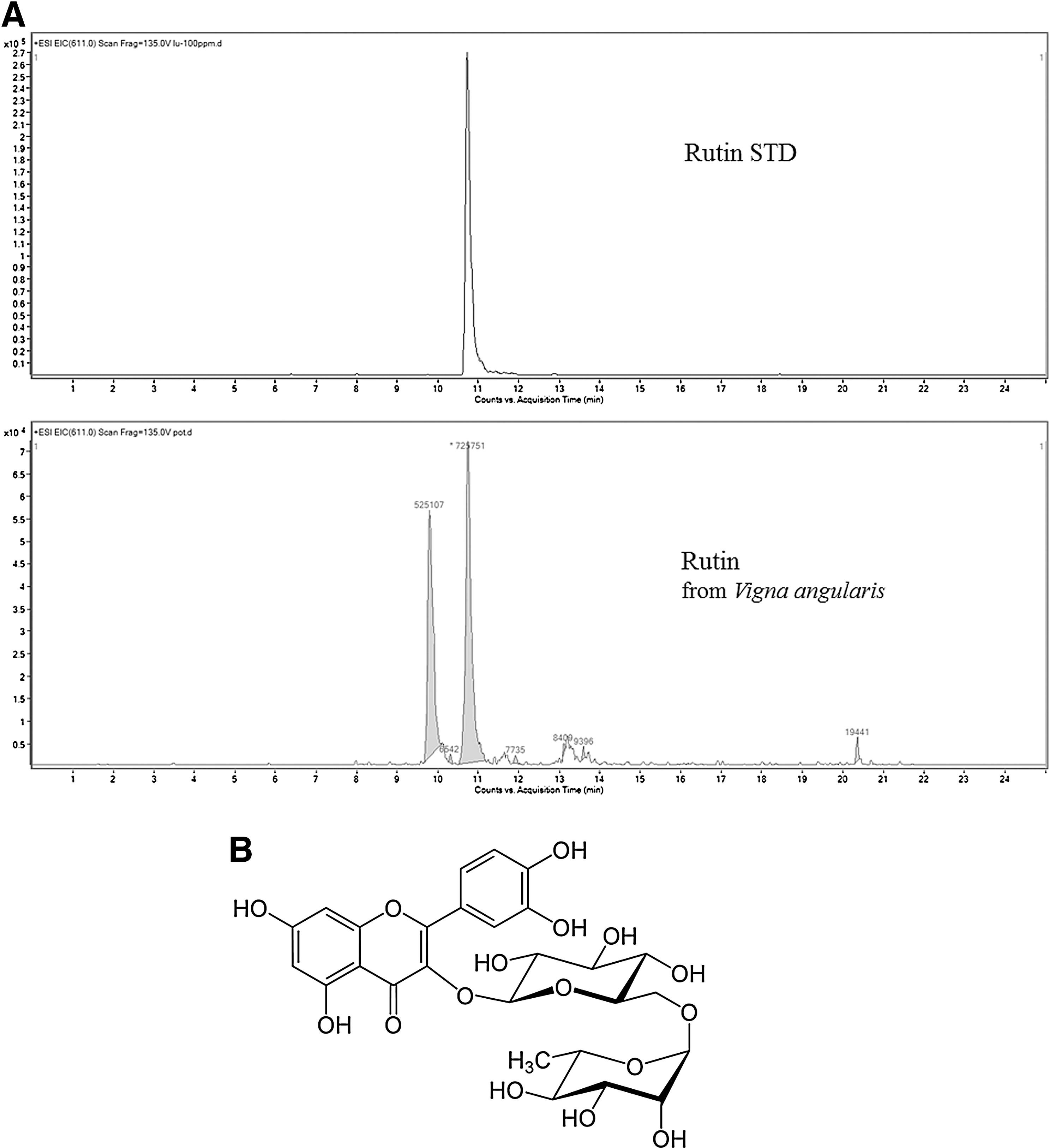
Liquid chromatography–mass spectrometry (LC/MS) results regarding VAE. LC/MS analysis was performed using 100 ppm of rutin and 2000 ppm of VAE ethyl acetate and butanol fraction
Antioxidative activities of VAE
The free-radical-scavenging activity of VAE and arbutin was determined by DPPH assay. As shown in Figure 2, free-radical-inhibition activity increased in a dose-dependent manner. Also, the free-radical-scavenging activity of VAE was considerably higher than that of arbutin used as a positive control.

Free-radical-scavenging activity of VAE and arbutin. Sample was treated with indicated concentrations of VAE and arbutin. Arbutin was used as positive control. Values are means±SDs. *P<.05 and **P<.01 indicate significant differences, respectively.
NHDFs have been used to investigate UVB-induced production of ROS with FACS analysis. Generally, UVB irradiation promotes ROS generation. As expected, UVB-irradiated cells evidently enhanced ROS generation compared with nonirradiated cells. From Figure 3, the increased ROS level was quenched by 43% VAE treatment at 25 μg/mL compared with the UVB-irradiated control (Fig. 3B).
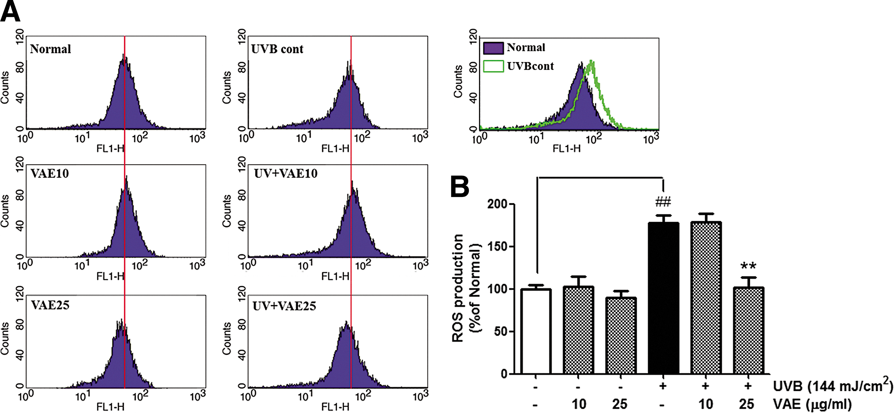
Influence of VAE on generation of reactive oxygen species (ROS) in NHDFs. ROS generation levels were determined after 24 h of UVB irradiation and sample treatment. Number of cells is plotted versus dichlorofluorescein fluorescence detected by FL-1 channel
CV and MMP-1 production in VAE- and rutin-treated, cultured human dermal fibroblasts
To confirm the cytotoxicity of VAE and rutin, MTT assay was performed in NHDFs. In the nonirradiated and UVB-irradiated groups, treatments of VAE and rutin exhibited little toxicity even at high concentrations (Fig. 4A, C). However, there was no significant difference between samples. UVB irradiation is known to induce MMP-1 expression. To investigate the effects of VAE and rutin on MMP-1 expression, a variety of VAE and rutin concentrations, from 1 to 25 μg/mL and 0.1 to 1 μg/mL, respectively, were applied to NHDFs with UVB irradiation. At their highest concentrations, VAE and rutin (25 and 10 μg/mL, respectively) decreased MMP-1 production by 73.2%±31.7% and 56.0%±1.7%, respectively, compared with nontreated cells (Fig. 4B). UVB exposure drastically increased MMP-1 secretion compared with nonirradiated cells. Treatment with VAE and rutin restored MMP-1 production by 90.4%±5.0% and 46.9%±5.7%, respectively, in UVB-irradiated NHDFs (Fig. 4D). These results indicate that VAE and rutin strongly diminished MMP-1 secretion in UVB-irradiated NHDFs.
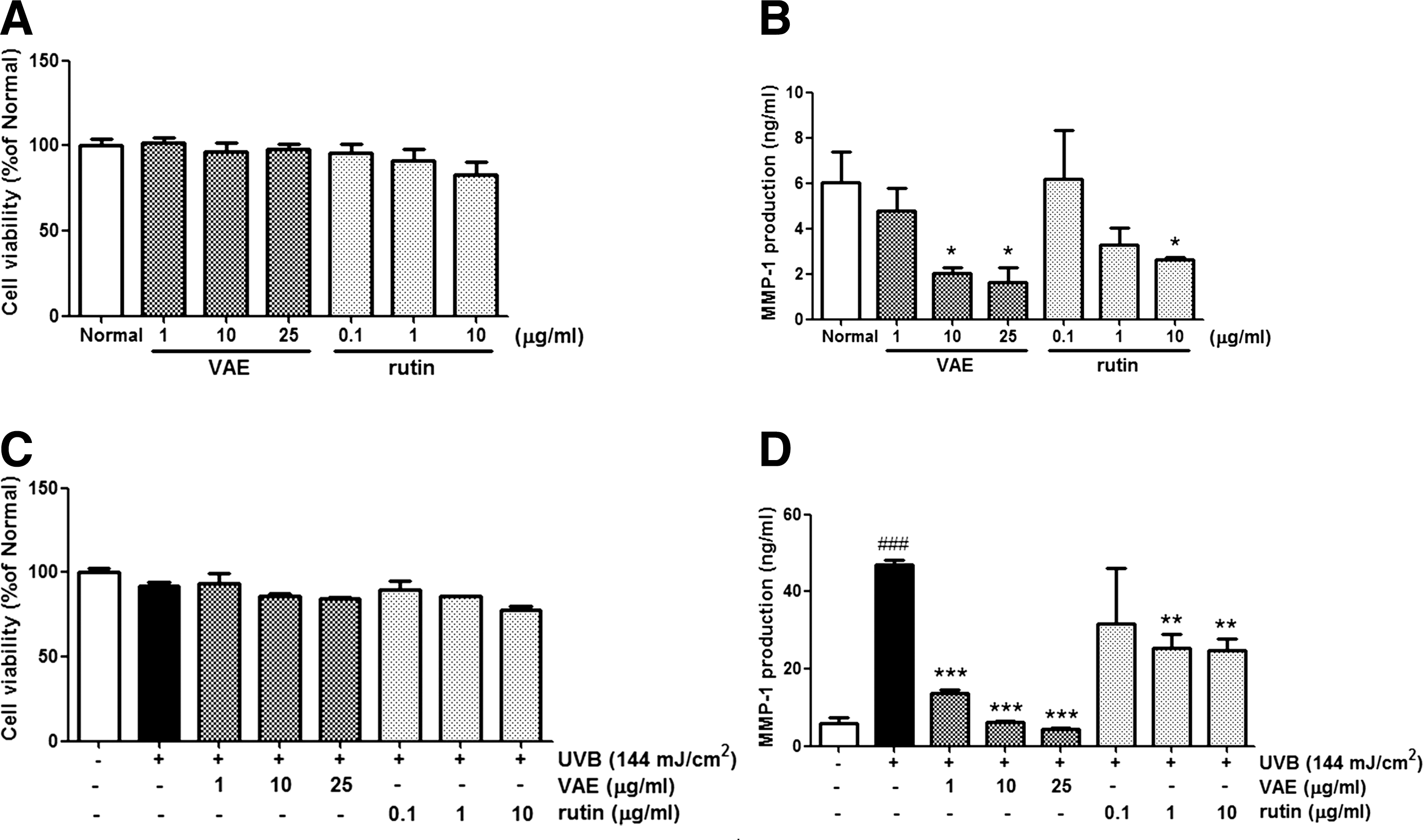
Cell viability
Effects of topical VAE application on UVB-induced skin photoaging in hairless mice
After 5 weeks of topical VAE application, wrinkle formation and physiological skin changes were determined using an analytical system. Figure 5 shows skin replicas and quantification of wrinkle formation in hairless mice dorsal skin. The UVB irradiation group exhibited deeper and wider wrinkles compared with the nonirradiated group (Fig. 5A). All arbitrary units showed higher values in UVB-irradiated controls than in the nonirradiated group. However, the VAE group had reduced wrinkle formation (Fig. 5B). The VAE 5% group had better protection against UVB-irradiated wrinkling than the VAE 1% group.

Photographs of replicas
Figure 6 shows SC hydration, TEWL, and EI. After UVB exposure, the SC hydration level decreased whereas TEWL and EI increased. The 5% VAE-treated group had elevated SC hydration levels compared with the UVB-irradiated control group (Fig. 6B). Both VAE 1% and VAE 5% groups quenched TEWL and EI (Fig. 6A, C).
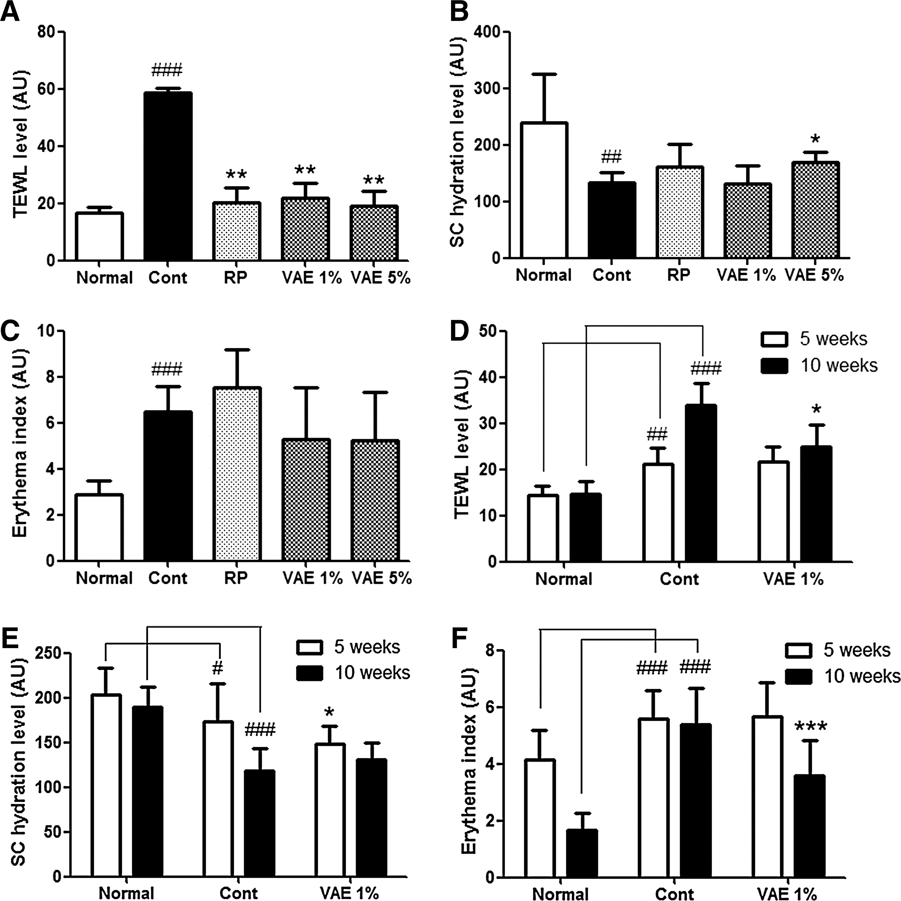
TEWL
To evaluate the epidermal thickness and collagen density of dorsal skin, H&E staining and Masson's trichrome staining were applied. As expected, UVB-irradiated controls showed increased skin thickness. However, the topical application groups of VAE 1% and VAE 5% had reduced epidermal thickness (Fig. 7A). As shown in Figure 7B, UVB irradiation disrupted collagen fibers. The tendency of decreased collagen fibers was reversed in topical application groups of VAE 1% and VAE 5%. VAE 5% had a stronger effect than VAE 1% (Fig. 7B).
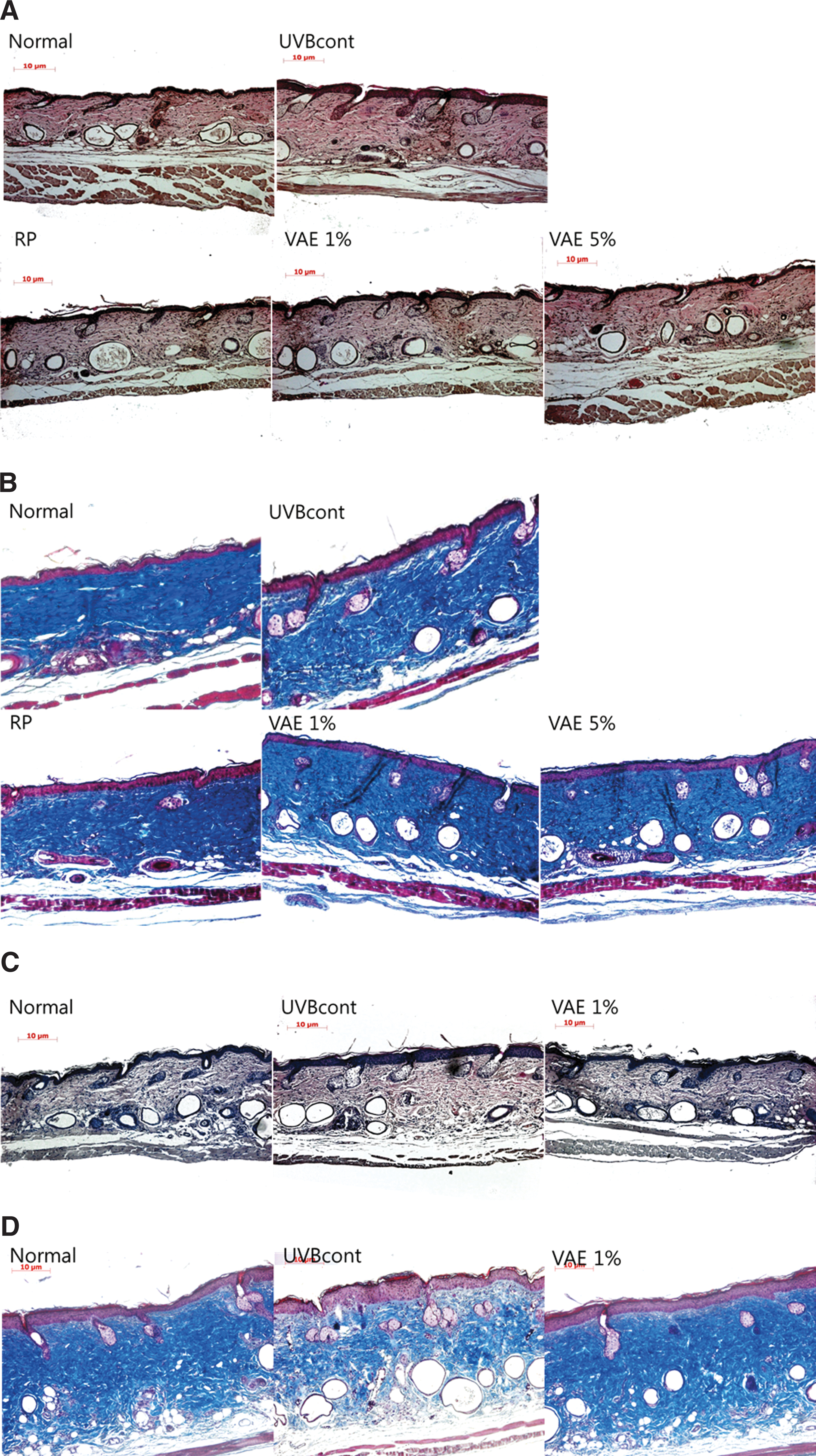
Photomicrographs of H&E-stained sections
Figure 8 shows the protein expression levels of MMP-1, elastin, procollagen type I, and TGF-β1. In the UVB-irradiated control group, MMP-1 expression was increased whereas elastin, procollagen type I, and TGF-β1 expression was decreased compared with the nonirradiated group. However, VAE-treated groups alleviated changes in protein expression caused by UVB irradiation. The VAE 5% group greatly increased expression of elastin, procollagen type I, and TGF-β1 (up to 118%, 156%, and 136%, respectively) compared with the UVB control group. These effects were more pronounced in the VAE 5% group than in the RP group. VAE 5% was more efficient at improving antiphotoaging effects compared with VAE 1%.
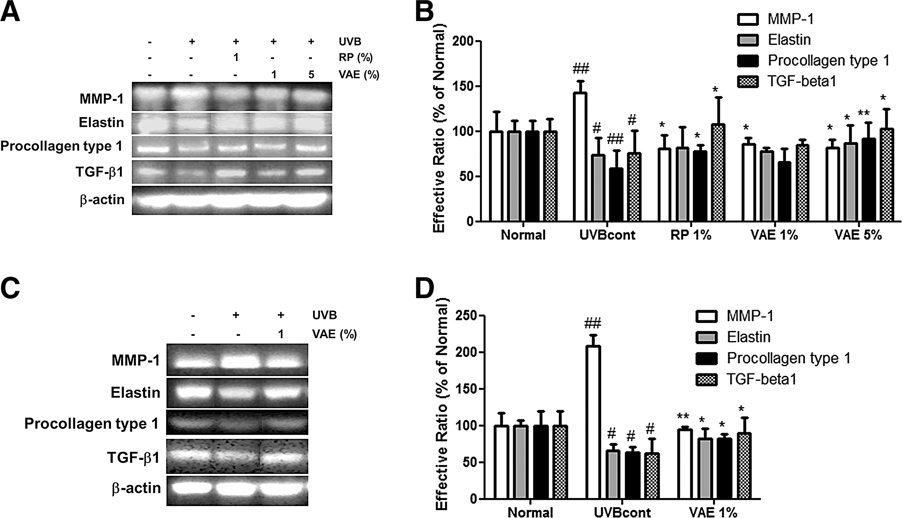
Protein expression of MMP-1, elastin, procollagen type I, and TGF-β1
Effects of dietary VAE on UVB-induced skin photoaging in hairless mice
During the experimental period, there were no significant differences in body weight between groups. And also the food efficiency rate was the similar among groups (Table 2). However, the food efficiency rate was lower in the UVB-irradiated control group (2.16±1.98) than in the normal and VAE 1% groups (2.37±2.23 and 3.41±2.46, respectively).
Group Normal: control diet only; group UVBcont: UVB irradiation+control diet; group VAE 1%: UVB irradiation+diet containing 1% VAE.
FER (food efficiency rate)=gain of body weight (g)/amount of food intake (g)×100.
Figure 5 shows replicas of the central dorsum of mice. In the nonirradiated group, skin appeared fine with thin wrinkle formation (Fig. 5C). Conversely, deeper and wider wrinkles were observed in the UVB-irradiated control group. The dietary VAE 1% group experienced reduced UVB-induced wrinkle formation (Fig. 2D).
Skin surface physiological changes, such as SC hydration, TEWL, and EI, were measured at the end of 5 and 10 weeks. As shown in Figure 6, the dietary group exhibited meaningful changes in SC hydration, TEWL, and EI. Compared with the 5-week values, all 10-week values were more significantly improved (Fig. 6D–F).
The histological structures of mouse skins were studied in the Normal, UVBcont, and VAE 1% dietary groups. H&E staining showed variation in epidermal thickness for each group. Figure 7C shows that epidermal thickness increased with UVB irradiation and was quenched by oral administration of VAE. Masson's trichrome staining verified cleavage of collagen fibers in the UVB irradiation group. Conversely, the VAE dietary group had abundant dense collagen fibers (Fig. 7D).
MMP-1, TGF-β1, elastin, and procollagen type I expression was detected using western blot analysis. Oral administration of VAE attenuated MMP-1 expression and promoted TGF-β1, elastin, and procollagen type I expression compared with the UVB-irradiated control group (Fig. 8C, D).
Discussion
Recently, there has been increasing interest in research on botanicals as a primary treatment against skin photoaging. Such botanicals offer potential protective effects against UV-radiation-mediated damage, referred to as “photochemo-preventive effects.” 28 The azuki bean (VA) is a common plant used in food and oriental medicine in East Asia. There are many reports on the components and clinical applications of VA. 19,29 –31 In particular, the antioxidant activity of VA has been established. 22 UVB is the main external factor that leads to oxidative stress, which is initiated by ROS and eventually results in premature skin aging. 3 The use of natural antioxidant products is a promising approach for skin photoaging prevention and treatment. 10,11 We confirmed that VAE has preventive effects on ROS production caused by UVB irradiation and protective effects against UV-induced photoaging. We found that VAE decreased UVB-induced MMP-1 secretion in a dose-dependent manner (Fig. 4). Further, VAE treatment at a high concentration (25 μg/mL) diminished the production of ROS more than in UVB-irradiated cells.
Based on the inhibitory effects of VAE on MMP-1 and ROS in UVB-exposed dermal fibroblasts, further animal studies were performed to determine whether VAE also protects against UVB-induced damage in vivo. After UVB irradiation, dietary and topical applications of VAE on the dorsal skin of hairless mice were investigated regarding skin wrinkle prevention. We also examined epidermal thickness and collagen fibers using histological staining. Following topical and oral administration of VAE, UVB-irradiated mice demonstrated reduced wrinkle formation, epidermal thickness, and collagen degradation. Our results suggest that VAE may prevent UVB-induced coarse wrinkles by inhibiting epidermal expansion and collagen breakdown.
Physiologically, photoaged skin may exhibit increased EI and TEWL with decreased SC hydration level. 32 We found that oral administration of VAE attenuated EI and TEWL while increasing SC hydration. Many studies have reported that topical administration of antiphotoaging materials influenced the level of TEWL. 33 Also, this study showed significant changes in TEWL level with topical application and dietary of VAE as well as EI and SC hydration. These results indicate that VAE has potent protective effect for preserving skin hydration.
UVB-induced activation of MMP-1 and reduction of elastin, procollagen type I, and TGF-β1 were recovered by topical and oral application of VAE. Moreover, the effects of topical VAE application were dose dependent. However, all mechanisms were not explored in this study. Further research is warranted to identify the VAE mechanisms responsible for suppressing UVB-induced skin damage.
Many studies have reported that rutin, a polyphenolic bioflavonoid, has antioxidant activity. 20 In this study, rutin was used as a standard compound in VAE. We examined whether rutin regulates MMP-1 production in UVB-irradiated NHDFs. Rutin showed significantly decreased MMP-1 secretion. However, we did not examine the effects of rutin on UVB-induced photoaging in vitro or in vivo, an issue to be investigated in future studies.
Free-radical damage caused by mental, physical, and environmental stresses can accelerate the skin's wrinkling and sagging. 3 Because the main function of the blood is to deliver nutrients and oxygen to different tissues, including the skin, overall health depends on how efficiently the blood circulates throughout the body. The body's inflammatory response can damage blood vessels, causing them to function less efficiently.
To maintain healthy blood circulation, it is imperative that the arteries and capillaries be kept as clean and strong as possible. Unfortunately, being so small, the capillaries are extremely susceptible to damage from free-radical assault. This can lead to rupturing and bleeding that is visible as “broken veins” on the surface of the skin. However, maintaining the health of the circulatory system can prevent this type of damage from occurring.
Rutin, and its partners, can fortify blood vessels against the inflammatory response, which can prevent the skin from getting its fair share of nutrients. Also, rutin has the capacity to regenerate vitamin C after it neutralizes a free radical, thus helping to restore its antioxidant potential. 34 Because vitamin C plays a critical role in the manufacture of collagen, an important component of capillary walls as well as the sustaining framework of the epidermis, this could have a dramatic impact on the health of your skin.
In addition, ability of rutin to provide microvascular protection, improve circulation, and defend against free-radical damage makes it an excellent ingredient in any natural skin care formulation for aging skin. 35 –38 Therefore, oral administration of VAE would give better effect than topical apply through maintaining the health of the circulatory system.
In summary, our study showed that VAE has antioxidant activity. VAE and rutin, an active component of VAE, decreased MMP-1 secretion in NHDFs exposed to UVB. We also found that topical and oral administration of VAE in UVB-irradiated hairless mice suppressed skin roughening, thickening, dryness, and wrinkling by stimulating TGF-β1 expression to increase synthesis of collagen and elastin. Therefore, VA has potential for use in reducing and preventing UVB-induced photoaging. These findings support that VA is valuable not only as a functional food but also a cosmetic product.
Footnotes
Acknowledgments
This research was supported by the Korea Institute of Planning and Evaluation for Technology in Food, Agriculture, Forestry and Fisheries (iPET, 810006-03-1-SB120), Republic of Korea.
Author Disclosure Statement
The authors declare that there are no conflicts of interest.