
Abstract
Probiotics and antioxidants have a definite improving effect in cardiovascular diseases. This study aims at mitigating doxorubicin toxicity on cardiac function through consuming a functional food. Five groups of adult male Sprague-Dawley rats were used along 22 weeks. Group I received 30 g/kg/day food enriched with yogurt, green tea extract, and carrots (80, 0.84, and 100 g/kg diet, respectively) from the first week, group II received carvedilol 30 mg/kg/day orally from week 17, group III received both carvedilol and tested food, and groups IV and V were +ve and –ve control groups, respectively. In week 17, cardiomyopathy was induced by i.p. injection of 2.5 mg/kg doxorubicin every 48 h for 2 weeks. Histopathological and electrophysiological examinations and biochemical analysis were done. Lipid peroxidation, antioxidant effect, heart failure compensatory mediators, and proinflammatory cytokines were assessed. Tested food normalized time between the start of Q wave and the end of T wave on electrocardiogram (QT interval) and heart rate compared to the doxorubicin group (P<.05). It also improved hypertrophy indicated by a significant (P<.05) decrease in heart/body weight ratio, angiotensin-II (Ang-II), and atrial natriuretic peptide (ANP) serum levels. Histopathological examination of cardiac sections from the tested food group revealed less marked vacuolization and low perivascular fibrosis percentage (0.7803±0.04). A significant (P<.001) decrease in serum creatine kinase-membrane bound, lactate dehydrogenase, triglycerides, cholesterol, low-density lipoprotein cholesterol, and tissue malondialdehyde (MDA) levels was observed in addition to an increase in serum Na+/K+ ATP1A1 and cardiac reduced glutathione (GSH) levels. Tested food also lowered the inflammatory cytokines tumor necrosis factor-α (TNF-α) and interleukin-6 (IL-6) serum levels significantly (P<.01). Probiotic food containing Lactobacillus acidophilus, green tea, and carrots can improve membrane integrity and cardiac contractility in doxorubicin-induced cardiomyopathy by decreasing TNF-α, IL-6, MDA, increasing GSH, and modulating compensatory mediators such as Ang-II and ANP.
Introduction
H
Many studies were carried out to prove therapeutic benefits of probiotics in many diseases, including heart diseases. 7 Probiotics are defined as live microorganisms that confer health benefits on the host when administered in adequate amounts. Contemporary knowledge concerning probiotics and their action is derived from traditional consumption of fermented milk products in addition to researches on lactic bacteria strains testing their beneficial harmless effects on health. 8 The most extensively studied and widely used probiotics are the lactic acid bacteria, particularly Lactobacillus and Bifidobacterium. Mechanisms mediating health effects of probiotics include production of antimicrobial compounds, reducing gut pH, improving immune function, and stimulate immunomodulatory cells, whereas the physiological circumstances for the activation of these mechanisms are still under research. 9
Since functional food that contains significant amounts of bioactive components can provide desirable health benefits beyond basic nutrition, this study was designed to apply consumption of food rich in the probacteria L. acidophilus and natural antioxidants in green tea and carrot in controlling HF. This functional food may represent a safe and commercially available treatment intervention for cardiac patients.
Materials and Methods
Animals
Adult male Sprague-Dawley rats (n=60, 300–350 g) were purchased from VACSERA (Giza, Egypt). Rats were allowed free water access. The experimental work complies with the ethical guidelines adopted by the Scientific Research Ethics Committee of Faculty of Pharmacy, Mansoura University.
Drugs and chemicals
Doxorubicin hydrochloride (Dox, CAS No. 25316-40-9) was purchased as Adriamycin® solution for injection 50 mg vial, Pfizer, Inc. (New York, NY, USA). Carvedilol (Carv, CAS No. 72956-09-3) was kindly supplied by Global Napi Pharmaceuticals (Giza, Egypt). 5,5′-Dithiobis (2-nitrobenzoic acid) was purchased from Sigma-Aldrich (St. Louis, MO, USA; CAS No. 69-78-3). Reduced glutathione (GSH, CAS No. 70-18-8) was purchased from FlukaChemie (Buchs SG, Switzerland). Other chemicals of best grades were purchased from available companies.
Induction of experimental cardiomyopathy
Cardiomyopathy was induced by cumulative injection of Dox as previously described by Lou et al. 10 Six doses of 2.5 mg/kg Dox were injected i.p. at 48-h intervals for a total period of 2 weeks to achieve a cumulative Dox dose of 15 mg/kg.
Tested food added components
Yogurt was purchased from the local market as semi-skimmed yogurt containing about 109 cfu/g of Lactobacillus acidophilus (unspecified strain, yogurt nutritional value, and ingredients are shown in Supplementary Table S1; Supplementary Data are available online at
Amounts calculated in the light of recommended daily amounts for human using Paget and Barnes formulas so that rats would receive yogurt, green tea extract, and carrot in amounts of 8, 0.084, and 10 g/kg weight, respectively.
Dissolved in saline to ensure uniform distribution.
Experimental design
The study involved five groups (n=15, except for the control group n=10). Schematic representation of study design is shown in Table 2. Carvedilol was used as a standard drug for HF treatment. By the end of week 22, animals in all groups were anesthetized with urethane (1.8 mg/kg, i.p.) and electrocardiograms were recorded. Blood samples were collected by orbital sinus vein puncture and centrifuged at 2000 g for 10 min at 4°C; serum was separated, divided into aliquots, and stored at −80°C. Animals' chests were opened and hearts were isolated and weighed. Cardiac apexes were cut and fixed in 8% (w/w) neutral buffered formalin solution for 24 h for histopathological evaluation. The rest of the hearts were used for the preparation of homogenates (10% w/w in phosphate buffer, pH 7.5).
Animals received free access to standard rodent food throughout the whole experiment.
□, Dox 2.5 mg/kg/2 days, i.p. injection; ○, carboxymethyl cellulose 1 mL/kg/day, 1% w/w aqueous solution, orally; ●, Carv 30 mg/kg/day, suspended in 1 mL 1% w/w carboxymethyl cellulose solution, orally;  , tested food 30 g/rat/day.
Electrocardiogram recording
Electrocardiograms were recorded from standard lead II limb leads using a single-channel ECG (Model: 501-B III; Fukuda ME Kogyo Co. Ltd., Tokyo, Japan). The electrocardiograph was standardized before each tracing to get sensitivity (1 mV pulse produces 20 mm height) and chart speed 50 mm/sec. Heart rate (HR) was calculated in beats/min by dividing 3000 over the number of millimeters (small squares) between two successive R waves. Time between the start of Q wave and the end of T wave on electrocardiogram (QT interval, msec) and QRS wave voltage (mV) was calculated to evaluate hypertrophy.
Histopathological examination and morphometry
Standard histopathological techniques were followed and quantitative analysis of collagen fiber deposition in trichrome-stained cardiac tissues was performed by morphometry. 11
Measurement of cardiac and renal function biomarkers
Serum levels of creatine kinase-membrane bound (CK-MB), alkaline phosphatase (ALP), lactate dehydrogenase (LDH), creatinine and urea/blood urea nitrogen (BUN), triglycerides (TGs), total cholesterol level, and high-density lipoprotein cholesterol (Spectrum Diagnostics, Cairo, Egypt) were analyzed.
Measurement of lipid peroxidation and antioxidant biomarkers
Lipid peroxidation was indirectly evaluated by measuring the malondialdehyde (MDA) cardiac tissue level (BioDiagnostic, Cairo, Egypt) in the tissue homogenate. The antioxidant biomarker GSH was measured chemically, as previously described by Moron et al., 12 with slight modification. In brief, 0.05 mL of 50% (w/w) trichloroacetic acid was added to 0.45 mL of liver homogenate to precipitate protein and then centrifuged at 1000 g for 5 min. 0.25 mL of supernatant was mixed with 1 mL of 0.2 M Tris–HCl (containing 1 mM EDTA, pH 8.9) and 0.05 mL of 0.01 M 5,5′-dithiobis-(2-nitrobenzoic acid) in absolute methanol and kept at room temperature for 5 min. The yellow color developed was measured spectrophotometrically at 412 nm. A standard curve (0–500 nmol/mL) was constructed and results were determined and expressed as nmol/g tissue.
ELISAs
Serum levels of angiotensin-II (Ang-II), atrial natriuretic peptide (ANP) (BlueGene Biotech CO., Shanghai, China), and Na/K-transporting ATP1A1 (CUSABIO, Hubei, China) were measured. Myocardium levels of the proinflammatory cytokines tumor necrosis factor-α (TNF-α) and interleukin-6 (IL-6) (eBioscience, San Diego, CA) were also determined according to the manufacturer's instructions.
Statistical analysis
Data are expressed as mean±SE in each experimental group. Statistical evaluations of the results were carried out by means of one-way analysis of variance, followed by the Tukey–Kramer multiple comparison test. Statistical tests were performed with GraphPad Instat V 3.05 (GraphPad Software, Inc., San Diego, CA, USA).
Results
Electrocardiogram recording
HR and QRS voltage were significantly (P<.001) increased by Dox besides significant prolongation of QT interval compared to the control group. Rats received Carv, and food combination with Carv showed a significant (P<.001) decrease in HR to normal rate. The QRS voltage was normalized in the group that received tested food alone and that received Carv in combination with tested food (P<.001 compared to Dox). However, the effect of Carv on QRS voltage was insignificant. The effect on normalizing QT interval was more significant (P<.001) in the group that received tested food and food in combination with Carv compared to the effect of Carv alone (P<.05) (Fig. 1, Table 3).
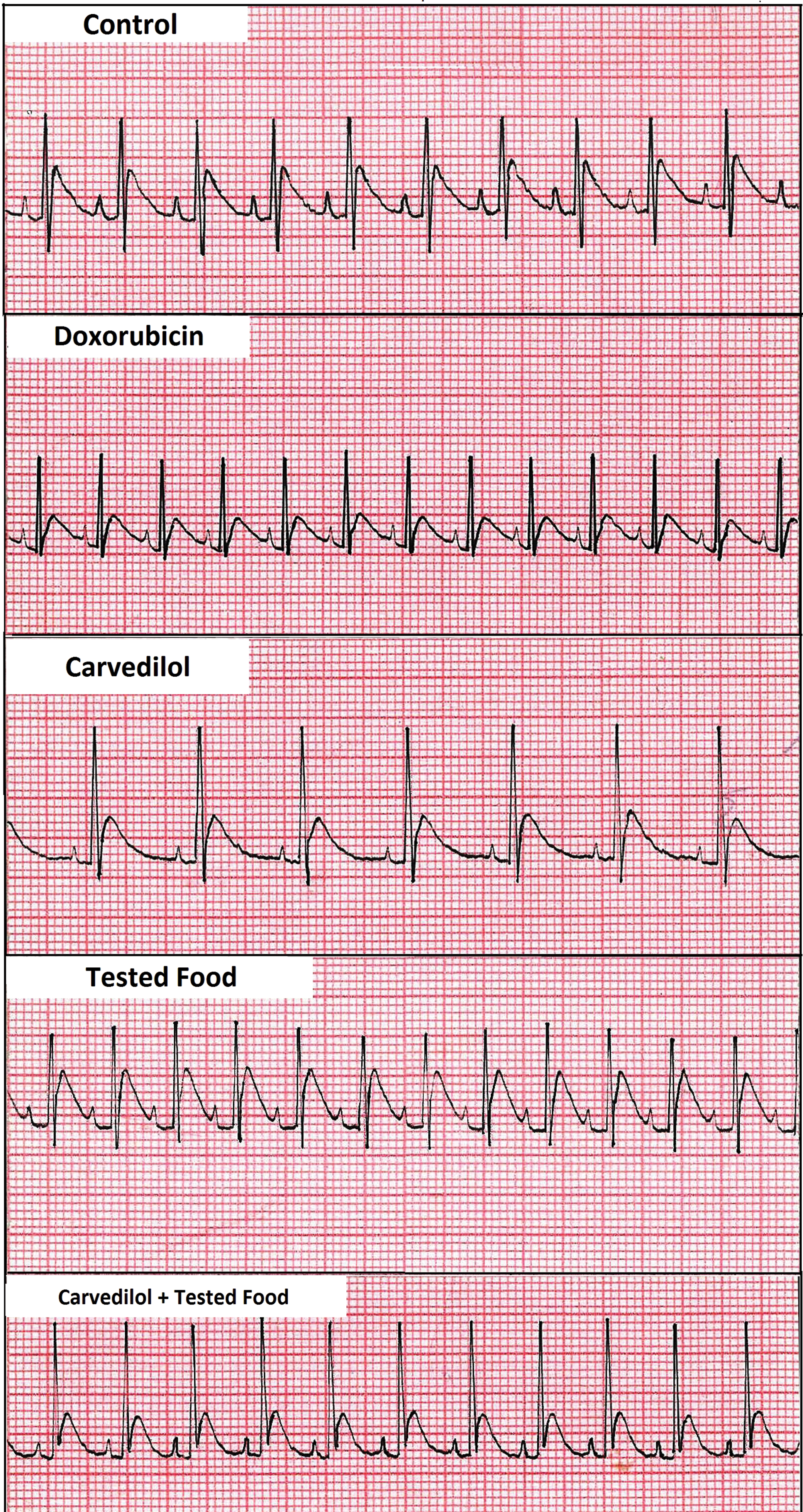
Rats electrocardiograph recordings showing effect of tested food (30 g/kg/day), carvedilol (30 mg/kg/day), combination of food and carvedilol on heart rate (HR), QT interval (msec), and QRS wave voltage (mV) in doxorubicin-induced cardiomyopathy model. Color images available online at
Data are expressed as mean±SE, n=8.
Significantly different (P<.001) from control.
Significantly different (P<.05, P<.001), respectively, from Dox.
Significantly different (P<.05) from Carv.
HW/BW, heart weight/body weight; HR, heart rate.
Mortality and heart weight to body weight ratio
There was a significant (P<.001) decrease in % mortality in groups that received tested food or in combination with Carv (8.34% in both groups) compared to the Dox group (58.34%) and to the group that received Carv alone (41.67%). The heart weight to body weight (HW/BW) ratio was calculated in g/kg. A significant decrease (P<.001) in the calculated ratio was observed in all treated groups. The best effect was noticed in the group that received tested food alone and that received the combination with Carv (Table 3).
Histopathological examination and morphometry
Doxorubicin administration produced marked histopathological changes. Tissue sections stained with Masson Trichrome (Fig. 2A, upper panel) revealed marked perivascular and interstitial fibrosis that was best improved by tested food. These results are consistent with fibrosis percentage (Fig. 2B) that was significantly (P<.001) lowered in groups that received tested food and food combination with Carv compared to the Carv and Dox groups.
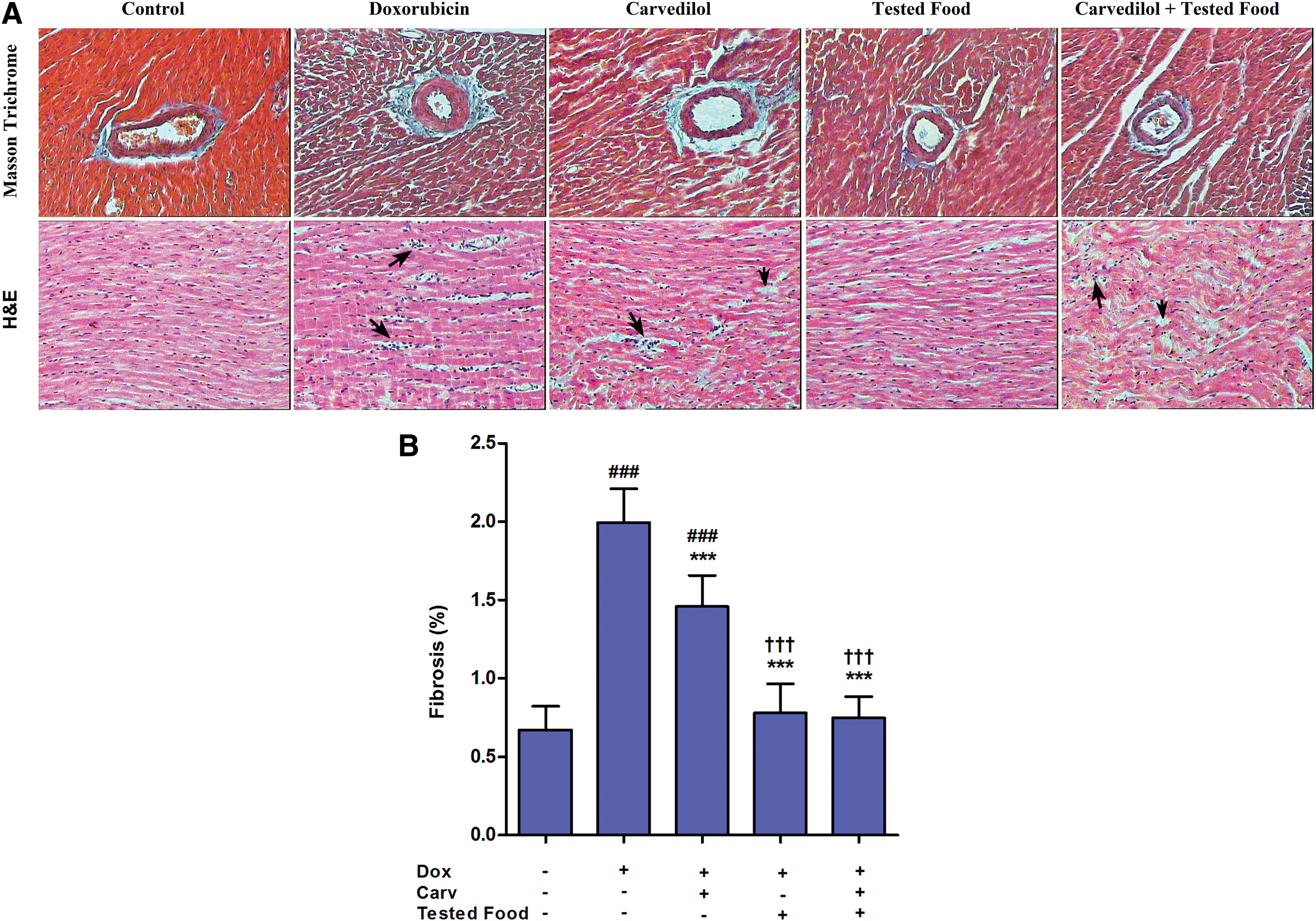
Sections stained with hematoxylin and eosin (Fig. 2A, lower panel) showed marked edema, vacuolization, and infiltration of interstitial tissue by inflammatory cells in Dox-treated rats with mild improvement by Carv compared to a higher improvement produced by tested food or a combination.
Cardiac function biomarkers and lipid profile
Doxorubicin resulted in significant (P<.001) elevation in measured cardiac and renal parameters; CK-MB, LDH, ALP, creatinine, and BUN. Carvedilol decreased these parameter levels significantly (P<.05) but still higher than control levels. Tested food and food combination with Carv normalized these levels to be comparable to the control group. The Na+/K+ ATP1A1 serum level was elevated to normal in all groups (P<.001compared to Dox group), with the best results in the combination group (significantly P<.05 higher than Carv group) (Fig. 3).
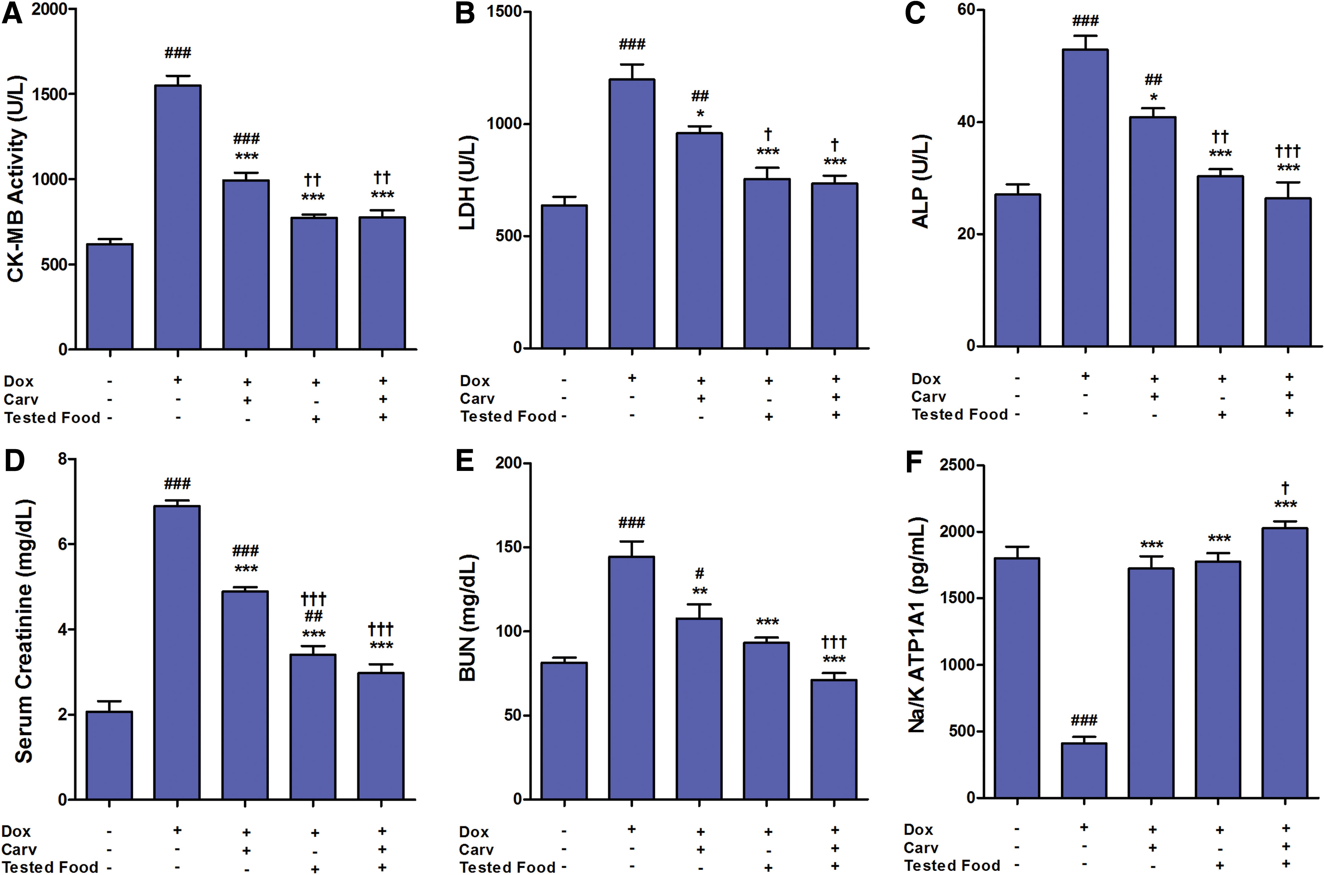
Effect of carvedilol (30 mg/kg/day), tested food (30 g/kg/day), combination of carvedilol and food on cardiac function parameters
Concerning lipid profile, tested food and food combination with Carv significantly (P<.001) decreased TGs, total cholesterol, and low-density lipoprotein (LDL)-cholesterol compared to the Dox group (Fig. 4). On the other hand, Carv showed less significant changes in these biomarkers (P<.05).

Effect of carvedilol (30 mg/kg/day), tested food (30 g/kg/day), combination of carvedilol and food on lipid profile parameters
Cardiac lipid peroxidation and antioxidant biomarkers
The GSH cardiac level was significantly (P<.001) increased by tested functional food. Carvedilol also increased the GSH level, but remained significantly (P<.001) lower than the control group level. The group that received both Carv and tested food showed a higher GSH level than Carv alone with no additional effect when compared to food alone. The MDA level was decreased significantly by tested food and food in combination with Carv (P<.001), while its level in the Carv group remained significantly (P<.001) higher than in the control group (Fig. 5).

Effect of carvedilol (30 mg/kg/day), tested food (30 g/kg/day), combination of carvedilol and food on antioxidant biomarkers
Cytokines and compensatory mediators
Levels of Ang-II and ANP were significantly (P<.001, P<.01, respectively) decreased by tested food compared to the Dox group levels. The combination of food and Carv limited the improving effect of food alone (P<.01, P<.05 compared to Dox group). The proinflammatory cytokines TNF-α and IL-6 levels were elevated significantly (P<.001) by Dox. These elevated levels were significantly lowered in groups that received tested food (P<.01) and that received food in combination with Carv (P<.001) (Fig. 6).

Effect of carvedilol (30 mg/kg/day), tested food (30 g/kg/day), combination of carvedilol and food on compensatory mediators and cytokines
Discussion
Medical therapies and lifestyle modification constitute general measures for HF management. Consuming healthy food is one of the lifestyle modifications that should be encouraged. In the present study, food enriched with natural antioxidants and probiotics showed a beneficial effect in attenuating cardiomyopathy induced by Dox and can be considered as a functional food. Electrophysiological and histopathological parameters were investigated besides analysis of cardiac parameters and plasma lipids. Proinflammatory cytokines and endogenous antioxidants were measured to elucidate possible underlying mechanisms for cardiac function improvement.
Hypertrophy of cardiac muscle was less apparent in rats fed the tested food as indicated by a decrease in the HW/BW ratio, QT interval, and QRS voltage. Tested food also improved cardiac function as it lowered serum levels of Ang-II and ANP. These neurohormonal mediators are principally involved in compensatory secondary mechanisms that result in left ventricular remodeling and subsequent cardiac decompensation. 13
During cardiomyopathy induction, Dox is reduced in the presence of free iron setting up a cycle for ROS generation. 14 These reactive species were indirectly detected in the study by measuring the cardiac MDA tissue level as a product of lipid peroxidation. Tested functional food decreased ROS release as indicated by a low MDA level; a result that may be contributed to the ability of L. acidophilus strains to scavenge MDA. 15 This effect would have a positive impact on cardiac function since ROS are involved in the development and progression of maladaptive myocardial remodeling as impairing contractile function, activating a broad variety of hypertrophy transcription factors and mediating apoptosis. 16
Administration of Dox elevated serum TGs, cholesterol, and LDL-cholesterol indicating a low lipolysis rate. Hyperlipidemia and lipid peroxidation are hallmarks in altering cardiac myocyte function in Dox-induced cardiomyopathy. 17,18 This alteration involves cardiac cell damage, change in cardiac cell membrane integrity, decreased contractility, and formation of atherosclerotic lesions. Tested food significantly lowered elevated lipid biomarkers and this hypolipidemic effect may account for its cardioprotective effect.
The increase in free radical production by doxorubicin is accompanied by a decrease in endogenous antioxidant enzymes 16 potentiating the harmful effects of ROS and aggravating the induced cell injury. The obtained results showed a decrease in GSH tissue level by Dox. This decrease was abolished by tested food administration; an effect that may be attributed to conversion of theanine (a component of green tea) into GSH using glutamate, as previously denoted by Sugiyamaa and Sadzuka. 19 Also, the tested probacteria may have the potential to induce GSH biosynthesis in cardiac tissue as in other tissues, for example, the pancreas and ileum. 20
Another consequence of Dox administration is oxidative damage to CK. This enzyme acts as a modulator of the energy reservoir by converting creatine to phosphocreatine using ATP as a substrate and it serves as a marker for myocardial damage. 21 The cardiac muscle activity and membrane integrity were evaluated by measuring serum CK-MB isoenzyme and LDH levels. Both enzymes were upregulated and released in serum by Dox, an effect that is strongly correlated to the unique heart vulnerability to oxidative stress underlying Dox-induced cardiomyopathy. 22 Improvement in cardiac membrane integrity by tested food administration was observed as indicated by the decrease in serum CK-MB isoenzyme level and by increase in the transmembrane enzyme Na+/K+ ATP1A1 level. In addition, histopathological examination of heart muscle revealed less vacuolization, edema, and fibrosis in sections isolated from rats fed tested food.
Cytokine release is an important compensatory mechanism involved in HF. Although proinflammatory cytokines are not constitutively expressed in the heart, a variety of clinical and experimental investigations have suggested that TNF-α and IL-6 may play a role in the pathophysiology of HF. A significant increase in these cytokines serum levels was observed in patients with congestive HF, 23 and both cardiac and infiltrating cells of the myocardium participate in their production. The degree of HF was reported to be directly related to expression of these inflammatory cytokines and their suppression can improve cardiac performance. 24 In this study, doxorubicin elevated serum levels of both TNF-α and IL-6 that coincides with Aluise et al. 25 TNF-α and IL-6 cause cardiac contractile dysfunction and contribute to both hypertrophy and apoptosis. In particular, TNF-α underlying maladaptive responses in HF include (i) induction of cardiac myocyte hypertrophy through increasing the myocardial renin–angiotensin system activity, 26 (ii) depression of myocardial contractility through inducing nitric oxide synthase that increases NO levels mediating myofilament desensitization to intracellular calcium, 27 (iii) induction of myocyte apoptosis through both intrinsic and extrinsic apoptotic pathways and loss of antiapoptotic proteins, 28 and (iv) remodeling of extracellular matrix by altering the balance in the activity of metalloproteinases and tissue inhibitor metalloproteinases leading to fibrosis. 29 The deleterious role of the pleiotropic cytokine IL-6 in HF is mediated by the same underlying pathways to that of TNF-α with different intracellular mechanisms and it does not participate in apoptosis.
Only tested food was able to reduce serum levels of TNF-α and IL-6 and hence can guard against its subsequent toxic effect on cardiac myocytes. The lowering effect on TNF-α may account for the decrease in Ang-II serum level by the tested food only and may be correlated to immunomodulatory effects of probiotics interestingly without eliciting a harmful inflammatory response. 9 Probiotic bacteria also mediate suppression of lymphocyte proliferation and cytokine production by T cells, presupposing their use as immune modulators. 30 The ability of probiotics to modulate the TNF-α level and IL-6 can improve inflammatory diseases such as inflammatory bowel diseases and ulcerative colitis 31 and proposes a similar effect in HF.
Conclusion
Consuming healthy diet is pivotal in controlling cardiovascular diseases and HF. The present study proved that enriching food with commercially available natural antioxidants such as green tea and carrots in addition to yogurt containing the probacteria L. acidophilus can help in maintaining cardiac function and myocardial membrane integrity besides ameliorating secondary damage by compensatory mechanisms. These effects are mediated by decreasing lipid peroxidation, increasing GSH, and decreasing proinflammatory cytokines TNF-α and IL-6. Before being introduced as a pharmacological intervention, further investigations on other models of cardiomyopathy should be directed. Possible mechanisms underlying the beneficial effect of tested functional food on cardiomyopathy are shown in Figure 7.

Schematic representation for possible mechanisms underlying the beneficial effect of tested food on doxorubicin-induced cardiomyopathy. Color images available online at
Footnotes
Author Disclosure Statement
No competing financial interests exist.
References
Supplementary Material
Please find the following supplemental material available below.
For Open Access articles published under a Creative Commons License, all supplemental material carries the same license as the article it is associated with.
For non-Open Access articles published, all supplemental material carries a non-exclusive license, and permission requests for re-use of supplemental material or any part of supplemental material shall be sent directly to the copyright owner as specified in the copyright notice associated with the article.