
Abstract
The three ayurvedic medicinal plants, Withania somnifera, Emblica officinalis, and Bacopa monnieri, were extracted by high-pressure static extraction using the Zippertex® technology. The extracts were mixed to reach quantifiable amounts of active compounds identified by high-pressure liquid chromatography-mass spectrometry (HPLC-MS) analysis. The mixture of extracts was incubated with resting cells of the fungus Beauveria bassiana ATCC 7159. The fermentation promoted the fluidization of the starting dense mixture, while HPLC monitoring evidenced the disappearance of glucogallin from E. officinalis extract and the concomitant increase in gallic acid content. Topical exposure of the chick embryo chorioallantoic membrane (CAM) to the nonfermented extract led to the extensive necrosis and destruction of the treated membrane. However, the fermented extract was shown to be free of any toxicity. Furthermore, compared with the untreated CAM, the fermented sample reduced CAM vascularization, suggesting its antiangiogenic potency. The innocuity of the fermented extract was demonstrated using the in vivo LD50 test, the morphological examination of internal organs of treated rats, as well as the evaluation of blood biomarkers of liver damage (aspartate aminotransferase and alanine aminotransferase). The fermented extract was developed as a nutraceutical antiangiogenic treatment of age-related macular degeneration and commercialized in an oral form named Ethnodyne-Visio™.
Introduction
A
Angiogenesis can be inhibited by a number of compounds, such as bevacizumab also named Avastin, 10 protamine, 11 fumagillin, 12 and glycoproteins such as interferon. 13 However, the insufficient therapeutic efficacy of these drugs, their associated toxicity or difficulty of administration, especially for protein factors, and their prohibitive cost limit their medical use. 14
Several reports have focused on plant extracts or natural compounds from Indian traditional medicine known as Ayurveda. 15,16 Long considered as an empirical medicine, Ayurveda benefits from indisputable scientific validation supported by high impact publications. 17 In particular, natural products are reported for the treatment or prevention of neovascularization promoted disorders. 18 Indeed, extracts of Withania somnifera (L.) Dunal 19,20 and Emblica officinalis Gaertner 21–22 exhibit antiangiogenic activity, whereas improvement of cognitive ability was reported for Bacopa monnieri (L.) Pennell. 23,24
In this article, we report the antiangiogenic effect of a biotransformed mixture of W. somnifera, E. officinalis, and B. monnieri extracts using chick embryo chorioallantoic membrane (CAM) bioassay. The combination of specific extraction procedures with the fermentation of the extract mixture by the fungus Beauveria bassiana ATCC 7159 offers the best technological and bioactivity compromise. The obtained fermented mixture did not exhibit any injury or toxicity against the CAM and significantly inhibits the vascular development. The fermented solution is developed as a nutraceutical antiangiogenic treatment of AMD and commercialized in an oral form named Ethnodyne-Visio™.
Materials and Methods
Plant material and extraction
Dry roots of W. somnifera were collected in India, powdered mechanically, and extracted according to high-pressure static extraction developed in our laboratory. The material (650 g) was extracted twice with 4 L of hydroalcoholic mixture (ethanol:water 60:40) at 40°C under static nitrogen pressure (100 bars, 30 min) in the 10-L cell of the Zippertex. The ethanol was evaporated under reduced pressure and the remaining aqueous fraction extracted with dichloromethane to remove withanolides reputed to be toxic. The organic layer was dried on sodium sulfate and evaporated to offer 5.15 g of light brown solid consisting mainly of withanolides as demonstrated by the high-pressure liquid chromatography-mass spectrometry (HPLC-MS) analysis. The aqueous phase was lyophilized to a brown powder (81.5 g, 12.5% overall yield), which was conserved at −20°C for further investigation.
Fresh fruits of E. officinalis were collected in India, lyophilized, and powdered mechanically. The material (512 g) was extracted twice with 3.5 L of ethanol at 40°C under static nitrogen pressure (100 bars) using the Zippertex extractor. The extracts were evaporated to give a dark yellow powder (166.5 g, 32.5% yield).
Dry aerial parts of B. monnieri were collected in India and powdered mechanically. The material (418 g) was extracted twice with 2.5 L of the hydroalcoholic mixture (ethanol:water 50:50) at 40°C under nitrogen pressure (100 bars) using the Zippertex. The filtered extracts were evaporated and then lyophilized to give a brown powder (77.1 g, 18.4% yield).
Cultivation of B. bassiana
Fungal strain B. bassiana ATCC 7159 (LGC Standards, Molsheim, France) was cultivated in a liquid medium consisting of (per liter of water) 10 g of corn steep liquor (Roquette, Lestrem, France), 0.5 g of KH2PO4, 1 g of K2HPO4, 1 g of MgSO4, 2 g of NaNO3, 0.5 g of KCl, 0.02 g of FeSO4, and 30 g of glucose. The final pH was 5.4.
Microorganisms were allowed to grow at 27°C in a New Brunswick biological shaker (150 rpm). After 3 days, the biomass was recovered by filtration. The wet biomass (65 g) was stored at −80°C until further investigation.
Fermentation of the plant extracts mixture
A plant extracts mixture was prepared containing the following per liter of water: 20 g of W. somnifera extract, 15 g of E. officinalis extract, and 15 g of B. monnieri extract. This mixture was incubated with 60 g of wet B. bassiana biomass, 50 g of glucose, and 10 g of NH4NO3 in a 2-L Erlenmeyer flask. The suspension was incubated at 27°C in a New Brunswick biological shaker (150 rpm). After 6 days, the mixture was filtered subsequently through 0.45 μm and then 0.22 μm membrane filters (AIT, Corbeil-Essonnes, France). Microscopic observations coupled to rich media Petri plates cultivation indicate that the final filtered solution could be considered sterile.
Analytical methods
The HPLC system consisted of a Waters device, including an Alliance® W2695 module, a photodiode array detector 2996, and an evaporative light-scattering detector 2424. The system is monitored by Waters Empower 2 software.
Samples were analyzed using a 3.5 μm, C-18 reversed-phase column (Waters Sunfire 150×4.6 mm) operating at 0.7 mL/min. We used a linear gradient for 40 min from water to acetonitrile, followed by an isocratic step with acetonitrile for 10 min. Both HPLC grade water and acetonitrile contain 0.1% formic acid.
HPLC-MS analyses were monitored on the same chromatographic system equipped with a Waters Micromass® ZQ mass spectrometer.
Monitoring of the fermentation of glucogallin by B. bassiana
E. officinalis extract (500 mg) was dissolved in water (4 mL) and purified by preparative HPLC (C-18 column Sunfire, 250×10 mm, from water to acetonitrile, both containing 0.1% formic acid). Detection was performed at 320 nm and 20 mg of pure glucogallin was obtained (according to nuclear magnetic resonance and mass data comparison with published results), and 13.5 mg of glucogallin, 1.5 g of glucose, and 0.6 g of NH4NO3 were incubated with 1.8 g of B. bassiana wet biomass in 30 mL of water in a 100-mL Erlenmeyer flask. The mixture was incubated at 27°C in a New Brunswick biological shaker at 150 rpm and monitored by HPLC.
Chick embryo CAM bioassay
Fertilized chicken eggs (Gallus gallus; EARL Morizeau, Dangers, France) were incubated at 37°C and 80% humidified atmosphere. 25 On day 4 of development, a window was made in the eggshell after punctuating the air chamber and sealed with Durapore tape. On day 7, plastic rings (made from Nunc Thermanox coverslips) were put on the CAM. On day 8, 40 μL of a 10 mg/mL solution of the biotransformed extracts mixture was applied to the CAM. A group of 12 eggs was used for each sample. Photographs of each CAM were taken daily under a stereomicroscope (Nikon SMZ 1500) using a digital camera (Nikon Sight DS-U1).
Toxicological study
Toxicological study was performed in partnership with INMPA laboratories, Morocco. Wistar rats (average weight 100 g) were divided into three experimental groups of five animals each (two females and three males) and a control group. Animals from group 1 and 2 were orally administered with a unique dose of 2 mL (1 g/kg, b.w.) and 1 mL (500 mg/kg, b.w.) of the biotransformed extracts mixture. Animals from group 3 were orally administered and received a daily dose of 0.1 mL (50 mg/kg, b.w.) during a period of 3 weeks. Animals from group 4 (negative control) were orally administered with water. Both viability and different physiological and behavioral effects (body weight, tumor appearance, edema, and limb paralysis) were monitored during the 3 weeks of the study. An autopsy was performed at the end of this period.
Results and Discussion
Ayurvedic plants W. somnifera (L.) Dunal, E. officinalis Gaertner, and B. monnieri were previously reported to inhibit angiogenesis 19 –22 or stimulate cognition. 23,24 According to our interest in developing alternative nutraceuticals with antiangiogenic properties, we screened innovative extraction technology coupled to the biotransformation of extracts mixture. Thus, the largely used maceration with hydroalcoholic or organic solvents was compared with the innovative high-pressure static extraction using the Zippertex prototype available in the ICSN Pilot-Unit facility. 26 –28 Zippertex is a 10-L extractor operating at high pressure, up to 100 bars, and temperatures ranging from −10°C to 180°C (Figs. 1 and 2). This equipment reached the subcritical water extraction at 150 bars and 180°C. A comparison shows that hydroalcoholic Zippertex extraction at 40°C and 100 bars offers higher yield of clean raw extracts compared with classical maceration and commercially available extracts. Besides the extraction efficiency, Zippertex offers the most economic and environmentally friendly conditions with low solvent supply, reduced extraction time, safe pneumatic operation, and combined extraction/filtration steps.
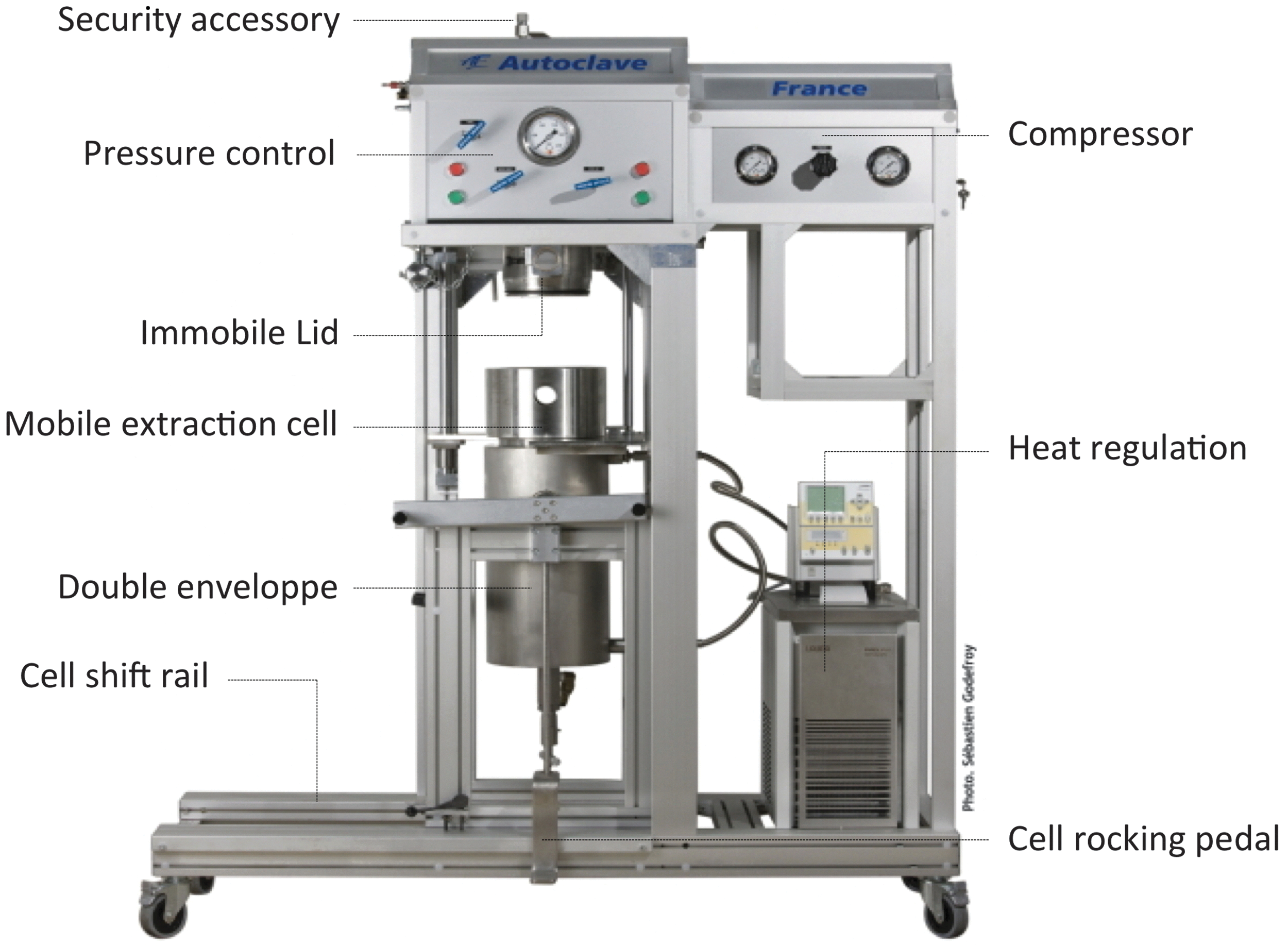
Zippertex was developed as a high-pressure static extractor; however, it offers a variety of extraction conditions and performances. Color images available online at

The extraction cell (left) requires only two paper filters, one for the bottom grid and one for the top grid. The mixture of solvent and plant is introduced between the two filters. The extraction takes 1 h, including filling of the extraction cell with the plant/solvent mixture
W. somnifera extract contains, among other families of compounds, withanosides and withanolide aglycones, the latter being regarded as toxic (Fig. 3A). After hydroalcoholic extraction on Zippertex and removal of ethanol under reduced pressure, the remaining aqueous fraction was further extracted with dichloromethane to remove the toxic withanolides. 29 As shown in Figure 3C, the withanolides (mainly withaferin A and withanolides A and B) were recovered in dichloromethane, while the more polar withanosides remained in the aqueous phase (Fig. 3B). E. officinalis and B. monnieri were similarly submitted to hydroalcoholic extraction on Zippertex. The ethanol was evaporated under reduced pressure and in the case of hydroalcoholic extraction, the water fraction was subsequently lyophilized. Finally, the three aqueous extracts from the three target plants were mixed in the appropriate proportions and submitted to the biotransformation step. The expected compounds from the extracts are represented in Figure 4.

Removal of potentially toxic withanolides from Withania somnifera hydroalcoholic extract by organic solvent extractions.

Major compounds in the selected plants (top, W. somnifera; bottom left, Bacopa monnieri; bottom right, Emblica officinalis).
Emerging evidence suggests the ability of fermentation to enhance the bioactivity and therapeutic potential of traditional medicines. Indeed, the fermentation was shown to increase the availability of the active molecules and to eliminate the undesired compounds. 30 –32 These fermentations were achieved by the endogenous microbial flora, excluding any control of the process and leading to heterogeneous final mixtures. 33 In our case, the fermentation involves a unique identified microorganism under controlled media and condition to make the process perfectly reproducible.
The chosen strain is the filamentous fungus B. bassiana known to exhibit a wide range of catalytic activities. 34 This strain is commonly used for the biological control of insects 35,36 and performs efficient biotransformation of natural compounds and xenobiotics. 37,38 Previously, W. somnifera, E. officinalis, and B. monnieri extracts were separately submitted to bioconversion using a panel of microorganisms in the resting fungus cells condition. While most of the microorganisms remain inactive, B. bassiana leads to a substantial modification of the HPLC initial profiles, mainly for the E. officinalis extract. This property is also observed when the mixture of extracts is submitted to B. bassiana bioconversion. In this case, the major constituent of E. officinalis extract glucogallin is converted into gallic acid (Figs. 5 and 6). Moreover, the fermentation leads to a drastic decrease of the peaks located between 11.5 and 16 min. However, their identification appeared to be difficult because of the intense overlapping as attested by HPLC-MS spectra. The withanosides located between 16 and 19 min are not transformed during the bioconversion by B. bassiana.

Conversion of the major constituent of E. officinalis extract glucogallin into gallic acid by Beauveria bassiana.
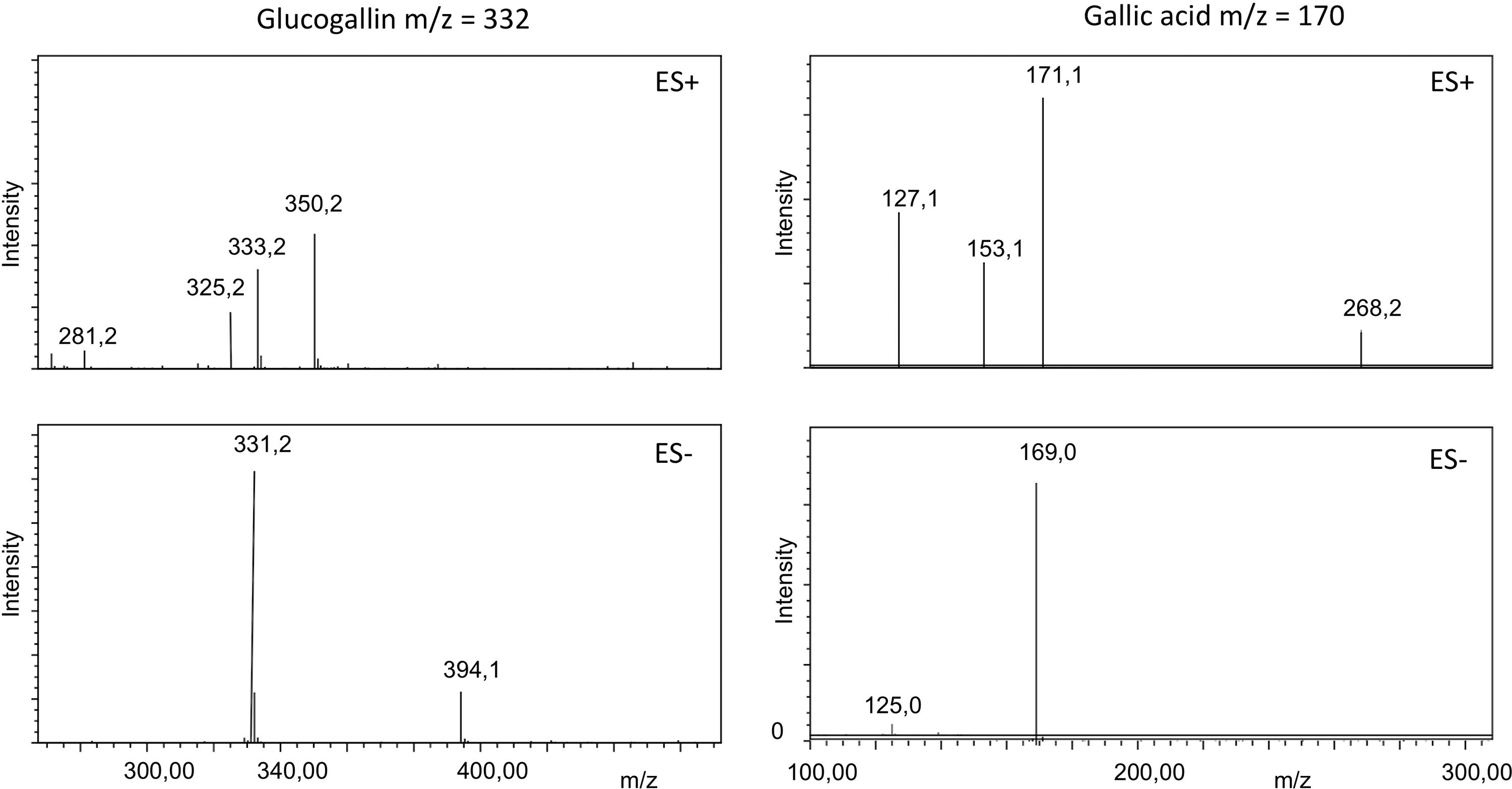
Mass spectrometry confirmation of the structures reported in Figure 5.
This characteristic was used as a marker to monitor the fermentation process by HPLC. We found that the period of 6 days was optimal; up to this period, gallic acid is degraded. The main compound glucogallin from E. officinalis extract (peak 1, m/z=332) has been purified and engaged in a bioconversion step with B. bassiana to confirm the formation of gallic acid. As shown in Figure 4, such transformation was observed after 3 days of incubation.
The CAM assay was used to investigate the ability of prepared botanical extracts to inhibit angiogenesis. 25 Forty microliters of a 10 mg/mL fermented and nonfermented plant extracts mixture was applied on a 7-day-old CAM (12 CAM per product). Pictures of the CAM were taken from the control untreated CAMs and compared with the treated ones 24, 48, and 72 h after treatment. These pictures serve to monitor the vascularization density and the morphology of the CAM. The nonfermented extracts mixture provokes important hemorrhages and total destruction of all treated CAMs after 24 h. In contrast, only 2 CAMs among 12 engaged were destructed after 72 h of treatment with the fermented mixture. More interestingly, as shown in Figure 7, the unaffected CAM treated with the fermented mixture exhibited a drastic reduction of the primary and secondary vascular network.
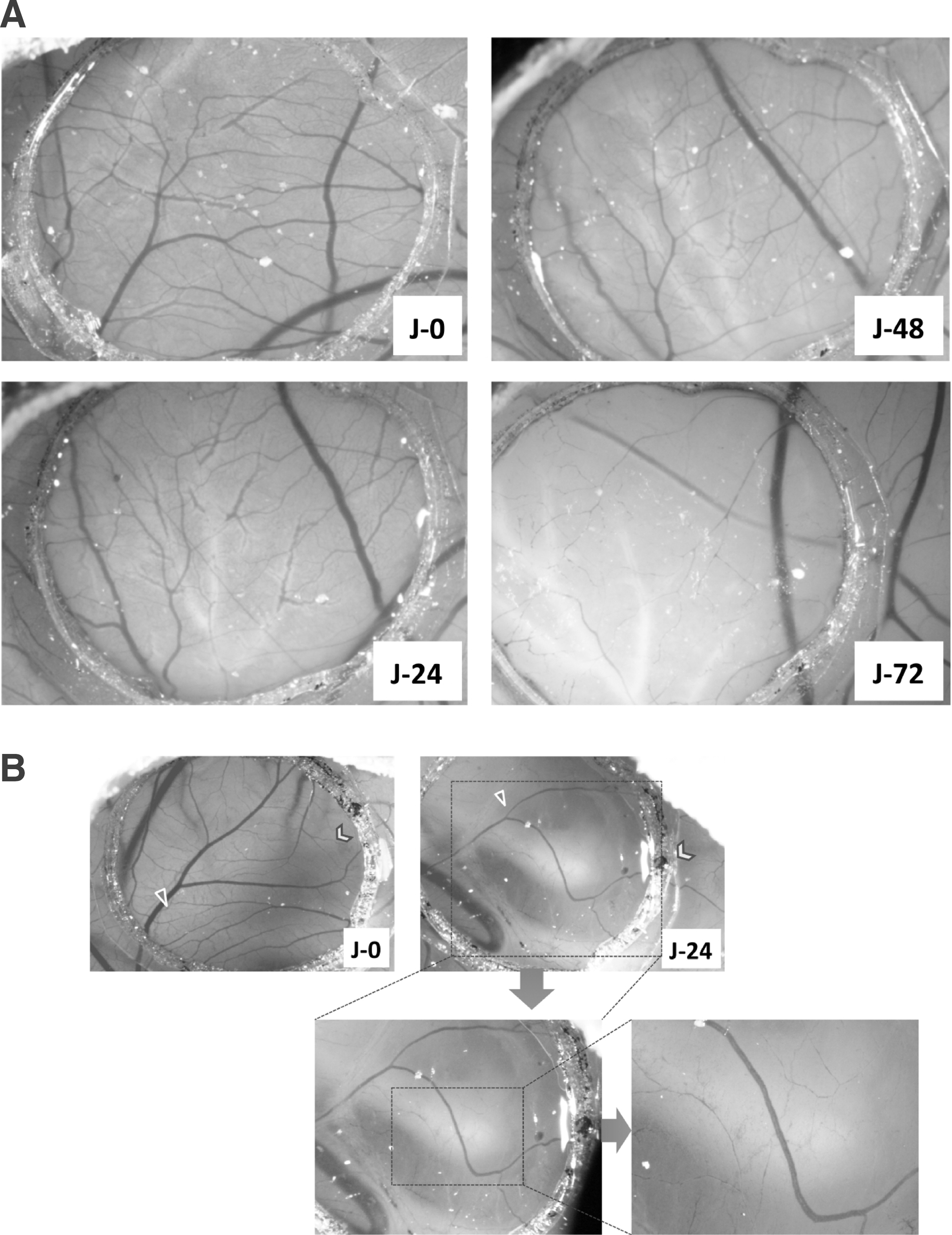
Pictures of the chorioallantoic membrane (CAM) treated with the fermented mixture.
These results confirm that the fermentation with B. bassiana leads to unexpected benefits. It reduces the toxicity of extracts, as demonstrated using the in ovo model of vascularized CAM, and revealed a significant and rapid antiangiogenic effect without apparent damage of the CAM. This activity seems to combine the inhibition of a new vascular vessel and the regression of the previously formed vessels. This result is of high applicable potential in a series of disorders due to uncontrolled vessel proliferation (AMD, diabetic retinopathy, keratitis, glaucoma, rheumatoid arthritis, atherosclerosis, psoriasis, and solid tumors development).
Toxicological in vivo and in vitro bioassays were undertaken to support the safety of the fermented solution. These evaluations consist of the determination of LD50, monitoring of rats' weight, and analysis of the morphology of their organs at the end of the treatment. Liver damage biomarkers were also investigated (aspartate aminotransferase and alanine aminotransferase activity). No deaths occurred during the 3 weeks of the study, and no particular adverse effect was observed after single or repeated administration. Autopsy of animals did not reveal any effect on the organs (spleen, stomach, kidneys, and liver).
The fermentation step was scaled up to 2 m3 volume and the fermented solution was formulated to a nutraceutical antiangiogenic treatment of AMD and commercialized in an oral form named Ethnodyne-Visio.
Footnotes
Author Disclosure Statement
J.O. is one of the inventors of Zippertex. No conflicts of interest exist for any of the other authors.