
Abstract
Solanum tuberosum L. cv Jayoung (JY) is a potato with dark purple flesh and contains substantial amounts of polyphenols. In this study, we evaluated the therapeutic effects of S. tuberosum L. cv JY in a mouse model of Dermatophagoides farinae body (Dfb)-induced atopic dermatitis (AD). The ethanol extract of the peel of JY (EPJ) ameliorated Dfb-induced dermatitis severity, serum levels of immunoglobulin E (IgE) and thymus and activation-regulated chemokine. Histological analysis of the skin also revealed that EPJ treatment significantly decreased mast cell infiltration. The suppression of dermatitis by EPJ treatment was accompanied by a decrease in the skin levels of type 2 helper T-cell cytokines such as interleukin (IL)-4, IL-5, and IL-13. The induction of thymic stromal lymphopoietin, which leads to a systemic Th2 response, was also decreased in the skin by EPJ. Nuclear translocation of nuclear factor-κB p65 was decreased by EPJ in Dfb-induced NC/Nga mice. The protein expression of filaggrin in the AD-like skin lesions was restored by EPJ treatment. These results suggested that EPJ may be a potential therapeutic tool for the treatment of AD.
Introduction
A
Steroids and antihistamines are examples of commonly available drugs that are used to treat AD. However, their use is accompanied by moderate side effects that may include hypertension, osteoporosis, iatrogenic Cushing's disease, dizziness, and blurred vision. 5 Recently, calcineurin inhibitors, such as tacrolimus and pimecrolimus, have been used topically and are considered quite safe. 6,7 Unfortunately, prolonged application of these drugs is also reported to cause skin cancer. 8 Therefore, the development of alternate therapeutic agents is required for long-term management of AD.
Some potato extracts are known to have potent biological activities, including anti-inflammatory properties and prevention of metabolic disease. 9 In addition, anthocyan in pigments was reported to inhibit inflammation. 10 Recently, potato cultivars (Solanum tuberosum L.) are a widely cultivated potato variety (commonly called colored potatoes), which were originally bred in the Republic of Korea during a joint program between the Highland Agriculture Research Center, the Korean National Institute of Crop Science, and the Rural Development Administration. Colored potatoes named Jayoung (JY) have dark purple flesh and contain substantial amounts of polyphenols, such as anthocyanin and phenolic acid. 11 Several studies have demonstrated that colored potatoes possess anti-inflammatory, antioxidant, antihypertensive, antimutagenic, and cytotoxic properties. 12,13 Recently, we reported the anti-inflammatory activities of the solvent fractions from the peel or tuber of colored and general potatoes. 11 Although the peel of JY has been reported to show various bioactivities, the effect of ethanol (EtOH) extracts of peel from JY (EPJ) as an antiatopic agent for AD remains unclear. In this study, we examined the inhibitory effect of EPJ on the development of AD in NC/Nga mice.
Materials and Methods
Animals and treatment
NNC/Nga male mice (14–22 g body weight, 4 weeks old) were purchased from Central Lab, Inc. (Seoul, Korea), a branch of Charles River Japan (Kanagawa, Japan). The mice were housed in specific pathogen-free conditions in a controlled environment (22°C±2°C; lighting 07:00–19:00 h; humidity 30–70%). Food and tap water were provided ad libitum. After acclimation for 2 weeks, the mice were randomly divided into three groups. The animal experiments in this study were approved by the Animal Care Committee of Sangji University (permission No. 2014-21). To induce AD-like skin lesions, the dorsal lesion and ears were topically treated with 25 or 150 μL of 10 mg/mL crude extract of Dermatophagoides farinae body (Dfb) (Biostir® AD; Biostir, Hyogo, Japan), respectively. Mite antigen application was repeated twice a week for 8 weeks (Fig. 1A). Barrier disruption was achieved by1 50 μL of 4% sodium dodecyl sulfate (SDS) treatment, 3 h before the application of Dfb ointment. The Dfb-induced dermatitis mice were divided into three groups: (1) the normal group with no Dfb application, (2) the control group with Dfb application, and (3) the EPJ-treated group (1 mg/mouse) with Dfb application for 4 weeks. Animals were sacrificed at 8 weeks after the first application of Dfb, and blood was collected from the orbital sinus. The dorsal skin tissues and ears of the mice were removed and used for histological examination.
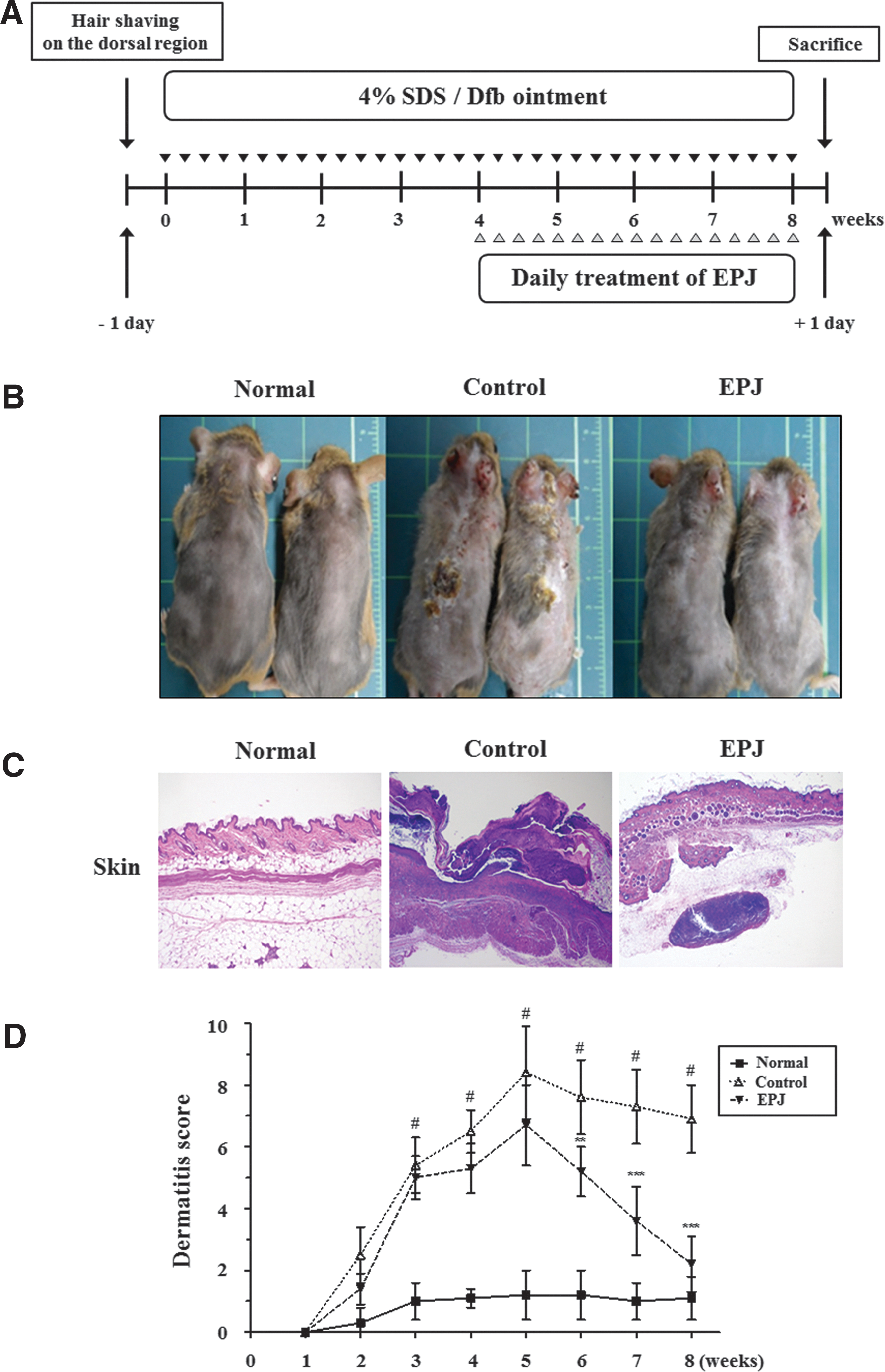
Effects of the EPJ on the development of Dermatophagoides farinae body (Dfb)-induced atopic dermatitis (AD)-like symptoms in NC/Nga mice.
Preparation and standardization of EPJ
The colored potato cultivar, JY, was supplied by the Highland Agriculture Research Center. The potatoes were cut into small pieces (<5 mm thickness); the samples were then freeze-dried and stored at −70°C. These slices (7 kg) were milled and extracted with 30 L of 70% aqueous EtOH thrice by maceration. The extracts were combined and concentrated in vacuo at 40°C to yield a 70% EtOH extract (641 g). From the EtOH extract, two phenolic acids (caffeic acid and chlorogenic acid) were isolated through repeated silica gel and ODS column chromatography and were identified by comparison with authentic samples. HPLC analysis was achieved using a Waters 2695 separation module (Waters, Milford, MA, USA) equipped with a Waters 2996 photodiode array detector (320 nm) and Shiseido Capcell Pak C18 (250×4.6 mm, ID, 5 μm; Shiseido, Tokyo, Japan) column. The mobile phase consisted of 0.4% phosphoric acid (solvent A) and methanol (solvent B), which were eluted at a flow rate of 0.7 mL/min with the following elution profile: isocratic elution 95% A/5% B, 0–5 min; linear gradient from 95% A/5% B to 50% A/50% B, 5–55 min; isocratic elution 50% A/50% B, 55–65 min; linear gradient from 50% A/50% B to 95% A/5% B, 65–70 min; isocratic elution 95% A/5% B, 70–80 min. A 2.5 mg sample of the EtOH extract was dissolved in a mixture of distilled water (2.5 mL) and ACN (2.5 mL). A 10-μL aliquot was injected after filtration into an HPLC system with a 0.45-μm membrane filter. Two phenolic acids, caffeic acid and chlorogenic acid, were eluted at 21.7 and 19.9 min, respectively. The content of caffeic acid and chlorogenic acid in the EPJ extract was determined to be 1.31 and 8.60 mg/g, respectively.
Evaluation of dermatitis severity
Clinical dermatitis severity was tested using the method described by Yamamoto and colleagues. 14 The severity of dermatitis was evaluated once each week. The development of erythema/hemorrhage, scarring/dryness, edema, and excoriation/erosion was scored as follows: 0, none; 1, mild (<20%); 2, moderate (20–60%); and 3, severe (>60%). The sum of the individual scores was used as the dermatitis score.
Histological analysis of skin lesions
Upon euthanasia, skin samples from the dorsal area and ears were collected. The samples were fixed in 10% buffered formalin, embedded in paraffin, sectioned into 4 μm slices, and stained with hematoxylin and eosin. Pathological changes (epidermal and dermal hyperplasia, parakeratosis, hyperkeratosis, dermal edema, vesicular formation, and inflammation) were evaluated and compared between the groups. Selected sections were stained with toluidine blue for evaluation of mast cell infiltration. Mast cell counts of 20 thin sections from each specimen were averaged to obtain mast cell density per square millimeter.
Measurement of ear thickness
The ear thickness was measured with a micrometer (Mitutoyo, Kawasaki, Japan), which was applied near the tip of the ear just distal to the cartilaginous ridges. The thickness was recorded in micrometers.
Measurement of IgE and thymus and activation-regulated chemokine concentration in serum
Blood from the orbital sinus was collected from each mouse at the end of the experiment. Serum was obtained by centrifugation at 1700 g for 30 min and stored at −70°C until analysis. The serum levels of total IgE and thymus and activation-regulated chemokine (TARC) were measured using an IgE enzyme-linked immunosorbent assay (ELISA) kit for mice (Shibayagi Co., Ltd., Gunma, Japan) and a TARC ELISA Kit (R&D Systems, Minneapolis, MN, USA), respectively, according to the manufacturer's instructions.
ELISA assay in dorsal skin
For measuring cytokines, dorsal skin samples of the inflamed region were homogenized in 1 mL of T-PER tissue protein extraction reagent (Pierce, Rockford, IL, USA) containing a protease inhibitor cocktail. Homogenates were then centrifuged at 12,000 g for 20 min at 4°C and then the supernatants were stored at −80°C. Levels of the cytokines IL-4, IL-5, and IL-13 in the supernatant were analyzed using Quansys reagents and instruments (Quansys Biosciences, Logan, UT, USA). Cytokine levels were normalized with a Protein Assay Kit and expressed as pg per mg total protein. The concentrations of TSLP were determined using an ELISA Kit (R&D Systems).
Measurement of relative mRNA expression level
Total RNA was isolated from the dorsal skin tissue by Easy Blue® Kits (Intron Biotechnology, Seoul, Korea). From each sample, 1 mg of RNA was reverse-transcribed by MuLV reverse transcriptase, 1 mM deoxyribonucleotide triphosphate (dNTP), and (dT12–18) 0.5 μg/μL. Polymerase chain reaction (PCR) amplification was performed using the incorporation of SYBR green. The oligonucleotide primers for TSLP designed from mouse were AGCTTGTCTCCTGAAAATCGAG (forward) and AGGTTTGATTCAGGCAGATGTT (reverse), for IL-4 designed from mouse were GGCATTTTGAACGAGGTCAC (forward) and AAATATGCGAAGCACCTTGG (reverse), for IL-5 designed from mouse were AGCACAGTGGTGAAAGAGACCTT (forward) and TCCAATGCATAGCTGGTGATTT (reverse), and for IL-13 were GGAGCTGAGCAACATCACACA (forward) and GGTCCTGTAGATGGCATTGCA (reverse). The oligonucleotide primers for β-actin used as a housekeeping gene designed from mouse were ATCACTATTGGCAACGAGCG (forward) and TCAGCAATGCCTGGGTACAT (reverse). Steady-state mRNA levels of TSLP, IL-4, IL-5, IL-13, and β-actin were determined by quantitative PCR using the A Step One Plus Real-time PCR System (Applied Biosystems, Foster City, CA, USA). The results were expressed as the ratio of optimal density to β-actin.
Preparation of the nuclear fraction
Dorsal skin lesions were homogenized by mortar and pestle and resuspended in a hypotonic buffer (10 mM 4-(2-hydroxyethyl)piperazine-1-ethanesulfonic acid [HEPES], pH 7.9, 1.5 mM MgCl2, 10 mM KCl, 0.2 mM phenylmethylsulfonyl fluoride [PMSF], 0.5 mM dithiothreitol [DTT], 10 mg/mL aprotinin). Tissues were then lysed by adding 0.1% Nonidet P-40 and vortexed vigorously for 10 sec. Nuclei were pelleted by centrifugation at 12,000 g for 1 min at 4°C and resuspended in a high salt buffer (20 mM HEPES, pH 7.9, 25% glycerol, 400 mM KCl, 1.5 mM MgCl2, 0.2 mM ethylenediaminetetraacetic acid [EDTA], 0.5 mM DTT, 1 mM NaF, 1 mM sodium orthovanadate).
Western blot analysis
Protein extracts from dorsal skin were prepared using a lysis buffer (50 mM HEPES, pH 7.0, 250 mM NaCl, 5 mM EDTA, 0.1% Nonidet P-40, 1 mM PMSF, 0.5 mM DTT, 5 mM NaF, and 0.5 mM sodium orthovanadate) and homogenated at 4°C. Tissue debris was removed by microcentrifugation followed by quick freezing of the supernatants. The protein concentration was determined using the Bio-Rad Protein Assay reagent according to the manufacturer's instruction. Protein from each group was electroblotted onto a polyvinylidene difluoride membrane following separation on 8–12% SDS–polyacrylamide gel electrophoresis. The immunoblot was incubated with a blocking solution (5% skim milk) for 1 h at room temperature, followed by incubation overnight with a primary antibody (1:1000 dilution in Tween 20/Tris-buffered saline [TBST]) at 4°C. Blots were washed four times with TBST and incubated with a 1:2000 dilution of horseradish peroxidase-conjugated secondary antibody for 1 h at room temperature. Blots were again washed thrice with TBST and then developed by an enhanced chemiluminescence detection reagent (Amersham Pharmacia, Piscataway, NJ, USA).
Statistical analyses
Data are presented as mean±standard deviation (SD). Data for the treatment groups were compared using one-way analysis of variance (ANOVA) followed by Dunnett's post hoc test. All statistical analyses were performed using SPSS v.13.0 statistical analysis software (SPSS, Inc., Chicago, IL, USA). The statistical significance of differences was accepted at the level of P<.05.
Results
Effects of EPJ on Dfb-induced AD-like skin lesions in NC/Nga mice
Pruritus, eczematous, inflammatory skin lesions, and ear thickening are common symptoms in NC/Nga mice with AD. To establish the house dust mite antigen-induced AD model in NC/Nga mice, Dfb ointment was applied repeatedly to the skin lesion and ears twice a week for 4 weeks. 15 As shown in Figure 1B, repeated application of Dfb ointment in control NC/Nga mice first induced skin dryness, followed by severe erythema, hemorrhage, scarring, dryness, edema, excoriation, and erosion. However, treatment with EPJ inhibited these skin symptoms. In addition, histological features of the skin lesions showed epidermal hyperplasia, edema, and accumulation of inflammatory cells in the dermis/epidermis of the control group compared with the normal group (Fig. 1C). However, the EPJ-treated group exhibited less of these changes. Skin conditions were evaluated once a week for 8 weeks using a dermatitis scoring metric. The dermatitis scores of the control group increased rapidly and were significantly different at 3 weeks, compared with those of the normal group. However, in EPJ-treated mice, the dermatitis scores were markedly lower at 6 weeks, compared with those of the control mice (Fig. 1D).
Effects of EPJ on Dfb-induced ear swelling in NC/Nga mice
After application of Dfb to the ears, swelling gradually increased. As shown in Figure 2A, the histological features of the ear included a thickening of the dermis and epidermis and accumulation of inflammatory cells in the control group compared to the normal group. However, decreased thickening of the dermis/epidermis and dermal infiltration by inflammatory cells were observed in EPJ-treated mice. At 7 weeks, a significant increase was observed in the ear thickness of the control group (48.6±4.68 mm) compared to that of the normal group (26.1±1.88 mm). Treatment with EPJ markedly attenuated the Dfb-induced increase in ear thickness to 39.25±3.08 and 37.78±4.42 mm at 7 and 8 weeks, respectively (Fig. 2B).

Effects of the EPJ on the ear thickness in Dfb-induced NC/Nga mice.
Effect of EPJ on Dfb-induced inflammatory cell filtration and release of IgE and TARC in NC/Nga mice
Mast cells are known to be key effector cells in IgE-mediated allergic disorders. Upon activation, mast cells undergo degranulation and release a variety of biologically active substances such as IL-4, IL-5, and IL-13, which play an important role in the development of AD. 16,17 An increase in serum IgE levels is an important component of AD. 18 TARC is a chemokine involved in lymphocyte migration. It has chemotactic activity specific to Th2 cells, in which the TARC receptor CCR4 is expressed. TARC is overproduced in the skin of patients with AD. 19 To evaluate whether inflammatory cells had infiltrated the skin after Dfb application, skin tissue sections were stained with toluidine blue to detect mast cells. The number of mast cells in the dermis markedly increased in the control group following Dfb application compared with normal mice, whereas EPJ treatment significantly suppressed the number of dermal mast cells (Fig. 3A, B). Application of Dfb in control mice significantly increased serum IgE and TARC levels compared with those of normal mice, whereas treatment with EPJ significantly decreased IgE and TARC levels (Fig. 3C, D).

Effects of the EPJ on inflammatory cell filtrations in Dfb-induced NC/Nga mice.
Effect of EPJ on Dfb-induced cytokine expression and production in NC/Nga mice
TSLP plays a key role in initiating systemic Th2 immunity favorable for the development of AD.
20
TSLP is associated with the induction of a Th2 immune response, TARC, and IgE.
21
Quantitative reverse transcription–polymerase chain reaction (qRT-PCR) and cytokine assay revealed that EPJ markedly downregulated Dfb-induced mRNA expression and production of TSLP in NC/Nga mice (Fig. 4A, B). Dysregulated Th1 and Th2 responses that are characterized by Th2-dominant allergic inflammation are thought to be central to the pathology of diseases such as AD.
2
Among the Th2 cytokines, IL-4 and IL-13 induce B-cell proliferation and immunoglobulin class switching for IgE production, and IL-5 plays an important role in eosinophil infiltration.
22
mRNA expression and levels of the cytokines IL-4, IL-5, and IL-13 markedly increased following Dfb application, but cytokine expression and production were lower in the normal and EPJ-treated groups (Fig. 4A, B). EPJ treatment, however, did not affect Dfb-induced downregulation of INF-γ (Th1-derived cytokine) (Supplementary Fig. S1; Supplementary Data are available online at
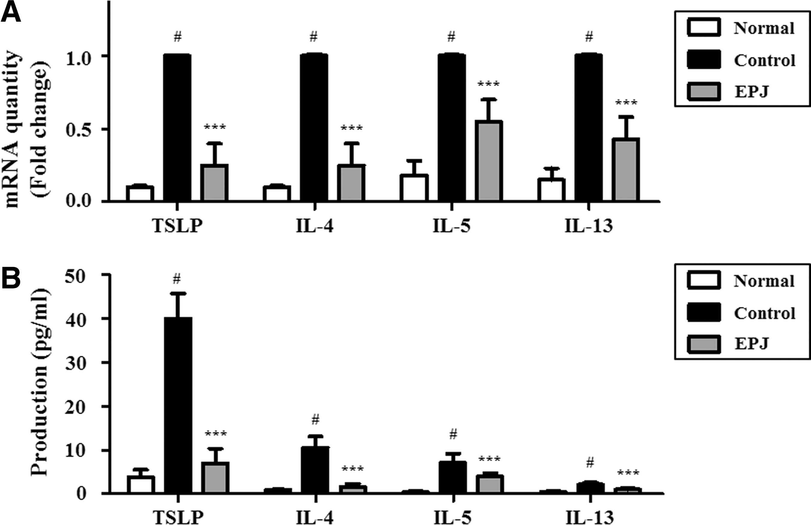
Effects of the EPJ on the mRNA expression and production of inflammatory mediators in Dfb-induced NC/Nga mice.
Effect of EPJ on Dfb-induced nuclear factor-κB activation and skin barrier dysfunction in NC/Nga mice
Nuclear factor-κB (NF-κB) is an important therapeutic target in AD, since it plays a crucial role in coordinated transactivation of key cytokines and adhesion molecules involved in the pathogenesis of this disease. 23 NF-κB has been identified as a common element in the signaling pathways from each of these stimuli. It is noteworthy that the NF-κB heterodimer p50/p65 can regulate TSLP gene transcription by binding to its promoter. 24 To investigate the inhibitory effect of EPJ on NF-κB activation, NF-κB was analyzed in the nuclear protein from skin tissues. EPJ treatment markedly suppressed the nuclear translocation of NF-κB compared to that in the control group (Fig. 5A).
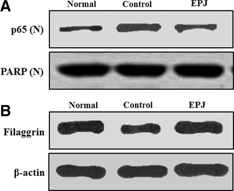
Effects of the EPJ on the translocation of nuclear factor (NF)-κB (p65) and expression of filaggrin in AD-like skin lesions.
The recurrence of a skin barrier defect is an important target of AD treatment. We investigated whether combination treatment changed the expression of proteins associated with the skin barrier defect in the Dfb-induced NC/Nga mouse. As shown in Figure 5B, filaggrin (a differentiation marker for keratinocytes) levels significantly increased in the EPJ group compared to the levels in the control group. These results suggest that topical application of EPJ might suppress the development of dermatitis by inhibiting mast cells through upregulation of filaggrin, presumably resulting in the inhibition of Th2-mediated immune responses.
Discussion
AD is associated with genetic, environmental, and psychological factors, immune system dysregulation, and epidermal barrier defects. 25 Although topical steroids (corticosteroids) or calcineurin inhibitors are the current standard treatment for severe AD, the use may be limited by potentially serious adverse effects. 26 Further research is needed to develop new treatments for patients suffering from AD. Therefore, natural product-based agents with a transcriptional mode of action, good efficacy, and low risk of side effects are promising substances for the treatment and prevention of AD.
Several studies have demonstrated that purple potatoes display various biological properties, such as antioxidant, antihypertensive, and anti-inflammatory effects. In our previous study, the chloroform fraction of the peel of JY (CFPJ) inhibited the lipopolysaccharide-induced production of nitric oxide and prostaglandin E2 in RAW 264.7 macrophages and the molecular mechanisms underlying the anti-inflammatory effects of CFPJ. It was found to inhibit inducible NF-κB activation and the subsequent induction of proinflammatory mediators and to protect mice from dextran sodium sulfate-induced colitis. 11 However, the biological effect of the peel of JY remains unclear. Therefore, this study investigated the inhibitory effect of EPJ on AD-like lesions in Dfb-induced NC/Nga mice.
In this study, the antiallergic effects of EPJ were evaluated following application of a mite antigen Dfb ointment to NC/Nga mice. Even though the severity of AD in humans might differ from those in the mouse model, in this study, as in previous studies, the clinical signs and symptoms of the NC/Nga mice were very similar to those of human AD. 27 Treatment with EPJ on the dorsal skin prevented the dermatitis score from increasing (Fig. 1D) and prevented ear swelling (Fig. 2B) in Dfb-induced NC/Nga mice. Histopathological examination also showed that EPJ treatment inhibited dermatitis-like skin lesions in the NC/Nga mice (Fig. 1C). Epidermal thickening and infiltration of inflammatory cells in the epidermis/dermis were observed in the control mice, whereas these changes were inhibited in EPJ-treated mice. These results indicate that EPJ suppressed the AD skin symptoms induced by Dfb in the NC/Nga mouse model.
The elevated IgE level is a hallmark of AD, and the expression of IL-4 contributes to this elevation. IL-4 stimulates IgE production in B cells. IgE released from B cells binds to mast cells. Mast cells degranulate and release various biological mediators involved in IgE-mediated AD. 16 TARC is overproduced in epidermal keratinocytes and is believed to attract Th2 cells into the skin from the circulating blood. 28 This may trigger or exacerbate the inflammatory process of AD. 28 Several studies have demonstrated that serum IgE and TARC levels are elevated in patients with AD. In this study, EPJ suppressed the Dfb-induced serum levels of IgE and TARC in Dfb-induced NC/Nga mice.
The T cell, according to cytokine and chemokine receptor expression, has been given a new Th cell classification, including Th1, Th2, Th17, and regulatory T-cell subsets that play important roles in allergic disorders. 29 AD is an allergic disease that results from dermal inflammation, a hallmark feature of which is a breakdown in the immunologic balance between Th1 and Th2 cells. 30 AD has been associated with Th2 responses that are driven by the production of IL-4, IL-5, and IL-13. TSLP is regarded as an important factor in AD, capable of enhancing the Th2 polarizing properties of DCs. 31 Acute AD skin lesions exhibit Th2-dominant responses characterized by dermal infiltration of CD4+ or CD8+ T cells, eosinophils, and increased skin expression of Th2 cytokines. Conversely, Th1 responses are characterized by production of the effector cytokines TNF-α and interferon (IFN)-γ. The chronic phase demonstrates a local Th1 response and tissue remodeling with increased deposition of collagen and dermal thickening. 32,33 In this study, mRNA expression and production of TSLP, IL-4, IL-5, and IL-13 markedly increased following Dfb application, but expression and production of these cytokines decreased in the normal and EPJ-treated groups (Fig. 4). These data indicate that EPJ suppressed Dfb-induced dermatitis in the NC/Nga mouse model by inducing dysregulation of the Th1/Th2 balance.
NF-κB is known to be part of the regulatory mechanism for TSLP activation. 34 NF-κB is a transcription factor that controls the expression of genes involved in apoptosis and inflammation, and activation of NF-κB plays a key role in inflammatory skin processes. 35,36 Thus, inhibition of NF-κB activation has been suggested as an anti-inflammatory strategy in AD. We examined the inhibitory effect of EPJ on the NF-κB signaling pathway in Dfb-induced NC/Nga mice. Nuclear extracts from AD-like skin lesions were evaluated in terms of NF-κB activation by western blot analysis. NF-κB was activated following Dfb application, but EPJ inhibited nuclear translocation of NF-κB p65 in Dfb-induced NC/Nga mice. These results suggest that EPJ markedly inhibited expression of TSLP through blockade of NF-κB activation in Dfb-induced NC/Nga mice.
AD skin is characterized by the infiltration of inflammatory cells and skin barrier defects. 37 In addition, mast cells are recognized as critical effector cells in IgE-associated Th2-type immune responses, because they are activated by cross-linking of FcɛR1. Th2 cytokines can downregulate filaggrin. 20 Filaggrin demonstrated a significant decrease in lesional AD skin compared to nonlesional skin. However, filaggrin was significantly higher in the EPJ group compared to the control group. Therefore, recruitment of inflammatory cells and restoration of the epidermal barrier are important objectives for the management of AD. We found that topical application of EPJ prevented infiltration of inflammatory cells and restored barrier function in the skin. These results suggest that topical application of EPJ might suppress the development of dermatitis by inhibition of mast cells through upregulation of filaggrin, presumably resulting in the inhibition of Th2-mediated immune responses.
The keratinocyte differentiation process is characterized by a series of biochemical events. 38 The switch in the expression of the proliferating keratins to differentiating compartment indicates that the changes occur in the keratin filament organization, and this change, in turn, influences the functional properties of the epidermis. 39 Filaggrin is a cationic protein presented in the cornified layer of the epidermis. During multistep processes of profilaggrin expression and degradation into free amino acid, several regulators, including activator protein-1 (Jun and/or Fos) 40 and PPAR-γ, 41 have been reported to enhance the profilaggrin expression during terminal differentiation of the epidermis. 42 Further pharmacological studies are needed to provide more detailed information on the structure of keratinocyte differentiation process exhibited by EPJ. We believe that further study may contribute to our current understanding of EPJ efficacy and, if we could confirm its effect in keratinocyte differentiation comparing to the AD treatment drug, EPJ could be a potent candidate for AD therapy.
Although several studies conducted on single phytocompound-based drugs have produced encouraging results, there is an increasing trend toward the use of whole plant extracts, since they offer synergistic effects of myriad secondary metabolites and provide multiple points of intervention for drug development. 43 In addition, mixtures of compounds present in plant extracts may provide the essential combinations that affect multitude targets and, thus, achieve clinical advantage beyond the scope of a single compound or solvent fractions. Considering all of these factors, we suggested that standardized EPJ with the phytochemical content is more advantageous compared to other extracts using different solvents in inflammation-associated diseases. Taken together, our results suggest that EPJ treatment improved Dfb-induced AD-like skin lesions in an NC/Nga mouse model by suppressing Th2-mediated cytokine levels, decreasing IgE levels, inhibiting infiltration of inflammatory cells, and improving skin barrier function. Considering all of these findings, it correlated with the observed clinical symptoms and we expect that EPJ could potentially be used as a safe and effective intervention for the treatment of AD.
Footnotes
Acknowledgment
This research was supported by the Rural Development Administration Grant (PJ009146), Republic of Korea.
Author Disclosure Statement
No competing financial interests exist.
References
Supplementary Material
Please find the following supplemental material available below.
For Open Access articles published under a Creative Commons License, all supplemental material carries the same license as the article it is associated with.
For non-Open Access articles published, all supplemental material carries a non-exclusive license, and permission requests for re-use of supplemental material or any part of supplemental material shall be sent directly to the copyright owner as specified in the copyright notice associated with the article.