
Abstract
Vascular function is mediated by various regulatory molecules, including endothelial nitric oxide (NO), which regulates the vasodilation of smooth muscle cells. We investigated whether standardized Houttuynia cordata extract (SHCE) could improve physical endurance performance by regulating the endothelial production of NO. For the standardization of Houttuynia cordata (HC) extract, its bioactive components were identified and quantified using ultraperformance liquid chromatography–mass spectrometry. Bioaccessibility and biological activity were measured by the in vitro digestion model system and free radical scavenging capacity, respectively. The vascular function in the endothelium was assessed by the phosphorylation of endothelial nitric oxide synthase (eNOS). A preliminary clinical trial was carried out to assess the physical endurance performance. HC extract was standardized to bioactive components, including chlorogenic acid, rutin, and quercitrin, with the concentration of 5.53, 6.09, and 16.15 mg from 1 g of dry weight, respectively. Bioaccessibility was 33.17%, 31.67%, and 11.18% for chlorogenic acid, rutin, and quercitrin, respectively. Antioxidant activities of SHCE were expressed as vitamin C equivalent antioxidant capacity in 55.81 and 17.23 mg/g of HC extract using ABTS and DPPH scavenging assay, respectively. In human aortic endothelial cells, insulin-mediated phosphorylation of eNOS was increased by SHCE in the presence of palmitate. However, the expression of blood pressure-regulating genes was not altered. The level of blood lactate concentration and the heart rate of subjects who drank SHCE were lower than those of subjects who drank plain water. Oxygen uptake from subjects drinking SHCE was slightly higher than that from those who drank plain water. This study demonstrated that SHCE decreased heart rate and blood lactate, increased oxygen uptake, and improved physical performance, presumably due to the increased NO production.
Introduction
H
Accumulating evidence suggests that HC is a potential source of natural antioxidant due to its contents, including various bioactive components, especially flavonoids.
7,16
The reports regarding HC have chemically identified flavonoids, such as quercitrin, isoquercitrin, rutin, hyperoside, chlorogenic acid, and quercetin in HC, but the profile of flavonoids varied depending on the origin of its source.
2,9,12,17,18
A variety of pharmacological effects of HC have been observed in either crude extracts such as water and organic solvent extracts, or single components. Quercetin and quercetin-3-O-β-
In this study, we prepared a standardized Houttuynia cordata extract (SHCE) and investigated its regulation of NO production in dysfunctional endothelial cells and the expression of blood pressure-regulating genes by SHCE. We also examined the effect of SHCE on physical endurance performance in preliminary clinical tests.
Materials and Methods
Chemicals
Standards (chlorogenic acid, rutin, and quercitrin hydrate) and digestive enzymes (α-amylase from human saliva, pepsin from porcine gastric mucosa, porcine lipase, pancreatin from porcine pancreas, and bile extract porcine) were obtained from Sigma-Aldrich (St. Louis, MO, USA). Dulbecco's modified Eagle's medium was purchased from Cellgro (Manassas, VA, USA). Fetal bovine serum (FBS) was purchased from Gibco (Introgen Corporation, Grand Island, NY, USA). Nonessential amino acids, gentamicin, and penicillin were purchased from Sigma-Aldrich. High-performance liquid chromatography grade solvents, acetonitrile and water, were obtained from J.T. Baker (Phillipsburg, NJ, USA), and acetic acid was purchased from Sigma-Aldrich.
Standardization of Houttuynia cordata extract
Extraction and concentration
Dried HC (40 kg) was extracted with 50% ethanol (700 L) at 80°C for 4 h. The residue was further extracted with 240 L of solvent for 2 h. The extract was filtered by a filter press. The filtrate was spray-dried and concentrated to 60% of solid content.
Identification and quantification of bioactive components in HC
Ultraperformance liquid chromatography, equipped with a photodiode array detector, Accela autosampler, and Accela 600 pump, was used to perform the identification of biomarkers (chlorogenic acid, rutin, and quercitrin) in HC. Chromatographic separation was performed on a Hypersil GOLD C18 column (50×2.1 mm, 1.9 μm; Thermo Scientific, San Jose, CA, USA) at a flow rate of 0.2 mL/min at room temperature. Mobile phases consisted of solvent A (0.1% acetic acid in acetonitrile) and B (0.1% acetic acid in water). The linear gradient mobile phase was used as follows: 0–2.19 min, 0–5% A; 2.19–3.26 min, 5–10% A; 3.26–7.29 min, 10–20% A; 7.29–12.00 min, 20–30% A; and 12.00–15.00 min, 30–0% A. The injection volume was 1 μL. The wavelength of UV spectrum was set at 280 nm. For further analysis, the mass spectrometer (LCQ fleet; Thermo Scientific) was operated with an electronic spray ionization (ESI) source and ion trap in negative ion mode. Analyses of samples were initially carried out using full-scan, data-dependent MS scanning from m/z 100 to 700. The capillary temperature was 275°C; the flow rates for sheath gas of nitrogen gas and auxiliary gas of helium gas were 43 and 5 mL/min, respectively; and the capillary voltage was 5 V. The tuning of the mass spectrometer was optimized by infusing a standard of each sample dissolved in the mobile phase.
Chromatographic peaks and mass spectrums in samples were identified using comparative retention times and molecular weights of pure standards. Quantitative analysis was conducted using standard curves.
Bioaccessibility of bioactive components in HC
The in vitro digestion model system simulating the human salivary, gastric, and small intestinal digestive processes was adjusted from a previous study with certain modifications
25
; 0.5% ascorbic acid was added to prevent oxidation during digestion. Five milliliters of 20 mM phosphate buffer was added into 20 mg of the samples to initiate a salivary phase. A 60 μL amylase solution (0.04 mg/mL) was mixed into each sample, and pH was adjusted to 6.9±0.5 with the addition of the 20 mM phosphate buffer. To mimic human gastric conditions, the pepsin solution (0.03 mg/mL of 100 mM HCl) was suspended in the samples reaching pH 2.0 by 0.1 M HCl. The sample was incubated in a shaking water bath (Precision™, ThermoScientific. Piscataway, NJ, USA) for 1 h at 37°C and 100 rpm, and then 0.1 M NaHCO3 was utilized to adjust the pH to 5.3. The pancreatic mixture (porcine bile:lipase:pancreatin, 1:1.5:1.5, v:v:v) was added into the sample. The sample was adjusted to pH 7.0 by adding 1 M NaOH, and the volume of the sample was equalized with 20 mM phosphate buffer, followed by incubation at 37°C for 2 h at 250 rpm in a shaking water bath. The supernatant was collected after centrifugation (1000 g, 4°C, 30 min) and stored at −70°C before analysis. Bioaccessibility after in vitro digestion was calculated using the following the formula:
Biological activity of free radical scavenging
Typically, 2,2′-azino-bis(3-ethylbenzthiazoline-6-sulfonic) acid radical (ABTS•+) and 2,2-diphenyl-1-picrylhydrazyl (DPPH) radical scavenging assay have been used to evaluate biological activity. 26 –28 AAPH (1.0 mM) was mixed with 2.5 mM ABTS in phosphate-buffered saline (PBS) solution (100 mM phosphate buffer [pH 7.4] containing 150 mM NaCl). The mixture was heated in a water bath at 70°C for 1 h and was agitated frequently. The concentration of the final blue-green ABTS•+ solution was adjusted to an absorbance of 0.650±0.020 at 734 nm by using PBS. A 20 μL aliquot of the sample was added to 980 μL of ABTS radical solution. The mixture was incubated in a water bath at 37°C for 10 min under darkness. The decrease in absorbance was monitored at 734 nm. A mixture of 20 μL of distilled water and 980 μL of ABTS radical solution was used for a control.
One hundred micromolars of DPPH• was dissolved in ethanol. The final concentration of DPPH radical solution was regulated to an absorbance of 0.70±0.05 at 517 nm. The sample (0.1 mL) was added to 2.9 mL of DPPH radical solution. The mixture was reacted at room temperature in the dark for 30 min while shaking. The absorbance values were measured at 518 nm. As a control, a mixture of 0.1 mL of ethanol and 2.9 mL of DPPH radical solution was prepared.
A vitamin C standard curve and absorbance decreases of ABTS and DPPH radicals were measured. Absorbance values of the samples were converted into vitamin C equivalent antioxidant capacity (VCEAC) using the vitamin C standard curves. The scavenging effect of the samples against ABTS and DPPH radicals was calculated in mg/g.
Measurement of vascular function in the endothelium
Cell culture
Human aortic endothelial cells (HAECs; BioWhittacker, Walkersville, MD, USA) were cultured in endothelial growth medium-2 (Lonza, Allendale, NJ, USA) supplemented with specific growth factors and 2% FBS. Cells were grown at 37°C in an atmosphere of 5% CO2/95% air. HAECs were grown to 70–80% confluency and treated with palmitic acid (500 μM) dissolved in media solution containing 2% fatty acid-free bovine serum albumin for 3 h. SHCE was administered with palmitic acid to examine its effect on phosphorylation of endothelial nitric oxide synthase (eNOS).
Immunoblot analysis
Cells were harvested and homogenized in PBS containing a protease inhibitor and phosphatase inhibitors (Roche, Indianapolis, IN, USA). Thirty micrograms of cell extracts were applied to SDS–PAGE and transferred onto nitrocellulose membranes. eNOS and phosphorylated eNOS were detected by human-specific antibodies (Cell Signaling, Danvers, MA, USA). Protein bands were detected by chemiluminescence analysis (Pierce, Rockford, IL, USA) and X-ray film exposure.
Gene expression
Total RNA for real-time polymerase chain reaction (PCR) was isolated using TRIzol according to the manufacturer's procedure (Invitrogen, Carlsbad, CA, USA). First-stranded cDNA was synthesized by reverse transcribing 1 μg of total RNA in a final reaction volume of 100 μL using the High-Capacity cDNA Archive kit (cDNA was synthesized by reverse transcription) according to the manufacturer's instructions. Measurements of mRNA expression were performed with SYBR Green PCR core reagents using 20 ng of synthesized cDNA (Applied Biosystems, Foster City, CA, USA). Incorporation of the SYBR Green dye into the PCR products was monitored in real time with an Mx3000 sequence detection system (Stratagene, La Jolla, CA, USA). All the PCRs were performed under the following conditions: 2 min at 50°C, 10 min at 95°C, and 40 cycles of 15 sec at 95°C and 1 min at 60°C in 96-well optical reaction plates (Applied Biosystems). Quantitative amounts were normalized by β-actin. The sequences of the primers used in this study are provided in Table 1.
Preliminary clinical trials for physical endurance performance
Subjects
Five healthy weight-stable men were voluntarily involved in this preliminary clinical trial. The study protocol was approved by the ethical committee of clinical research of Sejong University, Seoul, South Korea (Protocol Reference No. 14-05-7; approval date: June 5, 2014). Written informed consent was obtained from all participants. They kept their physical activity and weight stable for the entirety of the 3-week study. None smoked or had a history of recent serious acute or chronic disease, and all were judged to be healthy on the basis of routine screening physical examination: age, height, body weight, and body fat were 24.0±2.2 (year), 177.2±3.0 (cm), 71.8±9.1 (kg), and 19.8±5.5 (%), respectively. All subjects provided written informed consent. Five volunteers drank plain water in the first week of the study, and then they drank SHCE after a washing out period of 1 week. Subjects drank each treatment (plain water, SHCE) once per day and 10 min before the exercise and 10 min after the exercise. A crossover design was applied to this trial.
Protocol for physical endurance performance
The current study followed a Bruce protocol, the most widely used exercise protocol. It is a maximal exercise test consisting of seven stages with progressive increases of speed and incline every 3 min starting from 2.74 km/h at a 10% incline (Fig. 1). The length of time on the treadmill is the test score and can be used to estimate the oxygen uptake. During the test, the heart rate, blood pressure, and rating of perceived exertion were also recorded. The changing patterns of heart rate, concentration of blood lactate, and oxygen uptake were observed. The oxygen uptake and heart rate were measured continuously by cardiopulmonary exercise test and polar monitor, respectively. Blood samples for measuring blood lactate were collected before exercise, after intense (maximum level) exercise, and 2, 5, and 10 min after recovery. Blood lactate was measured by using portable lactate analyzer.
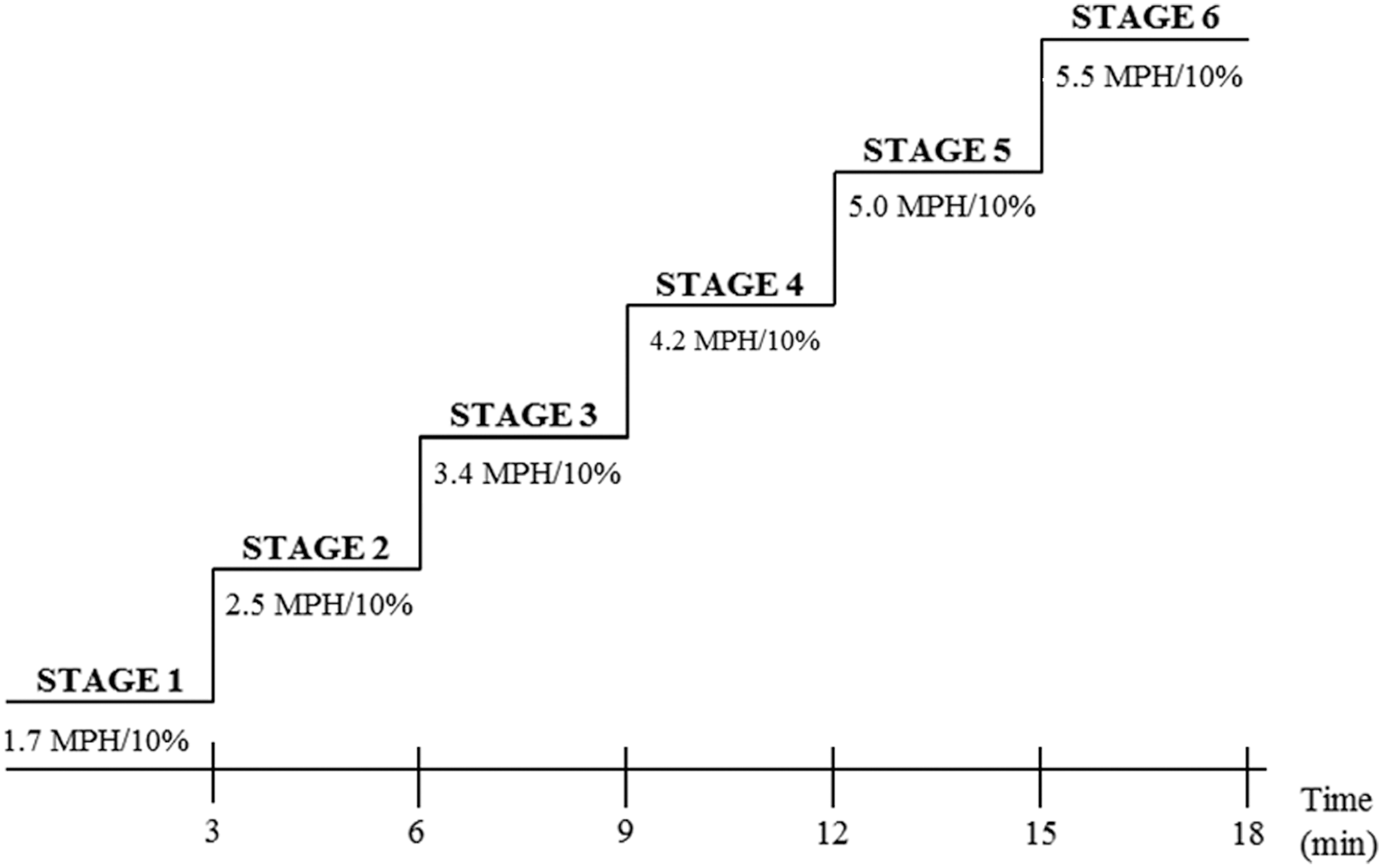
Bruce protocol is a maximal exercise test where the athlete works to complete exhaustion as the treadmill speed and incline is increased every 3 min. MPH, miles per hour.
Statistical analyses
Values are reported as mean±standard deviation from at least three different experiments. A one-way analysis of variance was carried out to measure significant differences among the groups at the significant level of P=.05. GraphPad Prism Version 3.0 (GraphPad, La Jolla, CA, USA) was used for all statistical analyses.
Results and Discussion
Standardization of HC extract
Plant extracts generally contain a complex mixture of organic chemicals such as secondary metabolites, including polyphenols, resulting in confusion in determining their precise biological activity in humans. 29 For utilization of extracts in the development of functional foods and pharmaceuticals, the most important challenge is the lack of complete standardization and maintenance of their quality. Thus, the current study was conducted for the standardization of physiologically bioavailable components in HC. First, the measurable marker compounds present in significant amounts in the HC extract were identified and quantified using LC-MS/MSn. Second, the bioaccessibility of bioactive components in HC extract was quantified using an in vitro biomimetic system simulating the human gastrointestinal tract (Table 2A). Then, the free radical scavenging capacities of biological components were measured as a quality control parameter for standardization (Table 2B).
Values are mean±SE.
DW, distilled water; HC, Houttuynia cordata.
As shown in Figure 2, detection at a wavelength of 280 nm showed the best abundance for target compounds with minimal noise within the chromatographic windows. Three peaks were eluted by the UV spectrum (Fig. 2A). Each empirical formula of major peaks (peaks 1, 2, 3) was identified by its mass fragmentation at each retention time (Fig. 2B, C). All samples showed more sensitivity and selective mass patterns in the negative ESI mode than the positive mode. Peak 1 eluted at the retention time (Rt) of 5.94 min was identified by its negatively charged molecular ion [M-H]− at m/z 353, while it produced an ion at m/z 162 on MS2 fragmentation, which corresponds to a 191 amu loss of quinic acid (Fig. 2B, C). Peaks 2 and 3 (Rt=8.59, 9.64 min) had [M-H]− at m/z 609 and 447, respectively. Further fragmentation revealed it to be an MS2 profile of ion at m/z 301, indicating a quercetin (Fig. 2B, C). This means that peaks 2 and 3 have the same backbone of quercetin. We confirmed that the resulting retention time and MSn fragments of peaks corresponded to those of the standard mixture (Fig. 2D). Therefore, peak 1 was identified as chlorogenic acid, which is the ester of caffeic acid and quinic acid (Fig. 2E). Peaks 2 and 3 were identified as rutin and quercitrin, respectively (Fig. 2E). Rutin, referred to as quercetin-3-O-rutinoside, is the glycoside between the quercetin and the disaccharide rutinose. Quercitrin is a glycoside formed from quercetin and deoxysugar rhamnose. Although both rutin and quercitrin have the same backbone, the retention time of rutin eluted before quercitrin (Fig. 2D). The molecular mass values were calculated from the difference between rutin and quercitrin. Its difference was 162 amu as sugar units such as glucose or galactose. 30 A previous study reported that the retention time decreased with increasing polarity, that is, increased the number of hydroxyl groups in the flavylium nucleus. 31 The presence of sugars shortens the retention time. In other words, diglycosides usually elute before monoglycosides.

UV absorption pattern and mass spectra of components in HC extract and chromatograms of UPLC-PDA peaks corresponding to a mix of standards.
For quantification of each bioactive component, the linear calibration curves were established from each standard. The regression equation was calculated according to y=ax+b, where y and x represent the peak area and concentration, respectively. High correlation coefficients and good linear calibration curves were obtained for each standard (R 2>0.994). Table 2 indicates that the concentration of chlorogenic acid, rutin, and quercitrin was 5.53, 6.09, and 16.15 mg from 1 g of dry weight, respectively. Quercitrin had the highest content in HC extract.
Bioaccessibility generally means the fraction of compounds released from its matrix in the gastrointestinal tract and thus becomes available for intestinal absorption. 32 –34 O'Sullivan et al. 35 reported that polyphenols are subject to degradation during digestion owing to the effect of pH and enzymes. In the same vein, the current study showed the low bioaccessibility of polyphenols in HC extract, resulting in 33.17%, 31.67%, and 11.18% for chlorogenic acid, rutin, and quercitrin, respectively (Table 2A). Chlorogenic acid is a phenolic acid, while both rutin and quercitrin are flavonoids. Chlorogenic acid was more bioaccessible than other polyphenols in the HC extract, presumably because of its low molecular weight and high solubility. Rodríguez-Roque et al. 36 found that the phenolic acids in blended fruit juice were less reduced than that of flavonoids during small intestinal digestion. Despite the same backbone of rutin and quercitrin, the bioaccessibility of rutin was ∼2.8 times higher than that of quercitrin, possibly due to the amount of added sugar. The previous study observed that attaching to more than one sugar made parent compounds have higher water solubility due to the presence of glycosyl groups, leading to improved bioavailability. 37
Measurement of biological activity such as free radical scavenging capacity is one way to standardize bioactive components in plant extracts. The reductions of ABTS and DPPH radical chromogens by antioxidants result in a decrease of absorbance at 734 and 517 nm, respectively. 27,28 The calibration curve showed a linear relationship (ABTS: R 2=0.9974, DPPH: R 2=0.9885) between vitamin C concentration and absorbance reduction at 734 and 517 nm, respectively (data not shown). Table 2B showed that the antioxidant capacity of HC was 90.59% and 89.75% for ABTS and DPPH radicals, respectively. The interaction of a potential antioxidant with DPPH depends on its structural conformation, and this structural requirement is correlated with the presence of hydroxyl groups on the flavonoids. As this electron becomes paired off in the presence of a free radical scavenger, the absorption fades and the resulting decolorization is stoichiometric with respect to the number of electrons taken up. 38,39 Our results showed that the ABTS assay yielded a higher value than the DPPH assay and also suggested that HC extract possessed a free radical scavenging activity of 55.81 mg/g for ABTS radicals and 17.23 mg/g for DPPH radicals in VCEAC. 40 These results indicate that the phenolic components in HC are beneficial due to their radical scavenging activity and have potential for development of functional food and medical products.
Measurement of vascular function in the endothelium
Obesity-mediated metabolic syndrome is associated with elevated free fatty acids (FFAs) in circulation. 41 In cultured endothelial cells and animal models, elevated FFAs disrupt NO synthesis and cause vasocontraction. 42 –44 Imbalanced NO production and degradation impair cardiovascular complications, including blood pressure increases. 45,46
HAECs were incubated with 500 μM palmitate in the presence of SHCE to resemble an in vivo obese condition. Palmitate treatment decreased the basal eNOS phosphorylation at Ser1177 in basal condition (Fig. 3). Insulin stimulates eNOS phosphorylation and palmitate exposure reduces phosphor-eNOS. Consistent with our expectations, SHCE activated eNOS phosphorylation in the presence of palmitate and increased sensitivity to insulin (Fig. 3). This result implies that SHCE has a stimulatory effect on eNOS phosphorylation and prevents obesity-mediated inhibition of eNOS phosphorylation. Therefore, increased eNOS phosphorylation in the endothelium by SHCE may have a beneficial effect on increasing blood flow in an exercise study.
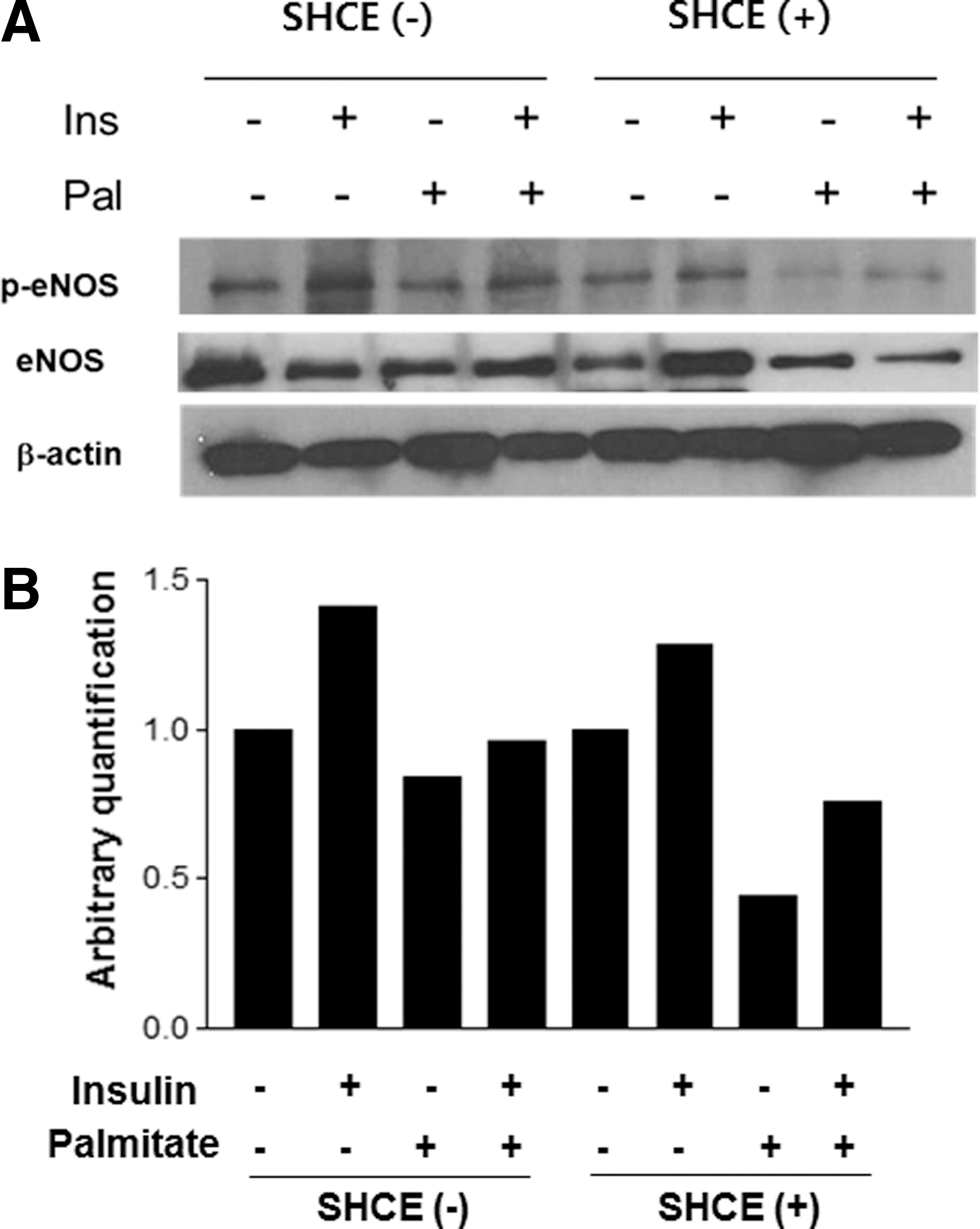
Effects of SHCE on phosphorylation of eNOS in HAECs; HAECs were preincubated with 50 μg/mL SHCE for 16 h and treated with 500 μM palmitate for 3 h. HAECs were treated with 100 nM insulin for the last 10 min of the 180-min treatment period. Immunoblot analyses for p-eNOS, eNOS, and β-actin were performed. The experiment was performed thrice and the representative immunoblot is shown
To examine the pharmacological effects of SHCE, the expression of genes regulating vasodilation was measured by real-time PCR. Overall, gene expression was suggesting no alteration of vasodilation in HAECs (Fig. 4). Human endothelial nitric oxide synthase 3 (NOS3) and SERPINE1 (plasminogen activator inhibitor type 1) were not altered by a combinational treatment of palmitate and SHCE (Fig. 4A). In contrast, the expression of ACE (angiotensin-converting enzyme) was upregulated by both palmitate and SHCE treatments. This result suggests that the aggravation of vasocontraction by SHCE occurred in addition to palmitate. Meanwhile, angiotensinogen (AGT) was upregulated by palmitate and/or SHCE and vascular cell adhesion molecule 1 (VCAM1) by cotreatment of palmitate and SHCE. Other genes, such as prostaglandin-endoperoxide synthase 2 (PTGS2), intercellular adhesion molecule 1 (ICAM1), superoxide dismutase 1 (SOD1), prostaglandin I2 synthase (PTGIS), endothelin (EDN), and endothelin receptor type A (EDNRA), were not affected by the combination of palmitate and SHCE (Fig. 4B–D). Overall, SHCE has no effect on vasculature-related inflammatory or ROS-related oxidative gene expression, except for an upregulated angiotensin-related gene (AGT). Thus, it is assumed that the expression of genes involved vessel contraction, and dilation was not modulated by SHCE treatment, except for angiotensin-related signaling.
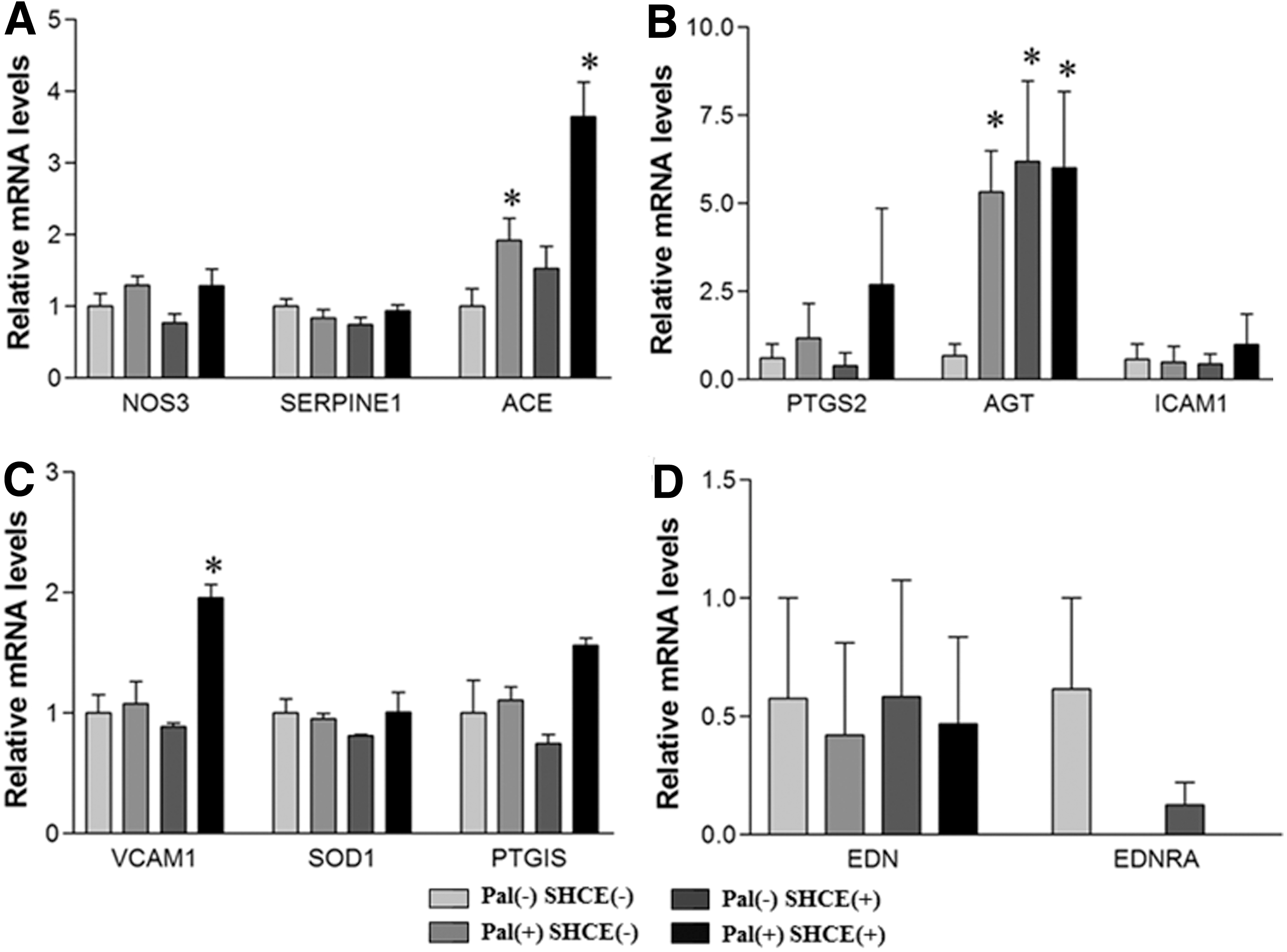
Effects of SHCE on blood pressure-related gene expression in HAECs; HAECs were incubated in the presence or absence of 500 μM palmitate or 50 μg/mL SHCE for 16 h. mRNA was extracted and cDNA was synthesized for real-time PCR measurement
Endothelial dysfunction is the first step in cardiovascular complications. Endothelial cells are located on the surface of blood vessels called intima. These cells act as a diffusion barrier and produce various signaling molecules such as inflammatory cytokines. Among them, endothelial cell-derived NO has vasodilatory, anti-inflammatory, and antiproliferative effects. 45,46 Impaired NO production inhibits vasodilation by smooth muscle cells, and limited bioavailability of NO leads to vascular dysfunction. 47 As a first step, we attempted to examine the pharmacological effects of SHCE on endothelial NO production and we found that activation of NO production by eNOS phosphorylation was improved. We therefore expect that endothelial dysfunction will be ameliorated by SHCE. The results of gene expression studies suggest that SHCE restores eNOS-mediated NO production by increased phosphorylation rather than inflammatory or regulatory cytokines (Fig. 4).
Preliminary clinical trials for physical endurance performance
A previous study revealed that polyphenols, such as quercetin, catechin, and resveratrol, could improve exercise performance by regulating oxygen uptake in vitro and in vivo. 48 Olthof et al. 49 found that the intake of chlorogenic acid induced biological effects in blood circulation. Rutin from buckwheat decreased capillary fragility. 50 Quercitrin was reported to enlarge capillary vessels. 51
Oxygen uptake (VO2) is a measure of the oxygen volume used by the human body to obtain energy (adenosine triphosphate, ATP) from food. 52 It is generally expressed as a relative rate in milliliters of oxygen per kilogram of body weight per minute (mL/kg/min). 53 Maximum oxygen uptake (VO2 max) is the maximum possible oxygen uptake that a given person can achieve. In addition, VO2 max measures the maximal ability of a human body to deliver and utilize oxygen and is related to the ability to perform prolonged exercise. Bassett and Howley 54 confirmed that endurance performance during exercise was partially affected by VO2 max.
Our study showed that VO2 from subjects who drank SHCE was slightly higher than that of subjects who drank plain water, although there was no significant difference between them (Fig. 5A). A previous study showed a similar result of higher level of VO2 from those who drank Prunus mume extract, as opposed to a control. 55 Short-term consumption of EGCG, a kind of catechin, also improved VO2 max as noted by Richards et al. 56

Effect of SHCE on physical endurance performance;
Heart rate refers to the speed of heartbeat, specifically the number of heartbeats per unit of time, expressed as beats per minute. 57 Figure 5B indicates that heart rates increased as subjects kept up with the exercise. Meanwhile, heart rates decreased during the recovery phase. The heart rates of subjects drinking SHCE were lower than that of a control with no significant difference. This result was similar to a report by Schmidt et al., 58 in which the heart rate in men with chronic heart disease decreased after the intake of flavonoids from hawthorn extract.
Blood lactate, an indicator of muscle lactate, increases with continuous work rate and incremental exercise, while it decreases immediately at the start of recovery. 59,60 It is generally expressed as a millimole per liter. As shown in Figure 5C, blood lactate concentration from subjects who drank SHCE was lower than that from those drinking plain water, indicating that drinking SHCE enhanced lactate clearance capacity in the blood. Park et al. 22 also demonstrated that Ecklonia cava polyphenol supplementation contributed to less production of lactate during intense exercise, resulting in improved endurance performance.
Alteration of blood flow comes from platelet activation. When a platelet is activated, serotonin, Ca2+, and thromboxane A2 are released to activate platelet aggregation or clotting. However, blood clotting interrupts circulation and causes thrombosis, a frequently fatal vessel clogging. This event can develop into coronary artery diseases such as myocardial infarction. Our exercise study demonstrated that oxygen uptake was increased and heart rate and blood lactate concentration were decreased by SHCE. Although these results are limited due to the small number of volunteers and lack of statistical significance, the result that one-time consumption exhibits a consistent pattern of blood flow improvement is worthy of further pursuit. Mismatch of a cell study and an exercise study might arise from the other mechanisms. A possible explanation for the mismatch between the cell culture study and exercise study might be an increase of vasoregulatory factors released from platelets. Blood serotonin and thromboxane A2 may be increased by consumption of SHCE and cause an improved response to exercise. Another possibility is an increase of oxygen uptake by red blood cells. Unfortunately, the mechanism of improved blood flow in a whole body is not clear at this moment and deserves further studies.
In conclusion, this study determined that the major bioactive components in HC extract were chlorogenic acid, rutin, and quercitrin with concentrations of 5.53, 6.09, and 16.15 mg from 1 g of air-dry weight, respectively. Bioaccessibility was 33.17%, 31.67%, and 11.18% for chlorogenic acid, rutin, and quercitrin, respectively. For biological activity, the free radical scavenging capacity of HC extract was 90.59% and 89.75% for ABTS and DPPH radicals, respectively. SHCE increased the insulin sensitivity of phosphorylation of eNOS in the presence of palmitate. However, the blood pressure-related gene expression in HAECs was not affected by SHCE. Consistent with the biochemical results, SHCE enhanced physical performance by improving the oxygen uptake and decreasing the heart rate and blood lactate in a preliminary clinical study.
Footnotes
Acknowledgment
This work was supported by the National Research Foundation of Korea (NRF) grant funded by the Korean government (MEST) (SMS, No. 2014R1A2A2A01007627) and the Gachon University Research Fund of 2015 (TSP, GCU-2015-0099).
Author Disclosure Statement
No competing financial interests exist.