
Abstract
Rhus verniciflua stokes (RVS) (Anacardiaceae) has been traditionally used as a folk remedy for gastritis, several cancers, and various metabolic diseases. The present study evaluated the anti-inflammatory effect of RVS extract standardized to fustin content using lipopolysaccharide (LPS)-stimulated rats. The rats were randomly divided into six groups and intragastrically administered 0, 100, 250, or 500 mg/kg body weight (bw) of RVS or 15 mg/kg bw of fustin for 14 days. LPS was intraperitoneally injected 18 h before sacrifice. The nitric oxide levels of RVS extract in either the serum or liver were significantly decreased compared to the LPS-treated rats (P<.05). The treatment with the RVS extract also blunted the rise of malondialdehyde levels in the liver (P<.05). The administration of RVS extract and fustin significantly prevented the elevation of interleukin 6 cytokine, iNOS, and COX-2 mRNA expression in the liver. Inflammatory cell infiltration was also significantly attenuated by the RVS extract or fustin supplementation. These results suggest that our standardized RVS extract has preventive effects on inflammatory reactions.
Introduction
L
Prolonged usage of anti-inflammatory agents is known to cause side effects such as fever, facial flushing, and aching muscles. Therefore, a natural product with a strong anti-inflammatory effect and less severe side effects would be a desirable alternative to the compounds currently used for this purpose. 8,9
Rhus verniciflua stokes (RVS) (Anacardiaceae) is one of the major medicinal edible plants in Korea, Japan, and China and is used as a folk remedy for gastritis, several cancers, and various metabolic diseases and as a foodstuff. 10 Furthermore, R. verniciflua has been found to possess a remarkable spectrum of biological activities, including antiproliferative, 11 proapoptotic, 12 antioxidant, 13 antimutagenic, 14 and anti-inflammatory activities. 15 The stem bark of R. verniciflua features a high urushiol content, which polymerizes to form a lacquer film by a radical-chain reaction and has been reported to contribute to the strong antioxidant and anti-inflammatory effects of this plant. 16 However, urushiols are also known to cause allergic reactions, such as irritation, inflammation, or blistering, in sensitive individuals. 17
Therefore, several attempts to remove urushiols have been made using heat treatment, solvent extraction, and enzymatic treatment. 18 –20 We previously prepared a urushiol-free fraction of RVS using a water and ethanol extraction to develop a standardized RVS extract preparation with low toxicity for use as a foodstuff or a food supplement. 21 Our preparation has been standardized to fustin, which was the most abundant flavonoid in the preparation, 22 and we confirmed that its antioxidant activity was retained. 23
Although RVS has been studied for its anti-inflammatory effect, to the best of our knowledge, there are no published studies examining the anti-inflammatory effects of our food-grade, standardized urushiol-free RVS extract. Therefore, the objective of this study was to investigate the protective effect of our standardized RVS extract against acute inflammation caused by LPS in Wistar rats.
Materials and Methods
Chemicals
LPS from the Escherichia coli serotype 055:B5 (L2880), 3-(4,5-dimethyl-2-thiazolyl)-2,5-diphenyl-2H-tetrazolium bromide, sulfanilamide, phosphoric acid, and naphthylethylenediamine dihydrochloride were purchased from Sigma-Aldrich (St. Louis, MO, USA). Formalin was purchased from Carl Roth (Karlsruhe, Germany). Cytokine ELISA kits for rat tumor necrosis factor α (TNF-α), interleukin 6 (IL-6), and interleukin 1β (IL-1β) were obtained from Uscn Life Science, Inc. (Wuhan, Hubei, China). Alanine transaminase (ALT), aspartate transaminase (AST), and nitric oxide (NO) assay kits were purchased from Bioassay Systems (Hayward, CA, USA). Malondialdehyde (MDA) assay kits were obtained from Cayman Chemical (Ann Arbor, MI, USA).
Preparation of standardized RVS extract
The RVS extract standardized with 3% fustin, and fustin was kindly prepared by Lifetree Biotech Co., Ltd. (Suwon, Korea) as previously described. 21 –23 Briefly, dried branches of RVS collected from the Gangwon Province (Korea) were cut into chips with a chip maker. The chips were added to water and extracted at 90–110°C for 4 h. The extract residue was re-extracted in the same way. The first and second extracts were mixed and filtered. After the impurities were removed, the extract was concentrated to over 15% solid content using a vacuum evaporator and treated with alcohol. The alcohol-treated extract was then freeze-dried. The yield was ∼4%. Fustin as the reference material, which was isolated from RVS using ethyl acetate fractionation and silica gel column chromatography, was also provided by Lifetree Biotech Co., Ltd. (Suwon, Korea). 22 The fustin content in RVS was quantified using a 1290 Infinity UPLC apparatus (Agilent, Santa Clara, CA, USA) as previously described. 21 Briefly, the chromatographic separation of the compounds was achieved using a Capcell Pak C18 (4.6 mm I.D.×150 mm, 3 μm; Shiseido, Tokyo, Japan) with column oven temperature maintained at 30°C. The mobile phase consisted of 0.1% formic acid (solvent A) and 90% acetonitrile containing 0.1% formic acid (solvent B). The mobile phase flow rate was 1 mL/min with gradient elution: 0–1 min, 10%; 1–15 min, 80%; 15–16 min, 10%; 16–25 min, 10% of solvent B. The injection volume was 10 μL, and the UV detection wavelength was set at 260 nm. A representative chromatogram of the fustin reference and its corresponding peak in the RVS extract are presented in Figure 1.
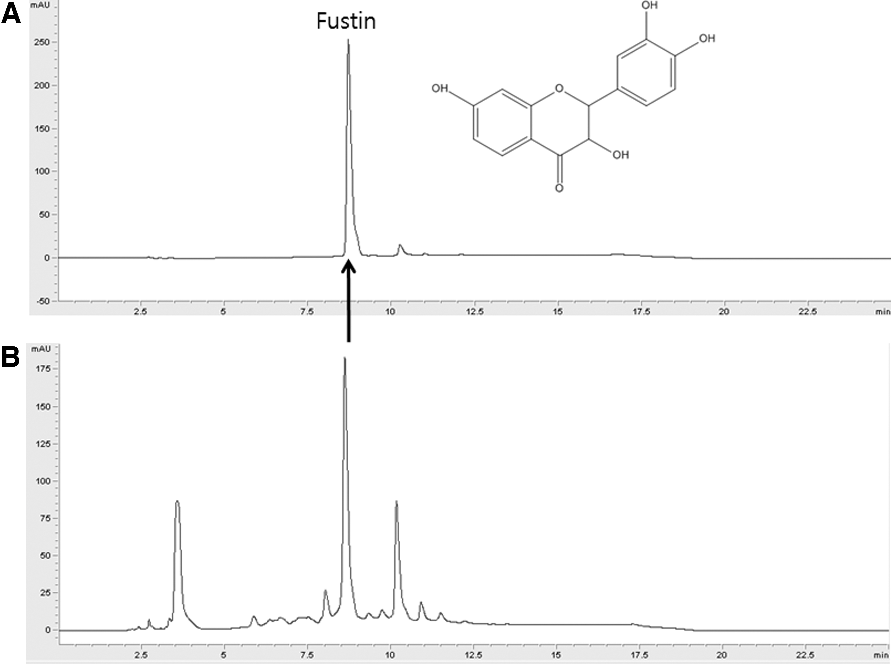
Representative HPLC chromatograms of
Animal care
Six-week-old male Wistar rats (Orient Bio, Seongnam, Korea) were used for the experiment. The rats were housed individually at 23°C±1°C with a 12-h light/12-h dark cycle and 45%±5% humidity. The rats consumed tap water and a standard chow diet for 19 days during the experiment. The animals were cared for in accordance with the Guide for the Care and Use Laboratory Animals of Ewha Womans University. After 7 days of acclimatization, the rats were randomly assigned to six groups of twelve rats each. The RVS extract and fustin were dissolved in sterile saline. The rats were intragastrically administered 0 (saline only), 100, 250, or 500 mg/kg body weight (bw) of RVS extract or 15 mg/kg bw of fustin for 14 days. To induce inflammatory stress, LPS (5 mg/kg bw) was intraperitoneally injected 18 h before sacrifice. The control group was injected with sterile saline instead of LPS. The animals were sacrificed on day 15 by exsanguination from the heart under an injection of intraperitoneal Zoletil (40 mg/kg bw) and Rompun (6.34 mg/kg bw). The livers were excised and weighed. Histopathological examinations were performed on the livers, and the biochemical biomarkers were determined. The study protocol was approved by the Institutional Animal Care and Use Committee (IACUC, approval #2013-01-030).
Biochemical evaluation of serum and liver
ALT, AST, and NO levels were determined using kits (Bioassay Systems), and hepatic IL-6 and TNF-α levels were measured using specific ELISA kits according to the manufacturer's protocol (Uscn Life Science, Inc.). Hepatic MDA levels were measured using a commercial kit purchased from Cayman Chemical. In brief, the livers were homogenized in 250 μL of radioimmunoprecipitation buffer containing a protease inhibitor and sonicated for 15 sec at 40 V. After centrifugation at 1600 g for 10 min at 4°C, the supernatant was collected to measure the MDA concentration according to the manufacturer's instructions.
Determination of hepatic iNOS and COX-2 mRNA expression levels
Reverse transcriptase polymerase chain reaction (RT-PCR) was performed to detect the expression levels of mRNA for iNOS and COX-2. The total RNA was extracted from the liver samples using the TRIzol protocol (Invitrogen, Carlsbad, CA, USA). The RNA concentration and quality were determined using a BioSpec-nano spectrophotometer (Mecasys Corp., Daejeon, Korea). cDNA (20 μg) was synthesized from total RNA using a high-capacity RNA-to-cDNA Kit (Hoffmann La Roche, Basel, Switzerland). Quantitative RT-PCR was performed using the SYBR Green method with the StepOnePlus RT-PCR system (Hoffmann La Roche, Basel, Switzerland). The primer sets for the target genes were as follows: iNOS, 5′-ACCTAGGAGCATCCCAAGT-3′, 5′-CAGCGCATACCACTTCAGC-3′; COX-2, 5′-ACCAACGCTGCCACAACT-3′, 5′-GGTTGGAACAGCAAGGATTT-3′; and β-actin (04686900001; Roche). Amplifications were performed starting with a 10-min template denaturation step at 95°C followed by 40 cycles at 95°C for 10 sec and 60°C for 30 sec. The relative amounts of mRNA were normalized to the amount of β-actin, and the relative amounts of RNA were calculated using the comparative CT method.
Hematoxylin and eosin staining
The central and left lateral lobes of the liver samples were immediately fixed with 10% phosphate-buffered formalin after necropsy. The fixed livers were dehydrated using a graded series of ethanol and embedded in paraffin according to standard procedures. Paraffin sections (5 μm thick) were deparaffinized in xylene, stained with hematoxylin and eosin (H&E), and examined using a microscope (Olympus Co., Tokyo, Japan).
Statistical analyses
All statistical analyses were performed using SPSS version 20.0 (SPSS, Inc., Chicago, IL, USA). The results are presented as the mean±standard error. The differences among all groups were analyzed by one-way analysis of variance with the post hoc Duncan's multiple range test. Statistical significance was defined as P<.05.
Results
Effects of RVS extract on LPS-induced liver dysfunction
The results for the hepatoprotective effect of RVS extract and fustin against LPS-induced liver damage in rats are shown in Table 1. The serum levels of AST and ALT were significantly higher in LPS-treated rats than in the saline control, providing evidence of hepatic injury caused by LPS. Compared with the saline control group, AST and ALT in the serum of LPS-treated rats were elevated by 10.6- and 6.6-fold, respectively. The activities of serum AST and ALT tended to be reversed by treatment with the RVS extract, regardless of dose, although the differences were not statistically significant. In addition, there was no effect on AST and ALT levels in the fustin-treated group.
Mean±standard error (n=12).
P values were calculated by one-way analysis of variance. Values with different superscript lowercase letters within the same line are significantly different at P<.05 by Duncan's multiple range test.
ALT, alanine transaminase; AST, aspartate transaminase; bw, body weight; Fustin, LPS+15 mg fustin/kg bw; LC, LPS control; LPS, lipopolysaccharide; NC, saline; RVS, Rhus verniciflua stokes; RVS_H, LPS+500 mg RVS extract/kg bw; RVS_L, LPS+100 mg RVS extract/kg bw; RVS_M, LPS+250 mg RVS extract/kg bw.
Effects of RVS extract on serum and hepatic NO levels
As shown in Figure 2, the LPS-treated rats showed a marked increase in NO levels in both the serum and liver (15.7- and 1.9-fold increases, respectively, versus the control group). When rats were administered with the RVS extract at 500 mg/kg bw, the serum NO levels were significantly decreased compared with those of LPS-treated rats. In addition, the effect of RVS extract on hepatic NO levels was investigated. Compared with the LPS-control group, the RVS extract 500 mg/kg group showed a significant decrease and the RVS extract 250 mg/kg group showed a trend for a decrease in hepatic NO levels. However, the groups receiving lower doses of RVS extract, and fustin did not show statistically significant decreases in serum and hepatic NO levels.
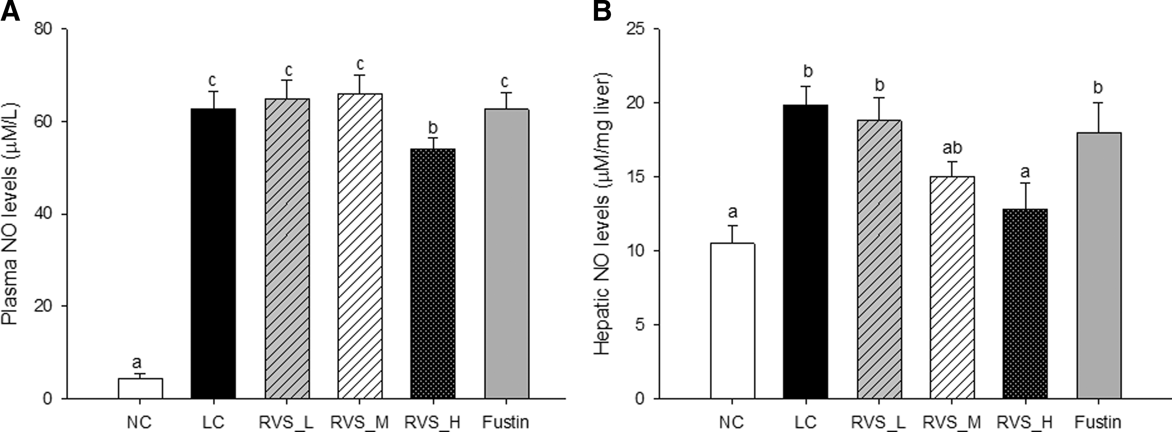
Effect of RVS extract or fustin on LPS-induced NO production in the serum
Effects of RVS on hepatic lipid peroxidation
MDA, an end product of lipid peroxidation, has been used as an index of oxidative stress. To examine the effect of RVS extract or fustin administration on LPS-induced oxidative stress in rats, the MDA content in liver homogenates was measured by the TBARS assay. As shown in Figure 3, LPS injection resulted in significantly greater production of MDA in liver homogenates by 1.18-fold compared with the control group. Treatment with the RVS extract blunted the rise in MDA levels in the liver. Compared with the LPS-treated group, RVS extract pretreatment at 250 and 500 mg/kg bw doses resulted in lower hepatic levels of MDA by 0.64- and 0.72-fold, respectively. However, pretreatment with fustin did not protect against lipid peroxidation.
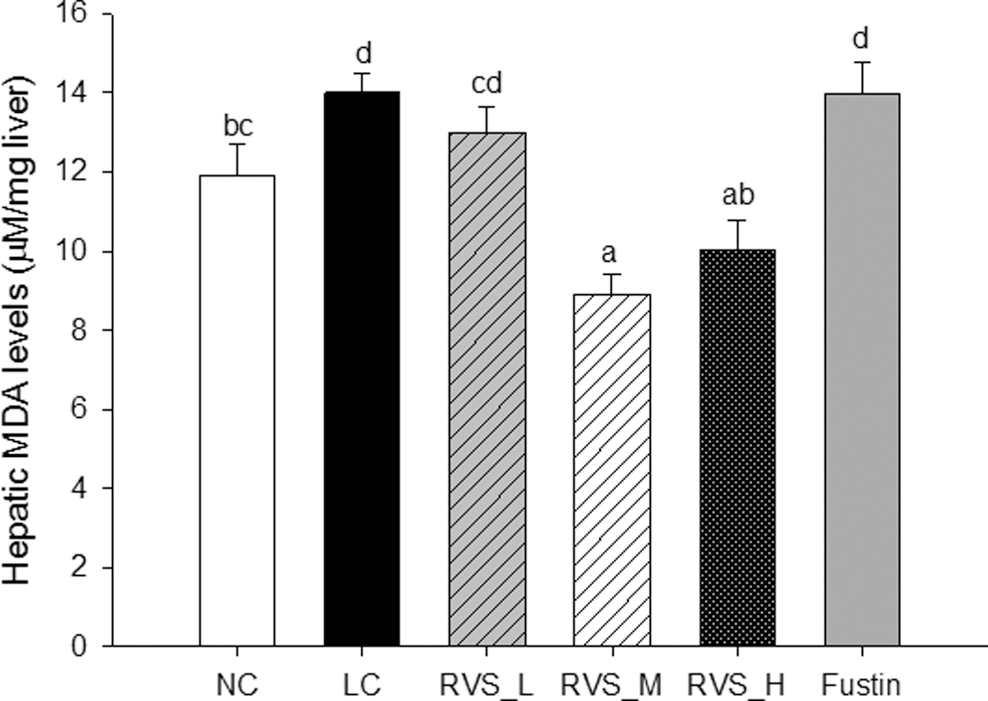
Effect of RVS extract or fustin on LPS-induced hepatic MDA levels. Hepatic MDA levels were determined using commercial kits as described in Materials and Methods. Each value represents the mean±SE (n=12). Different letters indicate significant differences between groups determined by one-way ANOVA followed by Duncan's multiple range test (P<0.05). MDA, malondialdehyde.
Effects of RVS extract on LPS-induced hepatic proinflammatory cytokines
The effects of RVS extract on LPS-induced changes in hepatic proinflammatory cytokines are shown in Figure 4. The presence of LPS caused a marked increase in the levels of the proinflammatory cytokines IL-1β, IL-6, and TNF-α compared with the saline control group. The administration of RVS extract at doses of 100, 250, and 500 mg/kg bw and fustin at a dose of 15 mg/kg bw significantly suppressed the elevation of IL-6 in the liver. Although IL-1β and TNF-α levels tended to decrease in the liver, there were no statistically significant differences. Only the group administered RVS extract 250 mg/kg showed significantly lower levels of TNF-α.

Effect of RVS extract or fustin on LPS-induced hepatic proinflammatory cytokines. Hepatic proinflammatory cytokine levels were determined using commercial kits as described in Materials and Methods. Each value represents the mean±SE (n=12). Different letters indicate significant differences between groups determined by one-way ANOVA followed by Duncan's multiple range test (P<0.05).
Effects of RVS extract on LPS-induced iNOS and COX-2 mRNA levels
To assess the inhibitory mechanism involved in LPS-induced inflammation, the mRNA levels of iNOS and COX-2 were determined using RT-PCR. The data showed that LPS markedly increased iNOS and COX-2 mRNA expression levels in the liver compared with the control group (Fig. 5). Furthermore, the hepatic iNOS expression level was significantly suppressed by fustin and the RVS extract, even in the lower dose group, and COX-2 expression level was inhibited by high-dose RVS and fustin. Thus, the RVS extract prevented LPS-induced inflammatory reactions, as indicated by decreased iNOS and COX-2 mRNA transcription.
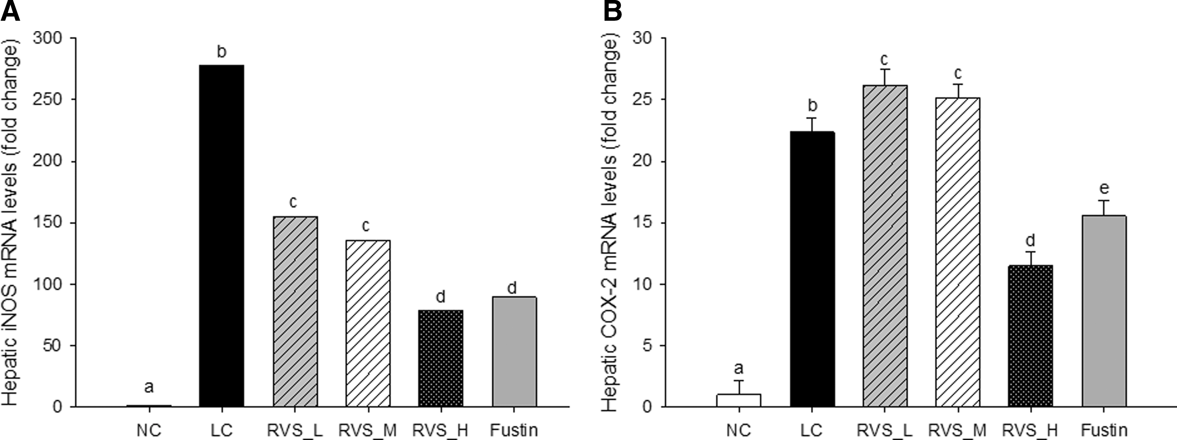
Effects of RVS extract or fustin on LPS-induced hepatic iNOS
Histological changes in liver tissue
Histopathological examination of liver sections from control animals revealed normal lobular architecture and cell structures (Fig. 6A). Compared with the normal liver tissue from the control group, there was significant evidence of injury with severe sinusoidal vascular congestion (white arrows), microvesicular steatosis, destruction of hepatic architecture, and focal necrosis with mild inflammatory cell infiltration in the livers of the LPS-treated rats (Fig. 6B). Although a slight increase in vascular congestion and necrosis relative to the control group was observed, significantly less congestion and necrosis were observed in the livers of all the RVS extract-treated groups (Fig. 6C–E) in a dose-dependent manner and in the fustin-treated group (Fig. 6F). Inflammatory cell infiltration was also significantly attenuated by the RVS extract or fustin supplementation. The RVS extract 500 mg/kg group, in particular, exhibited less vascular congestion, obstruction of sinusoids, and destruction of hepatic architecture than the LPS-control group. Moreover, some portions of the livers appeared normal, although high-dose supplementation with the RVS extract did not completely eliminate the LPS-induced liver injuries.
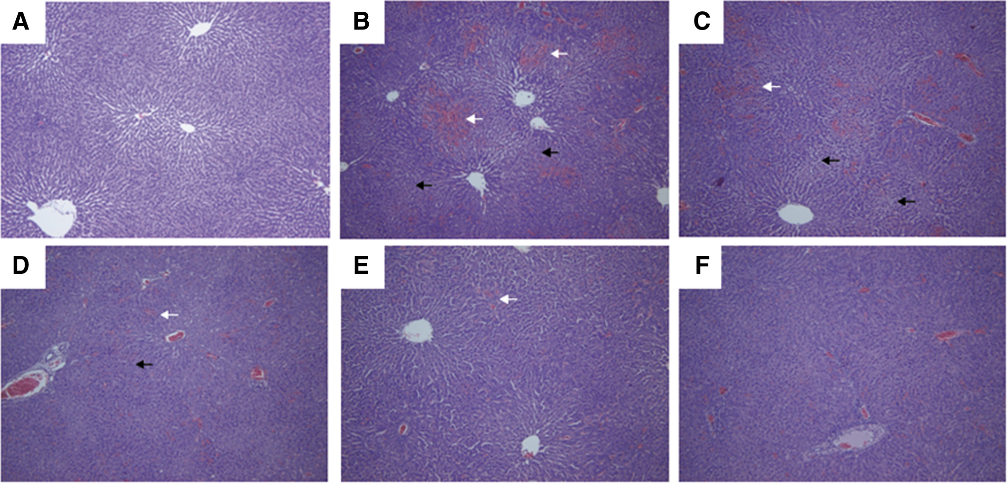
Effect of RVS extract or fustin on liver abnormalities induced by LPS (hematoxylin/eosin staining; magnification×200). Liver samples from
Discussion
In the present study, the anti-inflammatory effect of RVS extract standardized with fustin was confirmed using LPS-stimulated Wistar rats. RVS has been used for many years in traditional medicine and food in Korea, Japan, and China. However, its use is not supported by safety measures addressing the allergenic properties of urushiol. There have been several attempts to remove urushiol, while retaining the beneficial effects of RVS, such as fractionating the flavonoid-rich parts using n-hexane or ionic resin. 20,24 Safe manufacturing processes are required before its use as a food ingredient; therefore, a urushiol-free RVS extract standardized to fustin content has been developed using only water and ethanol as extraction solvents. 23 Although we previously reported that our food-grade, urushiol-free RVS extract had an antioxidant effect, 23 the retention of the well-known anti-inflammatory effect of RVS in our preparation required confirmation. 24 –26
LPS is a bacterial endotoxin that induces extensive damage to various organs, including the heart, gastrointestinal tract, kidney, liver, brain, and lung. 2,27 The use of flavonoids and other polyphenol compounds of herbal origin has been shown to be beneficial for preventing LPS-induced inflammatory responses and hepatotoxicity. 28,29 In the present study, the inhibitory effect of RVS extract supplementation on LPS-induced inflammatory stress and hepatotoxicity in Wistar rats was investigated. LPS induced the production of NO and proinflammatory cytokines by increasing iNOS and COX-2 mRNA expression levels. In addition, we also confirmed cytokine infiltration through H&E staining.
LPS is a potent stimulator of NO and increases oxidative stress. The treatment of endotoxemia was reported to increase the serum levels of nitrite by 4.5-fold. 30 The increased levels of NO resulting from LPS exposure can combine with superoxide anions (O2•−), resulting in the formation of the peroxynitrite anion (ONOO−). This highly toxic oxidant may increase tissue injury and organ dysfunction. 31 Furthermore, the peroxynitrite anion can also stimulate the production of TNF-α and IL-6 in human monocytes through activation of nuclear factor (NF)-κB. 32 NO also prompts hydrogen peroxide and O2•− production in the mitochondria, 33 which can cause oxidative damage to numerous tissues. In our study, LPS treatment for 18 h resulted in a 15.7- and 1.9-fold increase in the NO level in the serum and liver, respectively. However, after pretreatment with the RVS extract at 500 mg/kg, the increase in the NO level in the liver was dampened to a level similar to that of the normal control group. In addition to NO production, LPS also induced lipid peroxidation. MDA, as a marker of lipid oxidation, was analyzed to evaluate the protective effect of RVS extract on LPS-induced oxidative stress in Wistar rats. In agreement with the previous results for antioxidant effects, 23 the present data showed that the elevation of oxidative stress, as indicated by the MDA level, was prevented by the moderate and high RVS doses.
When LPS penetrates into the organs, it binds to TLR4, and this complex undergoes a series of biological cascades, ultimately activating the NF-κB signaling pathway and, thereby, inducing iNOS and COX-2. 34 A large number of cytokines produced by activated macrophages and neutrophils are intimately involved in many immune and inflammatory responses elicited by LPS. 35 Among cytokines, the secretion of proinflammatory cytokines, such as TNF-α, IL-1β, and IL-6, is especially strongly enhanced by LPS stimulation. 36,37 In the present study, although urushiol was removed, our standardized RVS extract prevented inflammatory stimulation by LPS, as confirmed by hepatic iNOS and COX-2 mRNA expression and proinflammatory cytokine secretion levels (TNF-α, IL-1β, and IL-6). In addition, inflammatory cell infiltration was significantly attenuated by RVS extract supplementation. In particular, the vascular congestion, obstruction of sinusoids, and destruction of hepatic architecture of the RVS extract 500 mg/kg group were limited relative to those of the LPS-control group, as determined by histological examination.
Previously, the flavonol-rich fraction of RVS was reported to have an anti-inflammatory effect in IL-1β-stimulated, rheumatoid arthritis fibroblast-like synovial cells and a rheumatoid arthritis mouse model. 20 The phenol-rich fraction obtained from ethanolic extraction and ionic resin columns significantly inhibited the production of proinflammatory material (such as NO, prostaglandin E2, and TNF-α) in LPS-induced RAW264.7 macrophages. 24 The major flavonoids in the phenol-rich fraction were identified as ρ-coumaric acid, fustin, kaempferol-3-O-glucoside, sulfuretin, butein, and kaempferol. Sulfuretin is known to be the most important flavonoid compound in RVS. 38 Sulfuretin was investigated for anti-inflammatory activity mediated through the regulation of the NF-κB signaling pathway and was confirmed to inhibit LPS-induced DNA binding and the transcriptional activity of NF-κB in the RAW264.7 cell line. 39 In addition, a few studies reported that the oral administration of R. verniciflua extract or sulfuretin prevented the development of rheumatoid arthritis in the Sprague-Dawley rat and DBA/1 mouse models of adjuvant-induced arthritis, respectively, by inhibiting the NF-κB signaling pathway. 38,40 Although sulfuretin is the most important flavonoid contained in RVS, the standardized RVS extract contains 2.74±0.28 mg/g of sulfuretin, but 43.08±3.55 mg/g of fustin. This relatively low sulfuretin content is difficult to standardize. 21 Therefore, in the present preparation, we standardized the RVS extract using fustin, which is present in sufficiently large amounts for analysis. In the present study, we compared the anti-inflammatory effect of RVS extract to that of fustin at the amount present in the highest dose of RVS extract. Similar to our previous report on the antioxidant effect of RVS, the effect of RVS extract was superior to that of fustin alone. A purified compound may lose its bioavailability or may not act according to the same pathway as the compound found in whole food. 41 In this study, pretreatment with the fustin single compound significantly prevented the elevated iNOS and COX-2 mRNA expression and proinflammatory cytokine production without decreasing the NO and MDA levels. Kim et al. reported that our RVS preparation contained gallic acid, fisetin, sulfuretin, as well as fustin, 21 which are assumed to be responsible, at least in part, for its anti-inflammatory effect. Further studies are needed to determine the exact cellular and molecular mechanisms and precisely identify the active compounds in our preparation.
In summary, the RVS extract standardized to fustin prevented LPS-induced NO production in both the serum and liver. This extract also prevented the elevation of the levels of hepatic proinflammatory cytokines, such as TNF-α, IL-1β, and IL-6, and hepatic MDA. We also confirmed the anti-inflammatory effect through histological examination. The analysis of mRNA expression for iNOS and COX-2 revealed that the anti-inflammatory effect of our standardized RVS extract might be mediated by the NF-κB signaling pathway.
Footnotes
Acknowledgments
We thank the staff of Lifetree Biotech Co. for the preparation of standardized RVS. This research was supported by the Ministry of Science, ICT, and Future Planning (Basic Science Research Program of the National Research Foundation of Korea Project No. 2013R1A1A1005859 and the Biosynergy Research Project No. 2012M3A9C4048761) and the Ministry for Food, Agriculture, Forestry, and Fisheries (Industrialization Support Program for Bio-Technology of Agriculture and Forestry), Republic of Korea.
Author Disclosure Statement
No competing financial interests exist.