




Abstract
Gastric ulcer is a common digestive disorder that results in considerable suffering. Hence, this digestive pathology has been the focus of a number of recent studies. Although numerous drugs have been developed to treat gastric ulcers, therapeutic approaches for many of the complications associated with these drugs remain to be identified. For this reason, many natural compounds have been explored as alternatives for these drugs. In this study, we have investigated the effectiveness of Areca catechu leaf ethanol extract (ACE) for treating ethanol-induced gastric ulcers in mice. We performed histological as well as immunohistochemical examinations to explore the therapeutic properties of ACE. We also examined the levels of inflammatory signaling molecules to confirm the anti-inflammatory effects of ACE. The histochemical data demonstrate that ACE can protect the mucosal epithelium as well as the vascular supply in the gastric tract. Furthermore, ACE significantly reduced the expression levels of tumor necrosis factor-alpha (TNF-α), interleukin-6 receptor (IL-6R), inducible NO synthase (iNOS), cyclooxygenase 2 (COX2), and nuclear factor-kappa B (NF-κB). Taken together, these data suggest that ACE administration may have the potential as an alternative treatment for gastric ulcer because of its cytoprotective and anti-inflammatory effects and ability to promote the rejuvenation and revascularization of the damaged gastric epithelium.
Introduction
T
In previous studies, excessive ethanol exposure was shown to be a risk factor for gastric ulcers because ethanol metabolism generates reactive oxygen species (ROS), such as superoxide (
Areca catechu, a member of the Palmaceae family, is a widely known plant in Asia. 12 Previously, we demonstrated that a crude extract of Indonesian Areca catechu leaves (ACE) had anti-inflammatory effects in vitro and in vivo. 13 However, its therapeutic effects on gastric disorders have not been explored. Antioxidants are commonly prescribed for gastric ulcers. 14 Phenolic compounds are potent antioxidants that exhibit a number of pharmacological properties, such as anti-inflammatory, antiproliferative, anticancer, and antiatherosclerotic effects. 15,16 This study explored the protective effects of ACE in an ethanol-induced gastric ulcer model in mice.
Materials and Methods
Chemicals
Cyclooxygenase 2 (COX2), iNOS, TNF-α, IL-6 receptor (IL-6R), and GAPDH antibodies were purchased from Santa Cruz (Santa Cruz, CA, USA), and all other reagents were purchased from Sigma (St. Louis, MO, USA).
Plant material and ethanol extraction
A. catechu was obtained from the Airlangga University Oriental, Indonesia. One hundred grams of the plant leaf was blended, and the crude powder was precipitated with 500 mL of 70% ethanol for 3 days. The aqueous extracts were lyophilized by freeze-drying at −60°C.
DPPH assay
A DPPH assay was performed as previously reported. 17 Briefly, a volume of 0.1 mL of aqueous ACE extract was mixed with 0.5 mL of DPPH solution (0.5 mM DPPH dissolved in absolute ethanol) and 0.4 mL of Tris–HCL (0.1 M, pH 7.4) for 20 min in darkness. Thereafter, the absorbance was evaluated at 517 nm. The percent inhibition is presented as (1 − [the absorbance of sample/the absorbance of sample DPPH] ×100). The results were recorded as an inhibition ratio of 50%. Vitamin C was used as a positive control.
Animal care and in vivo test
The six-week-old male ICR (CrljOri:CD1) mice were obtained from Orient Bio, Inc. (Seoul, Korea). Before the experiments, all animals were acclimatized to the new environment for 1 week (room temperature 24°C ± 2°C, humidity 50% ±15%, and 12-h light–12-h dark cycle). All experiments and animal care were conducted in conformity with institutional guidelines (IACUC-2014-004). Briefly, the mice were individually housed in stainless steel cages with ad libitum access to water and food. To test the gastroprotective effects of ACE, the animals were randomly divided into three different groups (n = 8 mice/group): control group (normal mice), ethanol-alone-treated group (injury mice), and ethanol- and ACE-treated group (test mice). Gastric injuries were induced by 50% ethanol that was orally administered for 3 days at a dose of 10 mL/kg. Crude extract (ACE; 5 mg/kg/day in normal saline) was administered orally every day for 3 days at a regular schedule after 5-h ethanol stimulation.
Tissue preparation and histochemical staining
The mice were anesthetized with sodium pentobarbital and killed after the ethanol treatment schedule. Their stomachs were removed and fixed in 10% formalin for 24 h. The gastric tissues were embedded in paraffin, and 6-μm-thick tissue sections were stained with hematoxylin and eosin (H&E). Some sections were used for periodic acid–Schiff (PAS) staining to observe the mucus production in the stomach.
Immunohistochemical staining
Some of the prepared sections were used for immunostaining assays. Samples were treated with 3% H2O2 for 5 min to inactivate endogenous peroxidase, blocked with 10% normal serum for 1 h at room temperature, and incubated overnight at 4°C with primary antibodies against iNOS (1:100), COX2 (1:200), TNF-α (1:50), and IL-6R (1:100). The following day, the sections were washed and incubated with the corresponding secondary antibodies for 1 h at room temperature. The Vectastain ABC Kit was used to implement the avidin–biotin complex interaction and was used in accordance with the manufacturer's instructions. Signal development was performed in a substrate solution of 0.05% DAB, and the slides were counterstained with hematoxylin. The sample sections were examined using a light microscope (Olympus BX50; Olympus Co. Ltd., Tokyo, Japan) at 200× magnification.
Statistical analysis
The results are expressed as mean ± standard error from at least three independent experiments. Statistical significance was analyzed using Student's t-test for each paired experiment with GraphPad Prism 4.0 software (Windows, San Diego, CA, USA). Values were considered statistically significant at P < .05.
Results
Total phenolic content in crude extract of Indonesian ACE
Total phenolic content of the aqueous extract, as determined by the Folin–Ciocalteu reagent method, 17 was expressed as micrograms of gallic acid (GA) equivalent per milligram. As shown in Table 1, the yield was 444.48 ± 0.60 μg GA eq/mg.
ACE, Areca catechu leaves; eq, Equivalent; GA, gallic acid; IC50, inhibit the reaction by 50%.
Antioxidant activity of ACE
To investigate the antioxidant activity of ACE, we measured the free radical scavenging activity using a DPPH assay to determine the concentration of ACE required to inhibit the reaction by 50% (IC50). Vitamin C (positive control) and ACE showed IC50 values of 4.05 ± 0.24 μg/mL and 7.76 ± 0.97 μg/mL, respectively (Table 1).
Protection against ethanol-induced gastric ulcers by ACE
To examine the protective effect of ACE against ethanol-induced tissue injury to the gastric mucosa, we performed histological analysis using H&E staining and PAS staining. The ethanol-treated group (10 mL/kg of 50% ethanol for 3 days) exhibited pronounced damage to the gastric mucosal epithelial lining compared to the untreated group. Samples from mice treated with ACE (5 mg/kg) showed morphological characteristics similar to the group not challenged with ethanol. As shown in Figure 1A, tissue samples treated with ethanol exhibited severe necrosis with vacuole formation in the gastric mucosa. In contrast, samples pretreated with ACE (5 mg/kg) exhibited similar morphologies as normal naive gastric mucosa compared to the untreated group. Next, to examine the effect of ACE on the ethanol-damaged gastric lining, we conducted post-PAS staining histological analysis. As shown in Figure 1B, the undamaged mucosal tissues in the ACE-treated group were similar to those observed in control animals.

The protective effect of crude extract of Indonesian Areca catechu leaves (ACE) on the gastric lining. The microphotographs of the gastric mucosal linings of the untreated group of animals (control panel) given normal saline only, treated with 10 mL/kg of 50% ethanol for 3 days (ethanol panel), or treated with ethanol after pretreatment with 5 mg/kg of ACE for 3 days at a regular schedule after 5-h ethanol stimulation (ethanol + ACE).
Effect of ACE on TNF-α and IL-6R expressions in ethanol-injured gastric tissue
As shown in Figure 2, the expressions of TNF-α and IL-6R significantly increased, up to 100%, in the injured tissue (ethanol-treated panels). However, after the ACE treatment, the expression levels of these proteins significantly decreased to 66.49% and 49.58% for TNF-α and IL-6R, respectively.
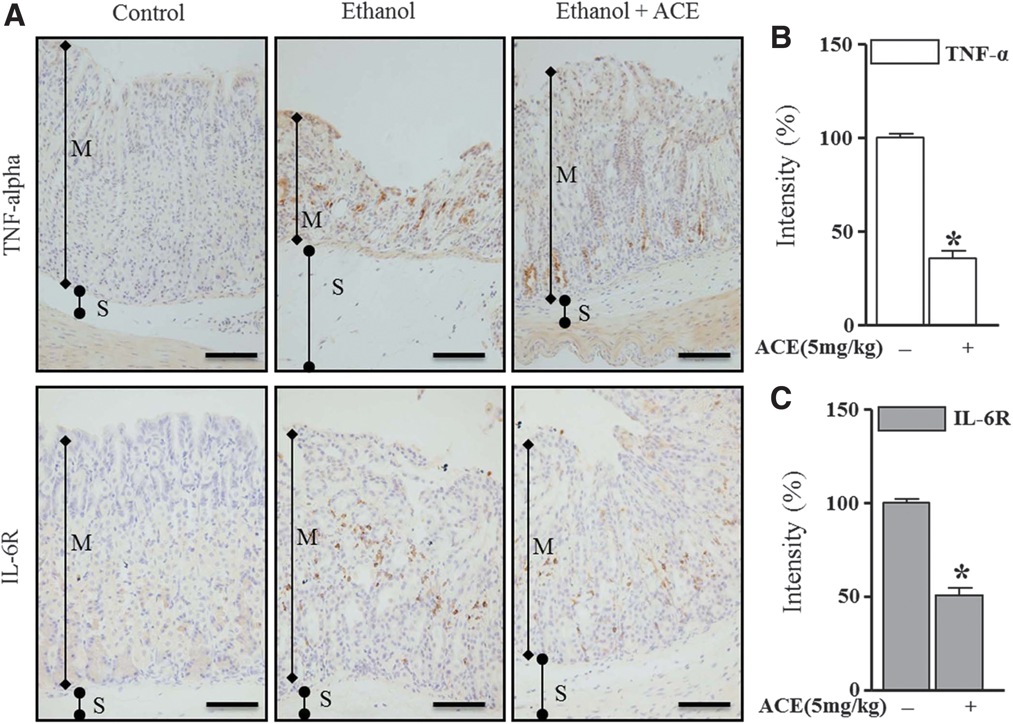
The effect of crude extract of Indonesian ACE on tumor necrosis factor-alpha (TNF-α) and interleukin-6 receptor (IL-6R) expressions in ethanol-induced gastric ulcer in mice. Immunohistochemical staining for TNF-α and IL-6R in gastric tissue was performed with cross sections obtained from the untreated group, the ethanol-alone group, and the ethanol- or ACE-treated group. The photomicrographs show a magnification 200× (bar = 100 μm). The graphs show the expression levels of TNF-α
Effects of ACE on COX-2, NF-κB, and iNOS expressions in ethanol-injured stomach tissue
To examine possible correlations between gastric protection and regulation of inflammation, we investigated the levels of expression of the inflammation-related signaling molecules, COX2, NF-κB, and iNOS, in the ethanol-induced gastric failure model. As shown in Figure 3, the expression levels of COX2, NF-κB, and iNOS significantly increased in the injured tissue (ethanol panels). We set the COX2, NF-κB, and iNOS expressions in the ethanol-induced gastric ulcer tissue to 100%. In contrast, the ACE treatment decreased the expression levels of COX2, NF-κB, and iNOS to 50%, 88%, and 76%, respectively.
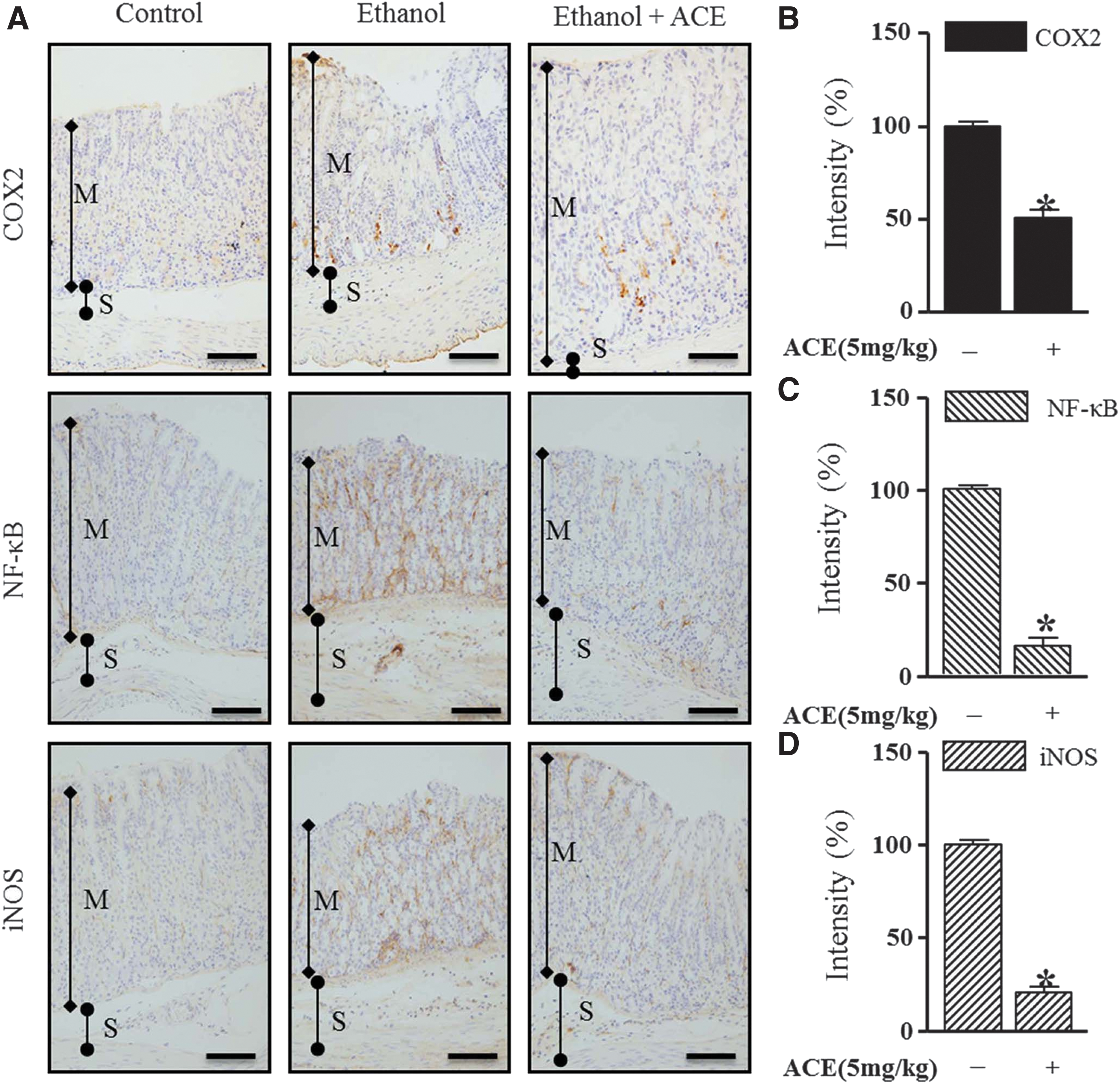
The effect of crude extract of Indonesian ACE on cyclooxygenase 2 (COX2), nuclear factor-kappa B (NF-κB), and inducible NO synthase (iNOS) expressions in ethanol-induced gastric ulcer. Immunohistochemical staining for COX2, NF-κB, and iNOS in gastric failure tissue was performed with cross sections obtained from the untreated group, the ethanol-alone group, and the ethanol- or ACE-treated group. The photomicrographs show a magnification 200× (bar = 100 μm). The graphs show the expression levels of COX2
Discussion
In the present study, we found that the natural compound, ACE, exhibited protective and therapeutic effects against gastric ulcer. Although NSAIDs have been established as effective anti-inflammatory medicines, they can alter the blood clotting mechanism in the gastric area and thereby induce the early phase of gastric pathogenesis. 3 Therefore, functional plant extracts as traditional drugs are potential alternatives that can promote anti-inflammatory effects while maintaining the normal physiological activities of the gastric epithelium. Previous studies have demonstrated that various natural compounds have beneficial effects on ethanol-induced gastric ulcer. 4 –7 Our results show that ACE is a valuable natural compound for gastric pathologies.
A. catechu nut is a traditional medicine in the Asian countries that has been widely used as a treatment for gastrointestinal problems. 18 It contains polyphenols, including catechin, caffeic acid, ferulic acid, and tannin. The average amount of tannins in A. catechu nut is 15%. 19 It is known that tannins can counteract carcinogenesis. 20
We have previously demonstrated the anti-inflammatory effect of ACE in vitro and in vivo. 13 However, its gastroprotective effect on ethanol-induced injuries had not been identified previously. In this study, we found that ACE has a cytoprotective effect on ethanol-induced gastric lesions, as shown in the histochemical assays. Previously, a study conducted by Kim et al. suggested that ethanol can induce gastric ulcers in mice. 7 Our data show that pretreatment with ACE through oral administration, before overdosing with ethanol, decreased ethanol-induced gastric mucosal degeneration and necrosis. The results suggest that ACE is a candidate for cytoprotection against gastric injury.
Next, we examined the molecular mechanism by which ACE provided protection against ethanol-induced gastric injury. Oxidative stress is a pathogenic factor for inflammatory responses in the injured stomach. 21 Therefore, we investigated the antioxidant effect of ACE against gastric ulcers. Inflammatory factors, such as TNF-α and IL, generate most of the ROS that are greatly influenced by ethanol metabolism. 7,22 We first determined the free radical scavenging effect of ACE on DPPH-induced free radicals. Interestingly, ACE exhibited high concentrations of phenolic compounds. Furthermore, it had an IC50 of 7.765 μg for DPPH scavenging compared to an IC50 of 4.5 μg for vitamin C. The result indicated that ACE has an abundance of active phenolic compounds for protection against gastric ulcer.
Numerous studies have reported that TNF-α contributes to the ethanol-related pathogenic mechanism. 9,23 Elevated production of IL-6 often mediates the pathological risk factors for tissue damage. 2 Our result shows that ACE can significantly decrease the expression levels of IL-6 and TNF-α. Moreover, ACE also significantly reduced iNOS, COX2, and NF-κB levels in ethanol-induced gastric injury, confirming the results of our previous study.
In conclusion, this study suggests that ACE is a potential alternative treatment against gastric ulcers, as shown by its cytoprotective and therapeutic effects against inflammatory molecules and ability to simultaneously promote regeneration of blood vessels in the damaged gastric epithelium.
Footnotes
Acknowledgment
This research was supported by the Cooperative Research Program (Project No. PJ009577), Rural Development Administration, the Republic of Korea.
Author Disclosure Statement
No competing financial interests exist.