
Abstract
Angelica sinensis (AS) is one of the most popular medicinal foods used as a hematopoietic herb and also traditionally applied topically for skin disorders. However, the effectiveness of AS on atopic dermatitis (AD) has not been reported yet. This study was conducted to evaluate the antipruritic and anti-inflammatory effects of AS on regulating AD-related mediators in DNCB (2,4-dinitrochlorobenzene)-induced mice. AS was topically applied to the dorsal skin of DNCB-challenged mice for 11 days. Alteration of skin thickness was measured for assessment of histological improvement. In addition, the number of mast cells, the level of serum immunoglobulin E (IgE), the counting of scratching behavior, and the expression of substance P were evaluated. Also, the expressions of cytokines, nuclear factor κB (NF-κB), phospho-IκBα, and mitogen-activated protein kinases (MAPKs) were measured for evaluating the improvement of skin inflammation. The repeated treatment of AS significantly inhibited the skin thickness, the number of mast cells, and the level of serum IgE. Moreover, AS significantly suppressed the increased scratching behavior and the expression of substance P compared to the DNCB group. Topical application of AS also reduced the level of cytokines (IL-4, IL-6, TNF-α, and IFN-γ) as well as the expressions of NF-κB, phospho-IκBα, and phospho-MAPKs in the dorsal skin. The results of our study suggest that topical application of AS might have efficacy for modulating pruritus and inflammation in AD. Further studies are required to further characterize the mechanism of actions of AS.
Introduction
A
Although the pathogenesis of AD is not fully understood, mediators of inflammation and pruritus, such as cytokines and neuropeptides, are known to play crucial roles in AD. In addition, nuclear factor κB (NF-κB) and mitogen-activated protein kinases (MAPKs) work as key mediators of inflammation in AD. 3 As in pruritus, neuropeptides, especially substance P, is reported to influence itchy sensation. 4
The current clinical treatment for AD is focused on solving clinical symptoms (especially inflammation and pruritus) using moisturizers, topical steroids, topical calcineurin inhibitors, systemic immunosuppressants, and phototherapy. 5 However, steroids have dose-dependent side effects, including skin atrophy, hypopigmentation, and adrenal suppression. 6 Exacerbations and rebounding of flares are common adverse effects after withdrawal of systemic medication. As a result, effective therapeutic tools without severe adverse effects need to be developed, and herbal medicine may have that potential.
Angelica sinensis (AS) (Oliv.) Diels (Apiaceae) has been used for the treatment of skin disorders as well as circulatory disorders in oriental medicine. The root of A. sinensis is known to have medical actions, for instance, anticancer, memory amelioration, radioprotective, neuroprotective, immunoregulatory, antioxidant, and other effects. 7 In addition, decursin, an active compound isolated from A. sinensis, inhibited the induction of inflammatory mediators from lipopolysaccharide-stimulated macrophages. 8 However, no studies have investigated the effects of A. sinensis on AD. In this study, we investigated whether A. sinensis can ameliorate AD symptoms, and its mechanism of actions on AD.
Materials and Methods
Preparation of AS
The dried radix of A. sinensis was purchased from Jung-do Herb (Seoul, Korea). A national licensed doctor of Korean medicine at Kyung Hee University authenticated the plant materials. A. sinensis (30 g) were cut into small pieces and extracted with 300 mL of 70% ethanol for 24 h at room temperature (RT). The filtrate was concentrated under reduced pressure using Whatman filter paper no. 3 (Whatman, Maidstone, Kent, England). The resultant solutions were concentrated in a rotary evaporator, freeze dried for 3 days, and stored in aliquots at −80°C. Upon removal of the solvent under vacuum, the ethanolic extract yielded 37.3% (w/w) for dry weight 11.27 g. A voucher specimen (#E70AS) was deposited in the herbarium of the Department of Convergence Korean Medical Science. The lyophilized powder was dissolved in phosphate buffered saline (PBS) before use.
AS was standardized based on the content of decursin using reverse-phase high-performance liquid chromatography (HPLC). HPLC was conducted on an Agilent 1100 series instrument and chromatographic separation was achieved on a SHISEIDO CAPCELL PAK C18 column (5 μm, 4.6 × 250 mm; Shiseido Co., Ltd., Tokyo, Japan). The mobile phase consisted of water–acetonitrile. A:B was as follows: 0 min, 80:20; 12 min, 70:30; 19 min, 50:50; 41 min, 50:50; 50 min, 50:50; the flow rate was 1.0 mL/min and the column temperature was maintained at 35°C. The detection wavelength was 230 nm.
Animals
The 5-week-old BALB/c mice (females, weighing 18–20 g) were obtained from SLC, Inc. (Hamamatsu, Japan). All mice were housed in an air-conditioned room at a temperature of 23°C ± 2°C. Food and water were supplied ad libitum. After the 10 day adaptation period, mice (n = 5) were randomized, divided into four groups: NOR (vehicle-treated group as a normal control group), DNCB (2,4-dinitrochlorobenzene-sensitized group as a negative control), DEX (dexamethasone-treated group as a positive control), and AS (AS-treated group). The animal procedures were reviewed and approved by the Review Board for the Care and Use of Laboratory Animals of the Kyung Hee University, Seoul, Korea (KHUASP [SE]-14–030).
Induction of AD-like lesions in mice
Induction of experimental AD was performed as previously described. 9 Hairs on the dorsal skin area were shaved 1 day before the experiment. The shaved dorsal skin was treated with an application of 100 μL of 1% DNCB solution (dissolved in a 4:1 mixture of acetone and olive oil) for sensitization (for 3 days). The same volume of acetone/olive oil vehicle was applied to the NOR group. After the first induction, mice were housed without any further treatment (for 4 days). In the secondary sensitization, 100 μL of 0.5% DNCB solution was applied to the dorsal skin for 11 days. The DEX group and the AS group were respectively treated with 100 μL of DEX (10 μM, dissolved in PBS) and 100 μL of AS (20 mg/mL) 4 h before DNCB application once a day, whereas NOR and DNCB groups were treated with PBS vehicle. After fasting for 12 h the mice were sacrificed on day 19 of the experiment. After sacrifice, the serum and the dorsal skin were collected for histological analyses, immunohistological and molecular indicators analyses.
Histological analysis
The dorsal skins were fixed in 10% buffered neutral formaldehyde for 24 h and embedded in paraffin wax. Four micrometer-thick sections of dorsal skin were stained with Hematoxylin and eosin (H&E) or Toluidine blue and observed by optical microscopy (Evos® XL; Life Technologies, Carlsbad, CA, USA). Thicknesses of the epidermis, dermis, and the number of mast cells were measured using Leica Application Suite (LAS; Leica Microsystems, Buffalo Grove, IL, USA). The magnification was × 100.
Measurement of scratching behavior
The scratching behavior of mice was measured after 1 h had passed, following the last application of DNCB (on day 18). Each mouse in all groups was videotaped for 20 min to assess the scratching behavior with a digital camera (Model NEX-C3; Sony, Tokyo, Japan) placed on the top of the cages. One bout of scratching was defined as an episode in which a mouse lifted its paw and scratched continuously for any length of time until the paw was returned to the floor.
Immunohistochemistry
The skin sections for immunohistochemistry were carried out in the same manner as histological analysis. After the deparaffinization and rehydration, the skin sections were boiled in 10 mM sodium citrate buffer for antigen retrieval. Slides were treated with 3% H2O2 for 30 min to reduce endogenous peroxidase and with normal goat serum (in PBS with 5% NHS, 5% fetal bovine serum, 2% bovine serum albumin (BSA), 0.1% Triton X-100) to minimize nonspecific binding. After blocking, the sections were incubated with antibodies against substance P (Santa Cruz Biotechnology Inc., Santa Cruz, CA, USA) overnight at 4°C. After washing with PBS, the sections were incubated with secondary anti-goat antibody (Santa Cruz Biotechnology Inc.) for 1 h at RT. Sections were then stained using the Avidin/Biotinylated Enzyme Complex (ABC) Kit (Vector Laboratories, Burlingame, CA, USA) and the substrate chromogen (3,3-diaminobenzidine; Dako, Glostrup, Denmark) mixture was prepared immediately before use. Immunoreactivity was viewed with an LAS (Leica Microsystems). The magnification was × 200.
Enzyme-linked immunosorbent assay
Immunoglobulin E (IgE) was measured in total serum samples (n = 5 per group) prepared from blood collected 24 h after DNCB application on day 19, and the level of IgE concentrations was detected by the mouse IgE Enzyme-Linked Immunosorbent Assay (ELISA) Kit (BD Pharmingen, San Jose, CA, USA). The cytokine productions (IL-4, IL-6, TNF-α, and IFN-γ) in dorsal tissue protein were measured using the mouse ELISA Kits. Each sample (100 mg, n = 5 per group) were weighed and homogenized in 1 mL of Tissue Protein Extraction Reagent (Pierce Chemicals, Rockford, IL, USA) containing a protease inhibitor cocktail (Roche, Indianapolis, IN, USA). Protein concentrations were requantified under identical conditions using a Protein Assay Reagent (Bio-Rad, Hercules, CA, USA). ELISA was performed according to the manufacturer's instruction and quantitation was done with ELISA reader (Molecular Devices, Downingtown, PA, USA) using 450 nm filters. Cytokine concentrations were calculated using a linear regression equation obtained from standard absorbance values.
Western blot analysis
Samples for western blot were prepared as described in the previous research. 9 Briefly, frozen skin dorsal tissues (100 mg) were homogenized in a cytoplasmic buffer containing the protease inhibitor cocktail to assess phospho-IκBα. To extract nuclear proteins, nuclear buffer containing the protease inhibitor cocktail was added to the pellet. The resultant homogenate was carefully removed without disturbing the pellet for assay of activated NF-κB. Levels of MAPKs (the extracellular signal-regulated kinases; ERK1/2, p38 kinases, and the c-Jun N-terminal kinases; JNK) were confirmed by total protein extracts using RIPA assay buffer containing the protease inhibitor cocktail. Protein concentrations were quantified with the BSA Protein Assay Reagent (Pierce Chemicals) using the Bradford protein assay. The denatured protein was separated by 15% SDS-PAGE. After electrophoresis, the proteins were transferred onto PVDF membranes and the membranes were blocked with 5% BSA for 2 h at RT. The membranes were incubated overnight with primary antibodies against β-actin, NF-κB, phospho-IκBα, ERK1/2, phospho-ERK1/2, JNK, phospho-JNK, p38, and phospho-p38 (dilution 1:1000 in TBS-T; Cell Signaling Technology, Danvers, MA, USA). After washing the membrane, anti-rabbit alkaline phosphatase-conjugated secondary antibody (diluted 1:2000 in TBS-T; Santa Cruz Biotechnology Inc.) was then added, followed by incubation for 2 h at RT. The proteins were then visualized using an enhanced chemiluminescence detection reagent.
Statistical analyses
All data are expressed as mean–standard deviation and represent one of three independent experiments. Statistical analyses were performed using Prism 5 (Graph Pad Software, San Diego, CA, USA). Treatment effects were analyzed using one-way analysis of variance followed by Dunnett's test. A value of P < .05 was used to indicate statistically significant differences.
Results
Phytochemical analysis of AS by HPLC
To ensure the quality and purity of AS, HPLC analysis was performed by measuring the content of known active compounds of AS, decursin. The result indicates that the contents of AS showed the upper value of the contents criterion (Fig. 1).
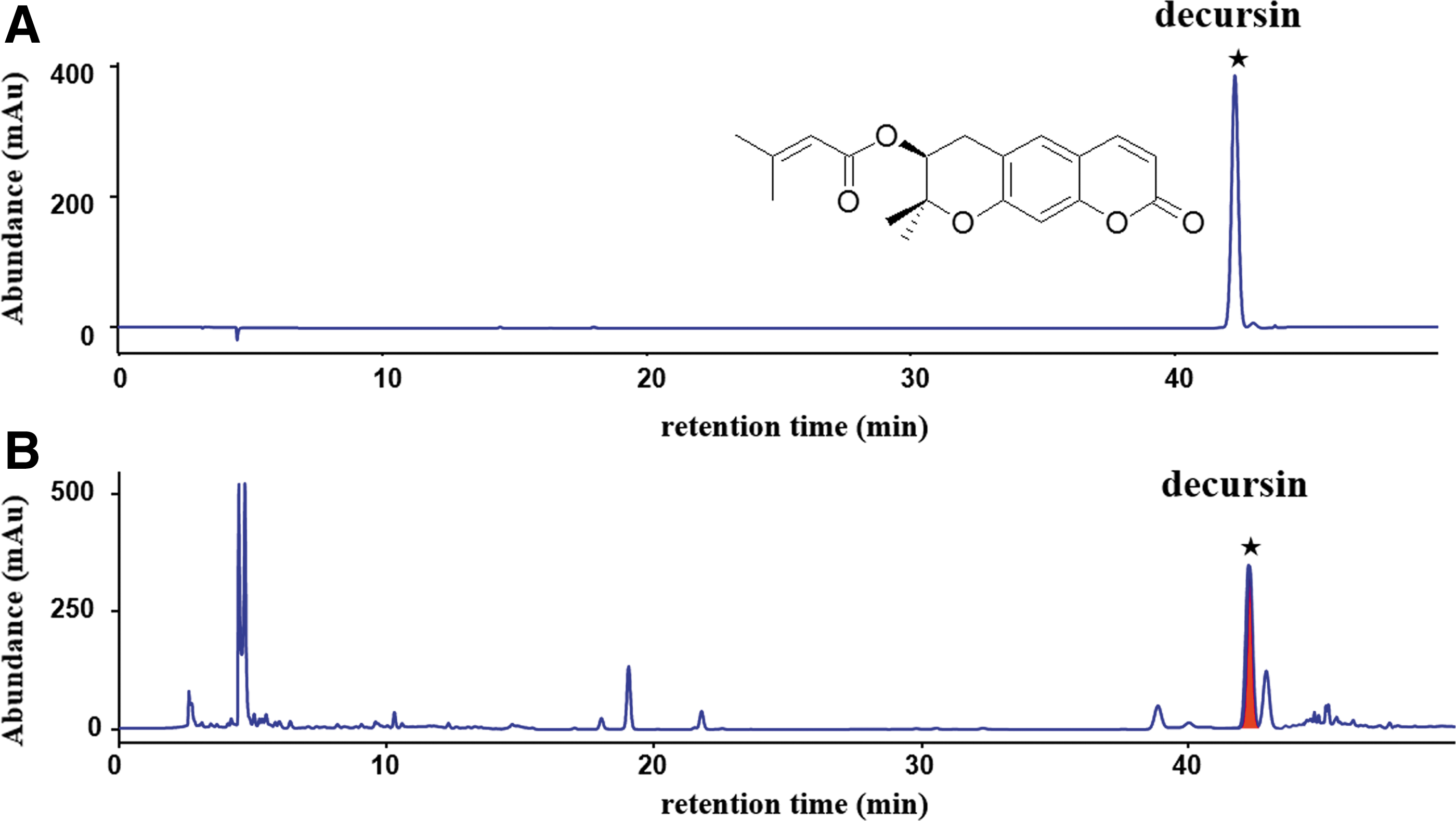
High-performance liquid chromatography (HPLC) chromatogram of the ethanol extracts from Angelica sinensis (AS).
Inhibitory effects of AS on histology
To assess the therapeutic potential of AS against contact dermatitis, mice were topically treated with AS extract during the DNCB induction and sensitization period. As shown in Figure 2, the DNCB group with fully developed skin lesions showed hyperkeratosis and hyperplasia compared to the NOR group. The increased thickness of the epidermis and the dermis were significantly suppressed by DEX. In addition, AS treatment significantly suppressed the increased thickness of the epidermis and the dermis (45.2% and 30.5%, respectively).
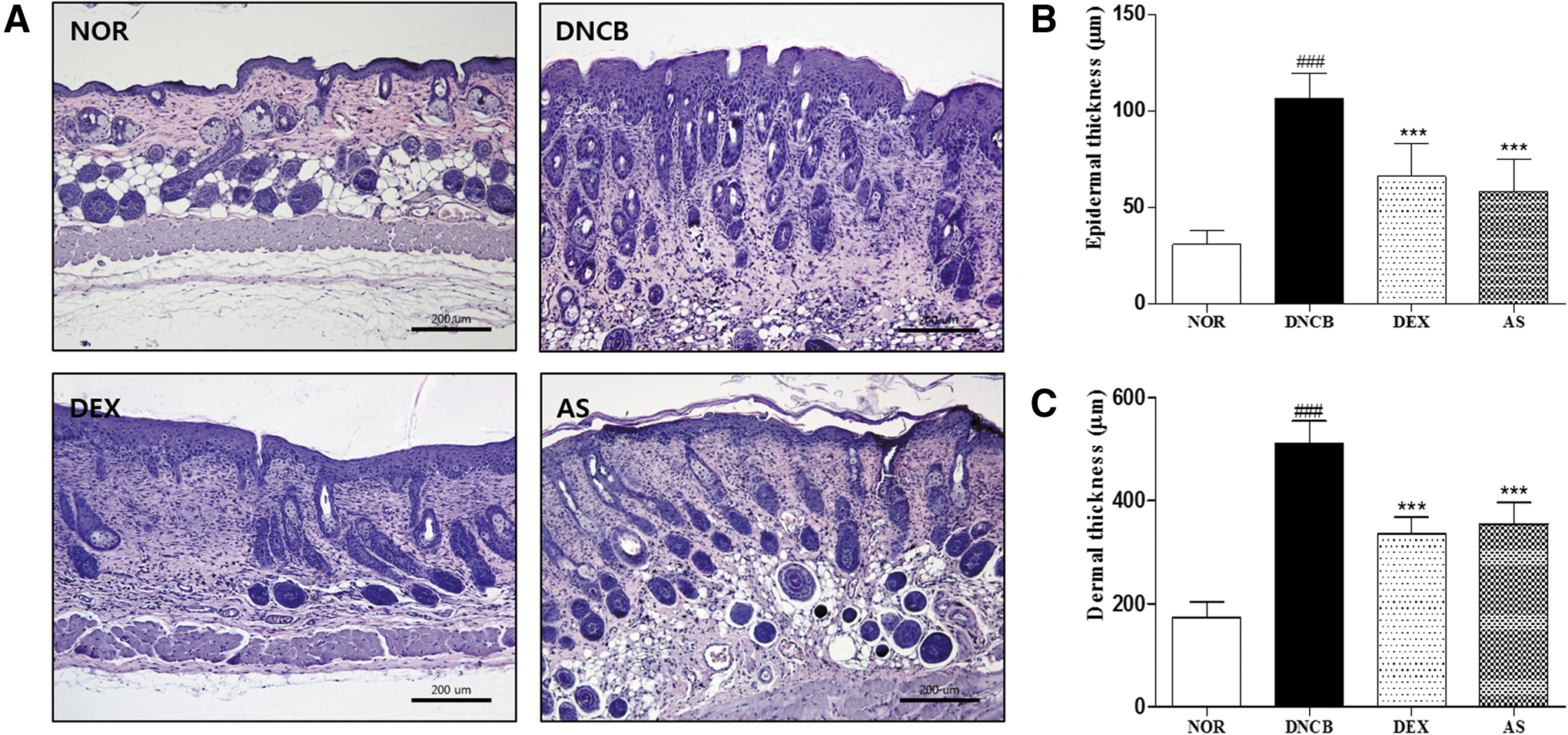
Effects of AS on DNCB-induced histopathological findings in mice.
Inhibitory effects of AS on mast cells
Using Toluidine blue staining, we also found that the number of severely infiltrating mast cells in the DNCB group was significantly higher than the NOR group, and DEX treatment. By treating AS, the numbers of mast cells in the dermis were decreased compared with the DNCB group (P < .001, Fig. 3A, B).
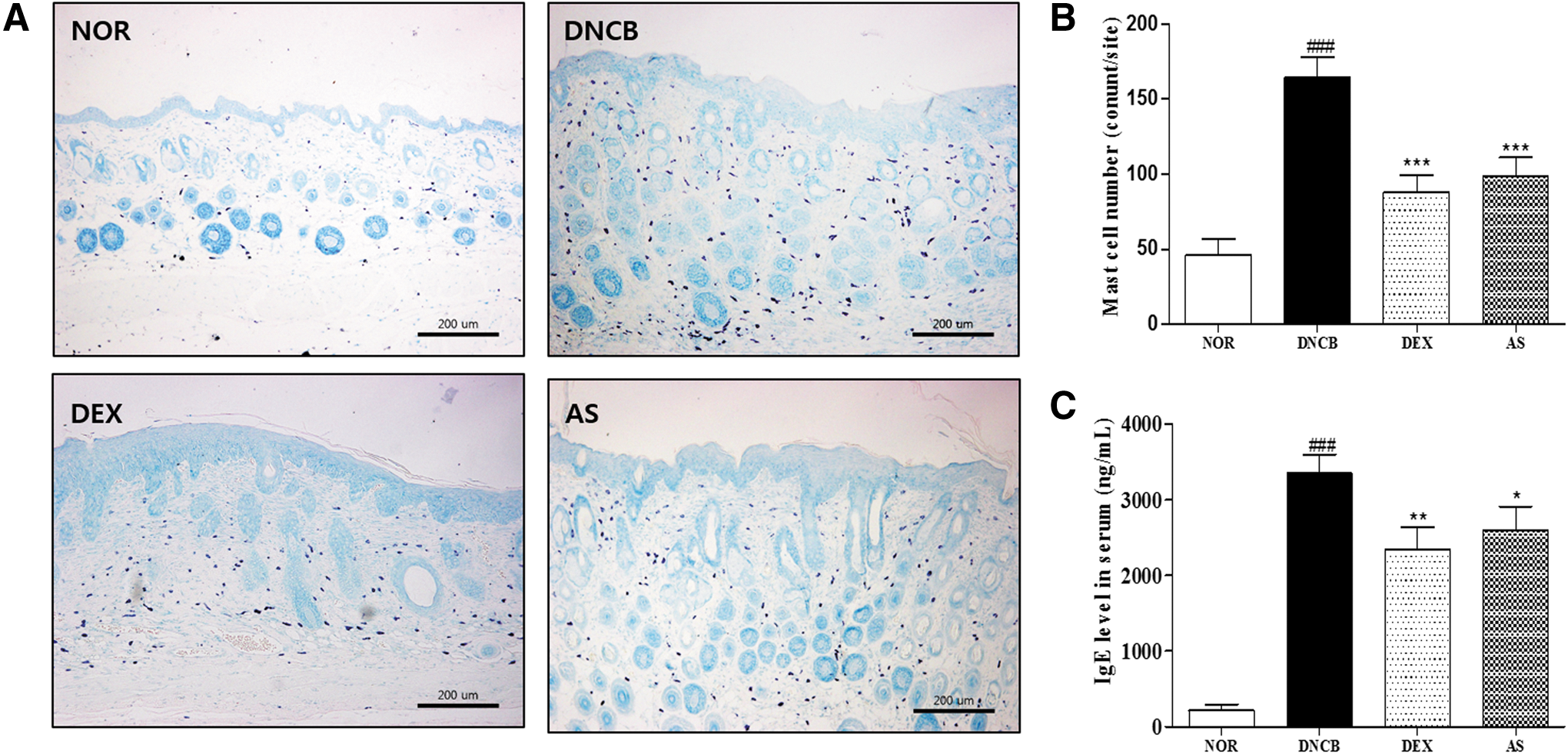
Effects of AS on DNCB-induced mast cells and serum immunoglobulin E (IgE) levels in mice.
Inhibitory effects of AS on IgE
To prove the therapeutic efficacy of AS, we measured serum IgE concentration in mice treated with AS versus DNCB-treated mice. In NOR group, serum IgE was present at a low basal level (146.7 ± 46.8 pg/mL). The total serum IgE level was significantly increased by repeated DNCB challenge (3,348.3 ± 247.1 pg/mL), and the application of DEX on AD-like skin lesions resulted in a significant decrease in serum IgE (2,337.9 ± 301.9 pg/mL). AS treatment also suppressed serum IgE levels compared to the DNCB group (2,593.0 ± 320.2 pg/mL, Fig. 3C).
Inhibitory effects of AS on the scratching behavior
To investigate the antipruritic effects of AS against DNCB-induced skin lesions, we measured the number of scratches (Fig. 4A). Repeated application of DNCB evoked a significant increase in the number of scratching bouts (146 ± 39) compared to normal mice (26 ± 6). Topical treatment of DEX attenuated these scratching behaviors compared to the DNCB group (87 ± 31). In addition, persistent application of AS led to a significant decrease of these scratching behaviors compared to the DNCB group (66 ± 16).
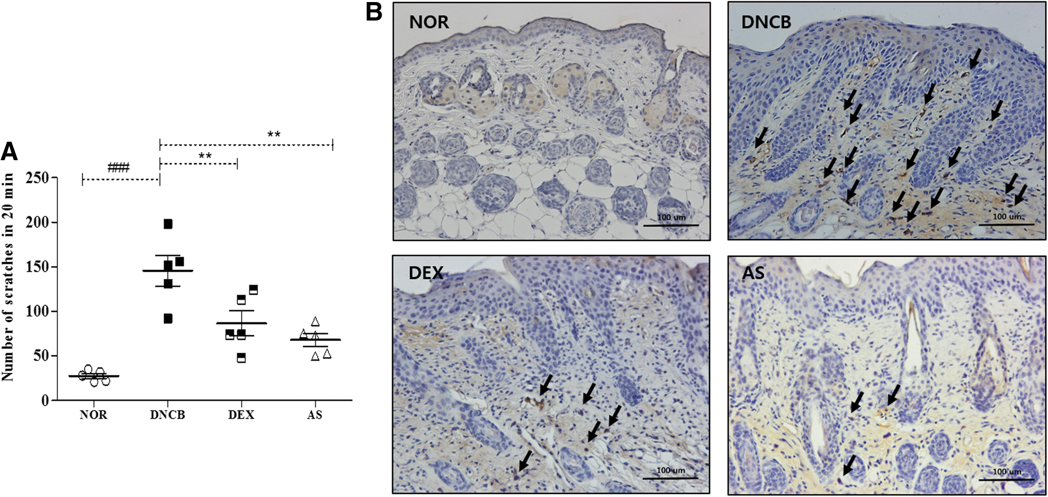
Effects of AS on DNCB-induced scratching behavior and substance P expression.
Inhibitory effects of AS on substance P expressions
The expression of substance P was correlated with the scratching responses. The DNCB-stimulated skin showed potent staining of abundant substance P. In contrast, repeated application of AS significantly suppressed the detection of substance P compared to the DNCB group. DEX treatment also significantly suppressed the increased expression of substance P (Fig. 4B).
Inhibitory effects of AS on cytokines
To assess the effect of AS on cytokine production, we measured IL-4, IL-6, TNF-α, and IFN- γ in skin homogenates. DNCB challenge increased the cytokine levels, such as IL-4, IL-6, TNF-α, and IFN-γ in the sensitized skin, whereas treatments with AS group and DEX group significantly ameliorated the increase in IL-4, IL-6, TNF-α, and IFN-γ levels compared to DNCB-induced mice (P < .001; Fig. 5).

Effects of AS on DNCB-induced cytokine levels: interleukin (IL)-4, IL-6, TNF-α, and IFN-γ in mice.
Inhibitory effects of AS on NF-κB and phospho-IκBα expressions
To determine whether AS suppresses the activation of inflammatory responses, we evaluated AS efficacy on the expressions of NF-κB and phospho-IκBα using western blot analysis. Quantitative evaluation showed that DNCB significantly increased the levels of NF-κB and phospho-IκBα. AS treatment significantly reduced nuclear protein levels of NF-κB and cytoplasmic phospho-IκBα protein levels compared to the DNCB group (by 63.4% and 44.4%, respectively). Especially, the levels of NF-κB and phospho-IκBα in AS-treated group were lower than DEX group (Fig. 6A).
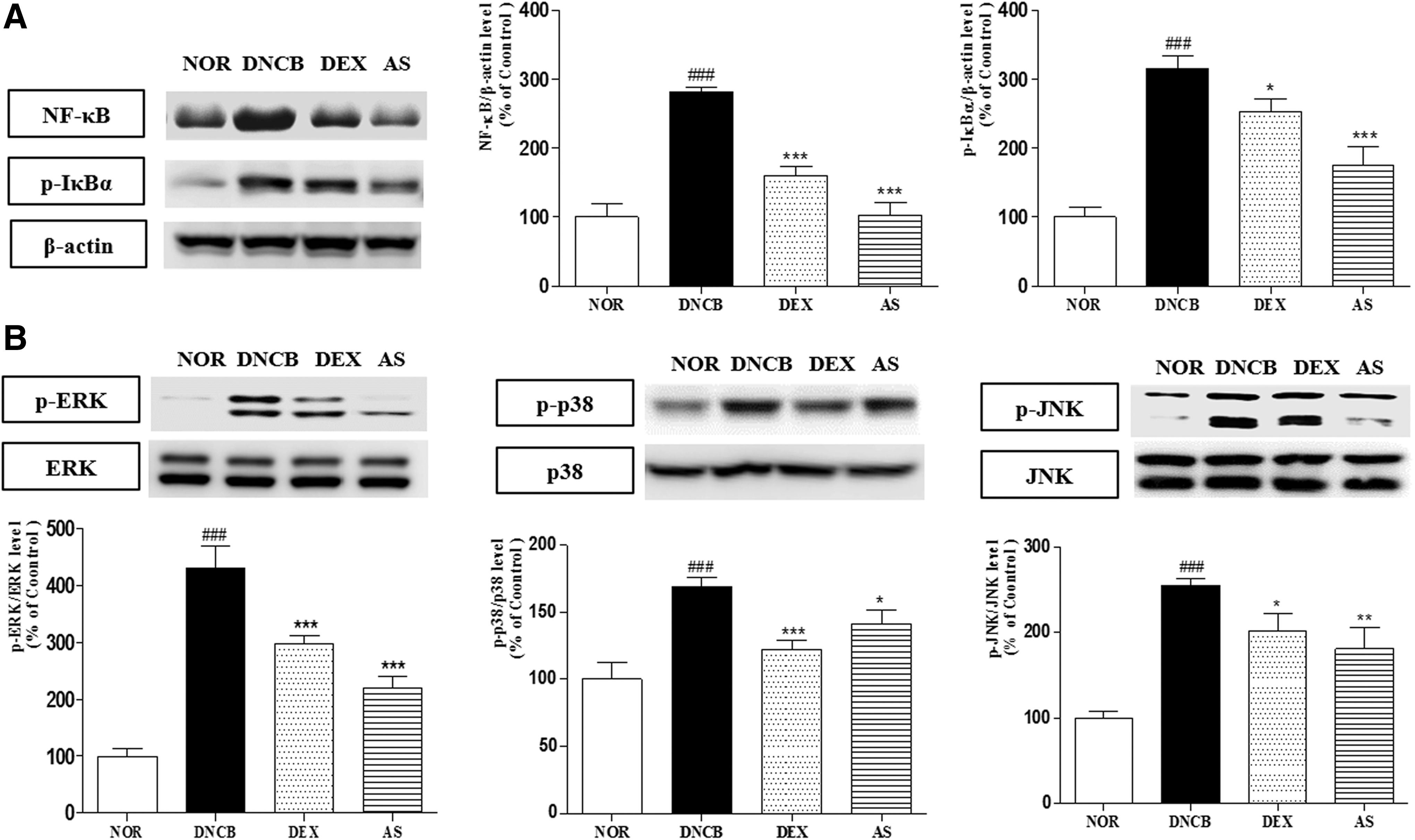
Effects of AS on DNCB-induced expressions of proinflammatory markers in mice.
Inhibitory effects of AS on MAPKs expressions
To further verify the mechanism of inhibition of inflammation, MAP kinase (ERK, p38, and JNK) levels in whole tissue lysates were evaluated. Figure 6B showed that DNCB application caused a significant elevation of MAP kinases, whereas phospho-ERK1/2, phospho-JNK, and phospho-p38 levels were decreased by DEX treatment. Topical application of AS restored phospho-ERK1/2 and phospho-JNK, and phospho-p38 levels decreased by 48.8%, 16.5%, and 29.1% (respectively).
Discussion
Skin thickness is a clinically and histologically visible symptom in AD patients, and more pronounced in lesional skin and also seen in nonlesional areas. 10,11 Epidermal thickness in AD leads to lichenification in the chronic phase and disturbed epidermal differentiation, often accompanied by increased proliferation, 10 and can cause an increase in the permeability of skin barrier. 12 Topical application of AS significantly reduced the thickness of epidermis and dermis compared to control group, demonstrating that AS might prevent lichenification and protect the skin barrier from dysfunction.
Pruritus is one of the symptoms that cause the most suffering in AD patients, in both acute and chronic phases of the disease, and the scratching behavior worsens the eczematous skin lesions in AD. 13,14 The mechanism of pruritus is known to be associated with substance P, mast cells, and IgE not only through histamine-dependent pathways, but also through the release of many other histamine-independent mediators. 15,16 In our study, the scratching behavior in the AS-treated group was significantly reduced compared with the negative control group. In addition, AS application significantly decreased the expressions of substance P and proliferation of mast cells, whereas reduction of serum IgE by AS was limited. From these results, topical application of AS might have antipruritic effects by regulating substance P and mast cells rather than serum IgE.
The productions of cytokine caused by allergen exposure is an important process in the pathogenesis of allergic skin diseases. 17 Th1/2 pathway has been proposed to play a major pathogenetic role in AD. 18 IL-4 and IL-6 are crucial factors for Th2-mediated immunity 19 and Th1 responses are characterized by the release of inflammatory and regulatory cytokines such as TNF-α and IFN-γ. 20 In this study, IL-4, IL-6, TNF-α, and IFN-γ increased in the DNCB-induced AD-like mice, but were downregulated by AS treatment. Accordingly, topical application of AS seems to exert anti-inflammatory effects by modulating both Th1- and Th2-related cytokines in AD.
Subsequently, we evaluated NF-κB, phospho-IκBα and MAPKs (ERK1/2, p38, and JNK), which are known to be associated with inflammatory cytokine secretion. NF-κB is a key transcription factor regulating Th2 cell differentiation and inflammation-related genes. 4,21 Phosphorylated and degraded IκBα induces activation of inflammatory cytokine gene expression by translocating NF-κB into the nucleus. 22 Phosphorylation of MAPKs (ERK1/2, p38, and JNK) have effects on the production of inflammatory mediators. In our study, NF-κB, phospho-IκBα, and phospho-MAPKs were significantly decreased in the AS-treated mice compared to the control group. Especially, NF-κB, phospho-IκBα, phospho-ERK, and phospho-JNK expressions were downregulated to levels lower than the positive control group. These results are consistent with a previous report demonstrating that decursin might contribute to a better understanding of anti-inflammatory potency of AS. 8
In conclusion, our study focused on the relationships between AS and mediators involved in the pathogenesis of pruritus and inflammation. AS treatment reduced the scratching behavior through regulating substance P and distribution of mast cells in the skin lesion. In addition, Th1- and Th2-related cytokines (IL-4, IL-6, TNF-α, and IFN-γ) were decreased by the application of AS. The expressions of NF-κB, phospho-IκBα, and phospho-MAPKs were also downregulated in AS-treated groups. Taken together, our findings suggest that topical application of AS on AD-like mice might have both antipruritic and anti-inflammatory effects. Further studies will be required to more precisely characterize the molecular mechanisms of AS as an intervention for AD.
Footnotes
Acknowledgment
This Research was supported by the 2014 KIOM Undergraduate Research Program funded by the Korea Institute of Oriental Medicine.
Author Disclosure Statement
No competing financial interests exist.