




Abstract
Many natural substances were screened to develop nutraceuticals that reduce menopausal symptoms. A complex of Cirsium japonicum var. maackii and Thymus vulgaris extracts, named MS-10, had significant positive effects. Under a low concentration of estrogen, which represents postmenopausal physiological conditions, MS-10 had beneficial effects on estrogen receptor-expressing MCF-7 cells by reversibly enhancing estrogen activity. In addition, in the ovariectomized rat model, changes in bone-specific alkaline phosphatase activity and osteocalcin, as well as low-density lipoprotein cholesterol and triglyceride levels were significantly decreased by MS-10. These results show that MS-10 protected bone health and reduced metabolic disturbances. Furthermore, in a clinical study, all menopausal symptoms, including hot flushes, parenthesis, insomnia, nervousness, melancholia, vertigo, fatigue, rheumatic pain, palpitations, formication, and headache, as well as colpoxerosis, were significantly improved by taking MS-10 for 90 days. Therefore, the evidence supports that MS-10 is an effective natural substance that can safely improve menopausal symptoms, including colpoxerosis.
Introduction
M
We previously screened a large number of natural substances, and their complexes, with the potential to have a positive effect on menopause. However, most of the substances did not show significant effects, even those materials exhibiting abundant phytoestrogenic, anti-inflammatory, antibacterial, and antioxidant effects. Therefore, it was difficult to identify natural substances that reduced menopausal symptoms. Fortunately, in our previous screening study, significant protective effects against menopause were identified with the natural substance MS-10, which is a complex of Cirsium japonicum var. maackii and Thymus vulgaris extracts.
Cirsium japonicum var. maackii with a pharmaceutical Latin name of Herba Cirsii is a member of the Compositae family and one of the most popular traditional medicinal herbs in Eastern Asia. 6,7 It has long been used as a herbal preparation for the treatment/control of various physical and mental disorders. 8,9 Furthermore, the water extraction of Herba Cirsii can be used to treat Weil's disease and chronic hepatitis. 7,10 Herba Cirsii flowers also have positive effects on dormancy and other beneficial effects. Pharmacological tests revealed that extractions of Herba Cirsii accelerate blood clotting, strengthen heart contractions, reduce cholesterol levels, and can be used to prevent the progression of tuberculosis. 11,12 The pharmacological effects of Herba Cirsii may be due to its rich resource of active compounds, including, but not limited to, linarin, pectolinarin, chlorogenic acid, rutin, acaciin, and luteolin. 6,13
T. vulgaris (thyme) is member of the Lamiaceae family, distributed throughout Southern Europe. Thyme leaves also possess various beneficial effects, including antiseptic, carminative, antimicrobial, and antioxidative effects. 14 –16 It also exhibits preventive and therapeutic activities against many diseases, including bronchial asthma, inflammatory diseases, hepatotoxicity, atherosclerosis, ischemic heart disease, cataracts, cancer, and insufficient sperm mobility. 17,18
MS-10 is a complex of Herba Cirsii and thyme extracts, which are botanical dietary supplements. These raw botanical herbs have gained much attention in recent years and were registered as nontoxic food materials by Korea's Ministry of Food and Drug Safety. They are well known and have been used as safe traditional herbal remedies for several hundred years in East Asia. However, the mixture of Herba Cirsii and thyme, designated MS-10, is the first identified nutraceutical for the improvement and prevention of menopausal symptoms.
In this study, we first examined whether MS-10 improves and prevents weight gain, abnormalities in lipid metabolism, and bone loss in ovariectomized rats as a preclinical model of menopause. We also report the curative and protective effects of MS-10 against menopausal and vaginal symptoms in a human clinical trial in South Korea. In addition, the safety of MS-10 was demonstrated using clinical biomarkers. Moreover, the molecular mechanisms underlying the antimenopausal effects were also investigated.
Materials and Methods
Preparation of MS-10
MS-10 was purchased from DaeHo, Inc. (Hwansung, Korea). Briefly, MS-10 was manufactured as follows: well-washed and dried whole plants of Cirsium japonicum var. maackii (Maxim.) Matsum. and the leaves of the thyme plant (T. vulgaris, Lamiaceae) were extracted with alcohol and purified, followed by a process of concentration and drying.
Cell culture
MCF-7 cells, estrogen receptor-positive human cells, were purchased from the Korean Cell Line Bank (Seoul, Korea). Cells were cultured in RPMI-1640 (GIBCO®; Thermo Fisher Scientific, Inc., Waltham, MA, USA) supplemented with 10% (v/v) fetal bovine serum (FBS; Biowest, Nuaillé, France) and 1× antibiotic–antimycotic solution (GIBCO, Thermo Fisher Scientific, Inc.) in a humidified atmosphere of 5% (v/v) CO2 and 95% air at 37°C.
Cell proliferation assay
MCF-7 cells were seeded on 96-well culture plates in a culture medium at a density of 8 × 10³ cells/well. After 24 h, the culture medium was removed and phenol red-free RPMI-1640 supplemented with 5% charcoal-dextran-stripped FBS (CD-FBS) was added. CD-FBS was prepared by treating FBS with dextran-coated charcoal (Sigma-Aldrich Co., St. Louis, MO, USA) to remove endogenous estrogens. To examine the effect of each extract on MCF-7 cell proliferation, MCF-7 cells were treated with 17β-estradiol (E2, β-estradiol; Sigma-Aldrich Co.), Herba Cirsii extract, thyme extract, or MS-10 for 72 h, and cell viability was assessed using the Alamar Blue proliferation assay. To assess cell viability upon cotreatment with MS-10 and E2, MS-10 was dissolved in distilled water and diluted with phenol red-free RPMI-1640 and then added at a volume of 100 μL/well. Cells were cotreated with various concentrations of E2 (0.001–1000 nM β-estradiol; Sigma-Aldrich Co.) and MS-10 at 37°C with 5% CO2. Cell proliferation was assessed after 72 h in culture, using an Alamar Blue proliferation assay. At appropriate times, cells were incubated with 10% (v/v) Alamar Blue (Invitrogen™; Thermo Fisher Scientific, Inc.) in the CD-FBS medium for 2 h in a humidified atmosphere (37°C in 5% CO2). Absorbance was measured at a wavelength of 570 nm using an enzyme-linked immunosorbent assay (ELISA) microplate reader (VERSAmax™; Molecular Devices, Sunnyvale, CA, USA). Background absorbance was measured at 600 nm and was then subtracted. Cell viability was defined as [(test sample count − blank count) ÷ (untreated control count − blank count)] × 100.
Western blotting
After treatment or withdrawal of MS-10, cells were washed twice with ice-cold phosphate-buffered saline (PBS) and then treated with lysis buffer, which comprised PBS containing 0.1% sodium dodecyl sulfate (SDS), followed by brief sonication. The protein concentration was determined by a bicinchoninic acid (BCA) protein assay (Pierce Chemical Co., Rockford, IL, USA). Thirty micrograms of total cellular protein was separated by 12% SDS-polyacrylamide gel electrophoresis and then transferred on nitrocellulose membranes. Blots were probed with antibodies specific to the following proteins: β-actin (1:5000; Assay Designs, Ann Arbor, MI, USA), estrogen receptor-α (ERα, 1:1000; Cell Signaling Technology, Danvers, MA, USA), and phospho-ERα (1:1000; Cell Signaling Technology). The bound antibody on each blot was detected with a horseradish peroxidase-conjugated secondary antibody and visualized by enhanced chemiluminescence (Western blot detection kit; Amersham Pharmacia Biotech, Piscataway, NJ, USA).
Animals and treatment protocol
Adult female Sprague Dawley rats (Shizuoka Laboratory Center, Inc., Shizuoka, Japan) aged 7 weeks were housed in an environmentally controlled facility at 25°C, 60% relative humidity, and a standard 12-h light/12-h dark cycle in groups of two or three per plastic cage. Rats were allowed free access to standard food and water. Rat body weight was measured on each Monday of the experimental period.
After acclimatization for 1 week, the 8-week-old female rats were anesthetized with isoflurane using a Matrx™ Quantiflex VMC® low-flow anesthesia system (Midmark, Versailles, OH, USA). Anesthesia was induced with 3% isoflurane in the same O2/N2O2 gas mix in a transparent acrylic chamber. A sham operation, where an incision was made and then sutured in the ventral area, was performed on the sham group (n = 10). In the ovariectomy group (OVX), a midline incision was made in the ventral area, and the ovaries were removed (n = 20). Rats were allowed to recuperate for 2 weeks.
Rats were assigned to the following three experimental groups (n = 10/group): sham (non-OVX rats, dextrin), OVX (OVX rats; 50 mg/kg dextrin once per day), and OVX-MS-10 (OVX rats; 50 mg/kg MS-10 once per day). Samples of MS-10 were prepared before use by dissolving in deionized water and administered orally (50 mg/kg) once per day for 8 weeks.
All experimental procedures involving animals were performed in accordance with a protocol that was approved by the Institutional Animal Care and Use Committee (IACUC) of Chung-Ang University (Approval No. 14-0010).
Blood and organ harvesting (urinary bladder, uterus, abdominal visceral fat, and femurs)
At the end of the treatment period, blood was collected by cardiac puncture, and the uterus and urinary bladder were excised from each rat and immediately weighed. Femurs were dissected and stored in 4% paraformaldehyde solution containing 0.5% KCl (pH 7.5) at −4°C for measurement of bone mineral density (BMD).
BMD measurements
Rat femur bones were evaluated in vivo after surgical removal. Femur BMD was measured by dual-energy X-ray absorptiometry using a GE Lunar PIXImus system (GE Healthcare, Madison, WI, USA) equipped with appropriate software for BMD assessment in small laboratory animals. Calibration of the instrument was conducted as recommended by the manufacturer. The BMD (0.0752 g/cm2) and percentage fat composition (18.3%) of the quality control phantom were also verified each time the instrument was switched on. All rats were placed in the same direction.
Serum analyses
Serum osteocalcin (OC) levels were determined using an OC ELISA kit (Biomedical Technologies, Inc., Stoughton, MA, USA) with a Spectra Max 190 (Molecular Devices) at a wavelength of 450 nm. Serum aspartate aminotransferase (AST), alanine aminotransferase (ALT), bone-specific alkaline phosphatase (ALP), triglyceride (TG), total cholesterol (TC), and high-density lipoprotein cholesterol (HDL-C) levels were assayed using an ELISA kit (Roche, Basel, Switzerland) and quantified with a Hitachi 7600 automatic analyzer (Hitachi Co., Tokyo, Japan).
Clinical experiments
Middle-aged women were recruited for the treatment groups and a matching placebo group at Chung-Ang University Medical Center, Seoul, Korea, during December 2012 through a posted advertisement. Females were excluded from the study if they presented with any of the following conditions: metabolic diseases, urethral stricture, neurogenic bladder dysfunction, cardiovascular events, thyroid disease during the past year, or the use of medications containing sex hormones.
Demographics and other clinical data were obtained through the Kupperman's Index (KI) questionnaire. The study protocol was approved by the institutional review boards of the participating hospitals. Volunteers were randomly distributed into one of three groups by a pharmacist who was unaware of any subject clinical information. Group 1 [flour-fed controls; n = 10] was the placebo group, group 2 (n = 10) received 300 mg of MS-10 daily, and group 3 (n = 10) received 500 mg of MS-10 daily. The color and shape of the placebo was the same as the test material. During the study period, the investigator, rater, and study participants were double blinded. KI data were compiled before the MS-10 regimen and after the 12-week regimen.
Statistical analyses
A one-way ANOVA was used to compare demographic variables between the groups treated with 300 mg of MS-10, 500 mg of MS-10, and placebo. The paired t-test was used to determine changes in cell viability, body weight, hormone levels, lipoprotein concentrations, ALT and AST levels, and KI before and after MS-10 administration. All statistical analyses were performed using SPSS 11.0 (SPSS, Chicago, IL, USA) with a (two-tailed) P < .05 considered statistically significant.
Results
Effects of MS-10 on MCF-7 cell proliferation
To evaluate the effects of MS-10 and its individual components (extracts of Herba Cirsii or thyme) on cell proliferation, MCF-7 cells expressing estrogen receptors were treated with various concentrations of estrogen, MS-10, or each component of MS-10. Treatment with 10 nM estrogen enhanced cell proliferation to 160%. However, treatment with MS-10 or its individual components did not affect cell proliferation (Fig. 1A).
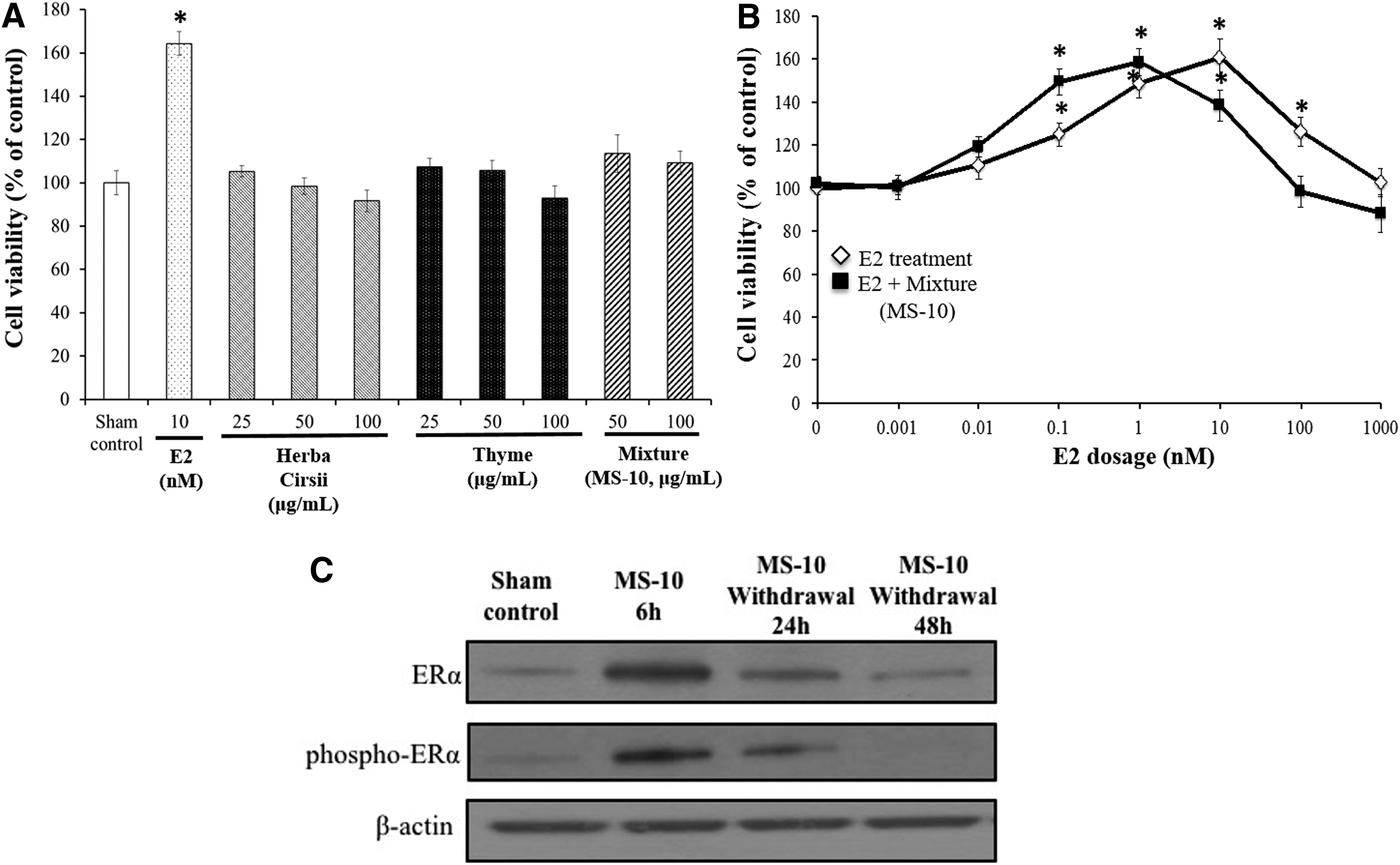
Growth-stimulating effects of extracts as measured by the Alamar Blue assay
Next, the dose-dependent effects of estrogen (0.001–1000 nM) on cell proliferation were investigated. Physiological concentrations of estrogen increased cell viability, whereas a high dose of estrogen inhibited abnormal cell growth. 19 Consistently, cell proliferation was increased dose dependently by treatment with estrogen up to a concentration of 10 nM and gradually decreased at higher concentrations (Fig. 1B). Therefore, it was reasonably assumed that a low estrogen concentration of around 0.1 nM represented physiological menopausal conditions and a concentration of about 1 nM represented premenopausal conditions in humans.
Interestingly, in the presence of MS-10 (50 μg/mL), even with a low concentration of estrogen (lower than 1 nM), cell proliferation was increased, and cell proliferation was decreased at a high concentration of estrogen (higher than 10 nM). These results showed that MS-10 facilitated the physiological role of estrogen when its concentration was low to normal, which represents the postmenopausal to premenopausal situation. The reduction in cell proliferation by MS-10 treatment when the concentration of estrogen was high demonstrates that MS-10 prevented the overproliferation of cells induced by an abnormal concentration of estrogen, such as arises when estrogen therapy is administered.
To evaluate the relevance of ERα in the mechanism underlying the increase in cell proliferation by MS-10, the expression levels and phosphorylation of ERα were assayed in MCF-7 cells. At 6 h after MS-10 treatment, expression of ERα was increased, and it steadily decreased to the control level over time (up to 48 h) after MS-10 was withdrawn (Fig. 1C). Similiarly, activation of ERα was detected upon MS-10 treatment. Phosphorylation of ERα was detected in the presence of MS-10, and this gradually decreased to the control level over time when MS-10 was washed out (Fig. 1C). These results showed that MS-10 facilitates the physiological role of estrogen when the estrogen concentration is low by increasing ERα expression and enhancing its activity in a reversible and safe manner.
Effects of MS-10 on body weight
Weight gain following sex hormone deficiency is a general characteristic of menopause. The body weight of normal control rats reached an average of 263.3 g after 8 weeks, whereas the body weight of OVX rats was 335.7 g. However, MS-10-treated OVX rats exhibited a significantly lower body weight (less than 290 g) than MS-10-untreated controls (Fig. 2A). Figure 2B shows the increase in body weight after 8 weeks of growth. After 8 weeks, in contrast to the ∼90 g increase in normal conditions, OVX rats exhibited a ∼170 g increase in body weight; thus an undesirable increase of ∼80 g was observed. However, a 140 g weight increase was noted in MS-10-treated OVX rats. Thus, the undesirable weight was significantly reduced to less than 50 g (Fig. 2B), equivalent to a ∼36% reduction in undesirable body weight.
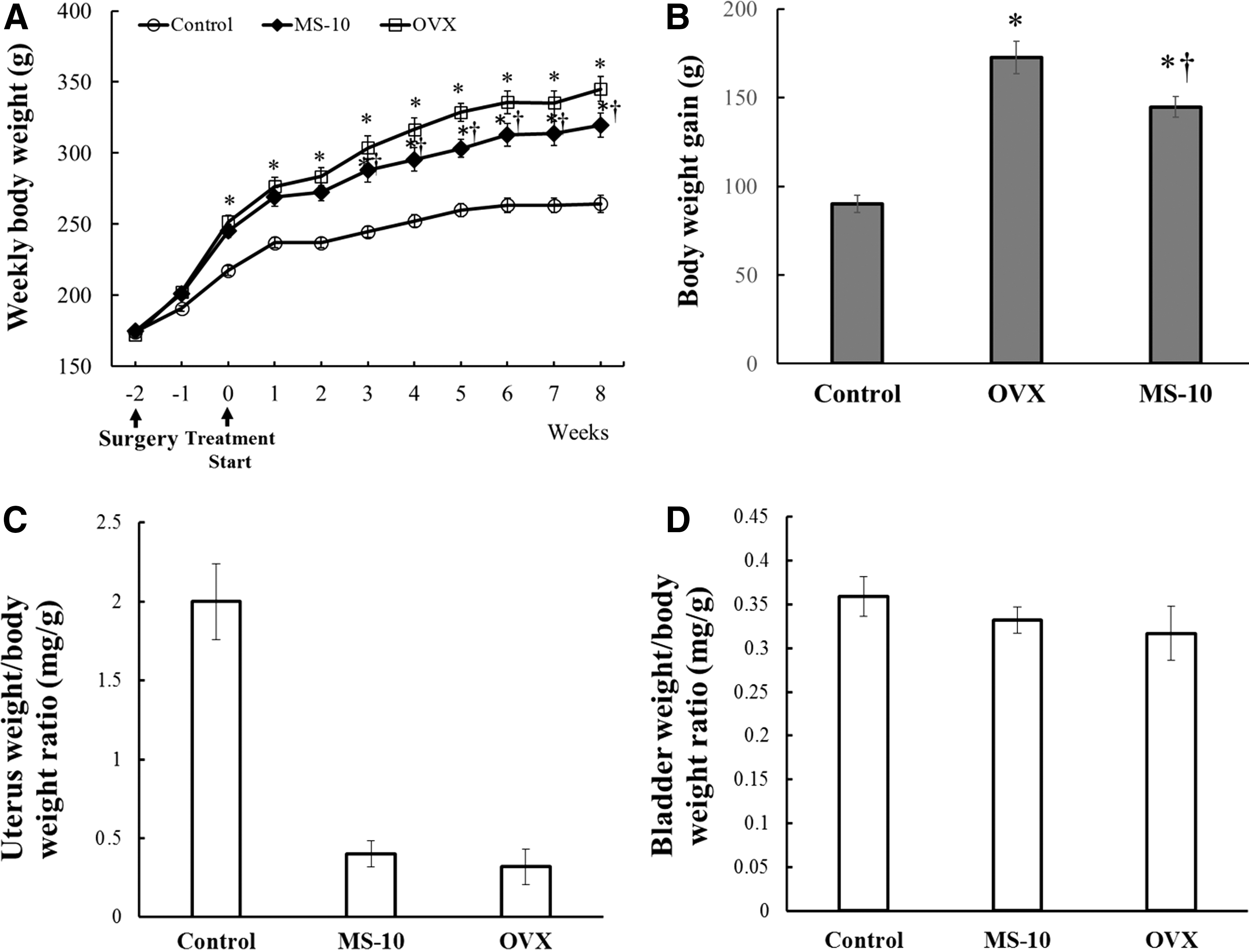
Effects of MS-10 on weekly body weight
Next, we weighed the uterus and bladder, which are among the organs that are most affected by an increased estrogen concentration. MS-10 treatment did not affect the uterus or bladder weight in comparison with untreated OVX animal model (Fig. 2C–D). These results indicate that MS-10 is safe and does not cause any abnormal physiological changes.
Effects of MS-10 on lipid concentrations
It is well known that serum lipid concentrations are increased during menopause. Thus, serum TG, TC, low-density lipoprotein cholesterol (LDL-C), and HDL-C levels were measured. Control and OVX rat TG levels were ∼80 and 100 mg/dL, respectively. The TG levels of MS-10-treated OVX rats were almost the same as those of the normal control (Fig. 3A), indicating that MS-10 prevented almost 100% of the undesirable increase in TG levels. Control and OVX rat TC levels were ∼92 and 113 mg/dL, respectively. The TC levels of MS-10-treated OVX rats were almost the same as those of the normal control (Fig. 3B), indicating that MS-10 prevented almost 100% of the undesirable increase in TC levels. LDL-C levels in control and OVX rats were ∼14 and 38 mg/dL, respectively, while those of MS-10-treated OVX rats were ∼20 mg/dL (Fig. 3C). Thus, MS-10 prevented almost 75% of the undesirable increase in LDL-C. These results indicate that in MS-10-treated rats, TG, TC, and LDL-C levels improved by ∼24%, 16.5%, and 47.5%, respectively, to those of the menopausal animal model's levels (Fig. 3D).
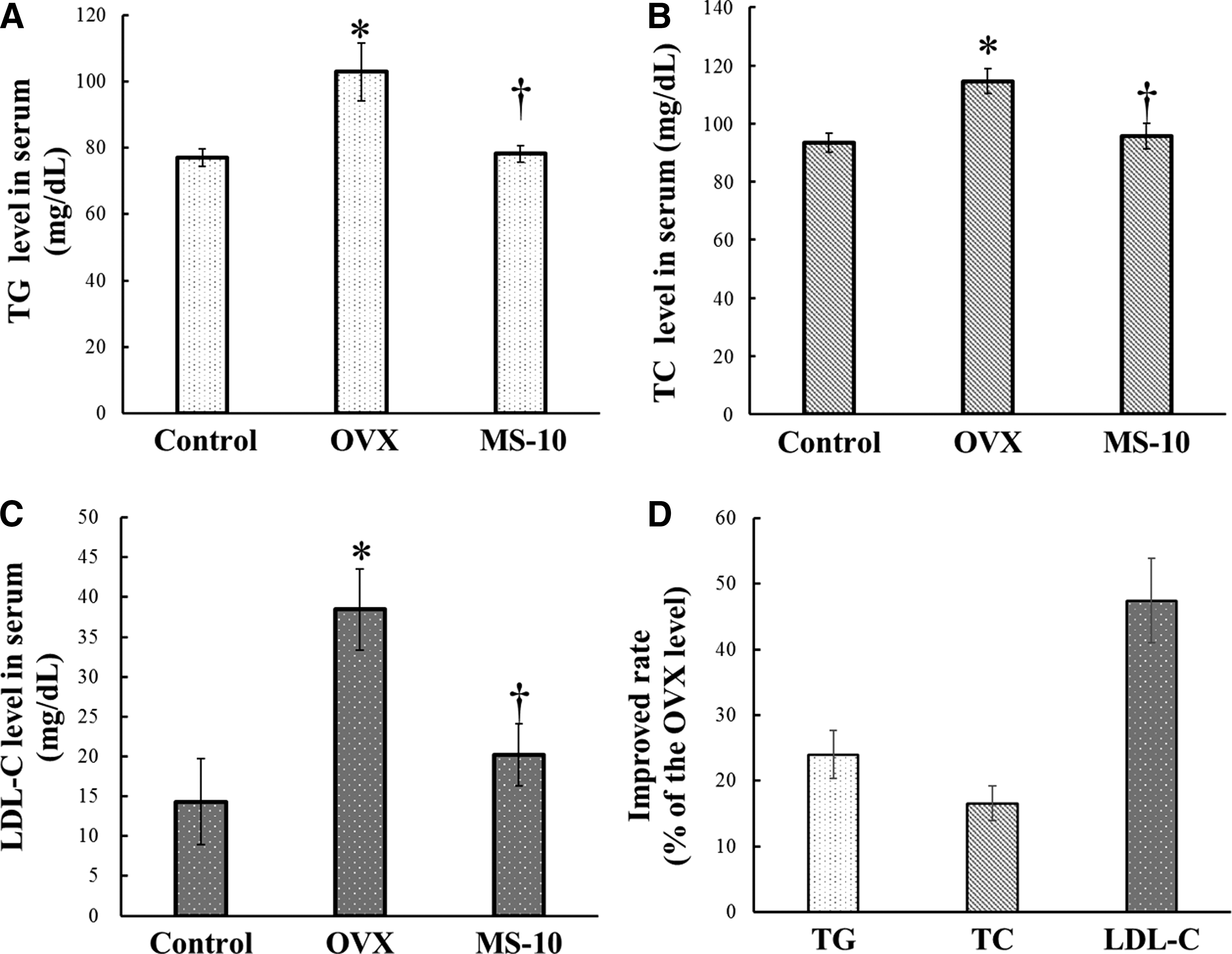
Effects of MS-10 on serum lipoprotein concentrations in OVX rats. Total lipoprotein concentrations were measured in the sham group, the OVX group, and OVX rats that had been orally administered 50 mg/kg/day MS-10 for 60 days.
In contrast to the results above, HDL-C concentrations did not show significant differences between MS-10-treated and MS-10-untreated OVX rats (data not shown).
Effects of MS-10 on serum AST and ALT levels
As shown in Figure 4, estrogen-deficient OVX rats exhibited a significantly higher liver enzyme activity (AST and ALT) than normal controls. AST and ALT levels were 65.00 ± 3.04 IU/L and 115.67 ± 13.11 IU/L, respectively, in the control group, but these levels were significantly higher at 123.56 ± 15.34 IU/L and 151.46 ± 14.67 IU/L, respectively, in the OVX group. However, AST and ALT levels were significantly lower in MS-10-treated OVX rats at 63.01 ± 2.79 IU/L and 99.00 ± 8.61 IU/L, respectively. These results indicate that MS-10 is relatively safe and may exert protective effects on the liver.
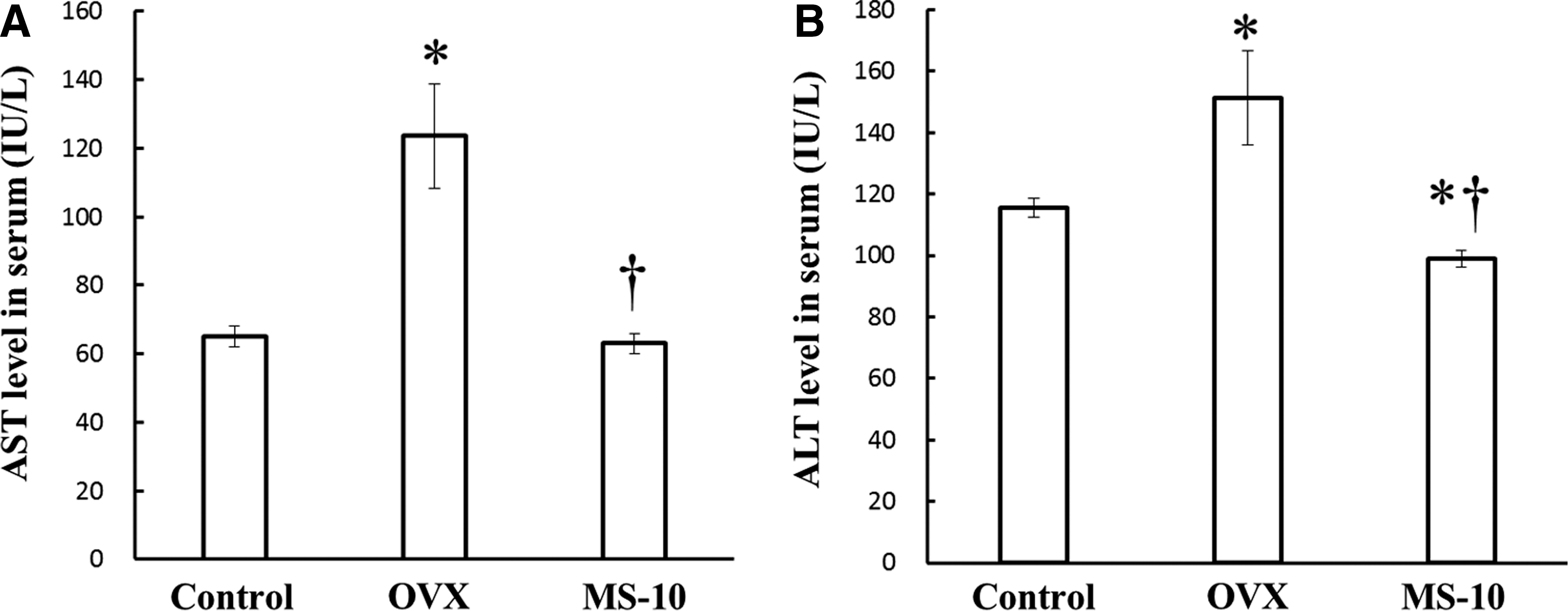
Effects of MS-10 on serum aspartate aminotransferase (AST) and alanine aminotransferase (ALT) concentrations in OVX rats. The concentrations of AST
Effects of MS-10 on BMD levels
It is known that BMD levels are reduced in menopausal individuals. To determine if MS-10 could ameliorate bone loss in menopause, BMD was examined. BMD levels in control and OVX rats were ∼0.25 and 0.215 mg/cm2, respectively. Interestingly, the BMD level of MS-10-treated OVX rats was, ∼0.26 mg/cm2 (Fig. 5). These results indicate that MS-10 prevented the OVX-induced decrease in BMD.

Femur bone mineral density (BMD) in each experimental group. The femur BMD was measured in the sham group, the OVX group, and OVX rats that had been orally administered 50 mg/kg/day MS-10 for 60 days. All data are the mean ± SE (n = 10 per group). †P < .05, versus OVX rats according to a one-way ANOVA with Bonferroni correction; *P < .05, versus sham control rats.
Effects of MS-10 on OC and ALP levels
In many previous reports, ALP and OC levels in OVX rats were significantly higher than those of normal controls. This indicates an increase in the bone turnover rate in OVX rats.
As expected, OC serum concentrations were markedly higher in OVX rats (∼85.71 ng/mL) than in control rats (41.45 ng/mL). However, MS-10 reduced the increase in OC levels in OVX rats to a concentration of 58.26 ng/mL (Fig. 6A). Thus, MS-10 treatment improved bone health by ∼32% compared with that of the OVX group (Fig. 6C).

Effects of MS-10 on
In a similar manner to OC, serum ALP concentrations were markedly increased in OVX rats (∼168 U/L) by more than thrice that of the control (∼50 U/L). However, MS-10 treatment reduced ALP levels to ∼83 U/L (Fig. 6B). Thus, MS-10 treatment improved bone health by ∼50.6% compared to that of the OVX group (Fig. 6C). These results strongly suggest that MS-10 may prevent the increase in bone turnover rate during menopause.
MS-10 improves menopausal symptoms in women
To determine whether MS-10 could reduce menopausal symptoms in humans, a clinical study was conducted with middle-aged women using the KI clinical questionnaire and self-reported vaginal symptoms (dryness) with a double-blind protocol for 12 weeks. The age, weight, and height of the subjects at baseline are shown in Table 1. There were no statistically significant differences among the three groups. In the placebo controls, the KI scores (menopausal symptoms) were unchanged. However, KI scores declined from 25 to 20 in the 300 mg/day MS-10-treated group and from 24.7 to 15 in the 500 mg/day MS-10-treated group (Fig. 7A). These results indicate that MS-10 improved menopausal symptoms dose dependently by as much as 20% (300 mg/day) and 40% (500 mg/day) (Fig. 7B). Moreover, 300 and 500 mg/day MS-10 effectively improved vaginal dryness (colpoxerosis score) by about 15% and 25%, respectively (Fig. 7C, D).

Improved Kupperman's Index (KI) and colpoxerosis scores following administration of 500 and 300 mg/day of MS-10. The KI questionnaire was completed to evaluate menopausal symptoms at 0 and 12 weeks. KI
Values represent mean ± standard error of mean.
Student's t-test, Paired t-test, Two-tailed P-value, pre versus after.
One-Way ANOVA, Two-tailed P-value, Placebo versus MS-10 300 mg/day versus MS-10 500 mg/day.
Discussion
Menopause causes unpleasant symptoms in most women and is associated with increased risk of metabolic syndrome, obesity, insulin resistance, diabetes, and osteoporosis. 20,21 Estrogen treatment can reverse the effects of menopause. However, estrogen therapy increases the risk of various cancers, including endometrial, breast, and ovarian cancers. 22 From this perspective, safe natural substances capable of reducing menopausal symptoms in aging women are required. Therefore, we screened a large number of natural substances for positive effects on menopause. However, most natural substances failed to meet sufficient safety and efficacy standards. Finally, we identified a safe natural substance with significant beneficial effects on menopause, termed MS-10, which is a combined extract of Herba Cirsii and thyme. This study is unique and impressive as it identified an effective treatment for menopausal symptoms.
Our in vitro results showed that MS-10 retained the positive effects of low concentrations of estrogen, which occur in perimenopausal and postmenopausal periods. Menopause is associated with a marked reduction in estrogen levels following aging of the ovary. Therefore, an increasing estrogen activity with MS-10 may be a safe and effective strategy for improving menopausal symptoms.
Similarly, MS-10 may also reduce the possible undesired negative effects of high concentrations of estrogen. Temporary abnormal increases in estrogen can occur during estrogen therapy because exogenously supplied estrogen can disturb the hormonal balance. Our data suggest that MS-10 may be helpful for reducing the effects of excess estrogen by increasing expression and phosphorylation of ER.
OVX is a well-known experimental model of menopausal symptoms and mimics estrogen deficiency in females. 23 This animal model exhibits a marked increase in body weight, while administration of estrogen prevents this effect.
In the present study, OVX-control rats exhibited a significantly higher body weight than sham-operated rats 8 weeks after surgery, whereas OVX-MS-10-treated rats exhibited a marked decrease in body weight compared to that of the OVX-control group. Notably, there were no significant differences in food intake between OVX-MS-10, OVX-control, and sham-operated rats. These results suggest that administration of MS-10 may also have beneficial effects on obesity caused by estrogen deficiency.
Consistent with previous reports, we noted undesirable lipid metabolic disturbances (increased TC, LDL-C, and TG, and decreased HDL-C levels) in OVX rats. Lipid disturbances are a major factor underlying metabolic syndrome and vascular disease. Notably, MS-10 effectively ameliorated these lipid metabolic disturbances. To the best of our knowledge, this is the first report of a positive effect of a natural substance (MS-10) on lipid metabolism in a menopause model.
Bone loss caused by estrogen deficiency in both experimental animals and humans is primarily due to an increase in osteoclastic bone resorption. 24,25 Decreased bone mass (osteoporosis) is the main risk factor for fractures. Femur mass and BMD were decreased in the menopause model, and MS-10 suppressed the decrease in femur mass and BMD. Since MS-10 increased femur BMD, MS-10 may also increase femur calcium and inorganic phosphorus content. BMD loss was accompanied by a significant increase in ALP and OC levels. The ALP activity is a biomarker of osteoblastic activity and bone remodeling, 26 –28 and serum OC levels are a marker of bone formation. 28,29 Our results strongly suggest that MS-10 has a positive effect on bone resorption during menopause and may promote bone health.
In the clinical study, a positive effect of MS-10 was identified. Notably, all menopausal symptoms, including hot flushes, parenthesis, insomnia, nervousness, melancholia, vertigo, fatigue, rheumatic pain, palpitation, formication, and headache were improved. Furthermore, vaginal dryness was also significantly improved.
In conclusion, our results clearly demonstrate that MS-10 improves menopausal symptoms. Therefore, this study provides convincing evidence that MS-10 is an effective nutraceutical agent for menopausal symptoms in women.
Footnotes
Acknowledgment
This research was supported by the Agriculture, Food and Rural Affairs Research Center Support Program (714001-07), Ministry of Agriculture, Food and Rural Affairs.
Author Disclosure Statement
No competing financial interests exist.